El proceso de formación del hueso se llama osificación. Los LOS Neisseria 2 tipos de osificación son la osificación intramembranosa, en EN Erythema nodosum is an immune-mediated panniculitis (inflammation of the subcutaneous fat) caused by a type IV (delayed-type) hypersensitivity reaction. It commonly manifests in young women as tender, erythematous nodules on the shins. Erythema Nodosum la que el hueso se desarrolla directamente a partir de las células del mesénquima, y la osificación endocondral, en EN Erythema nodosum is an immune-mediated panniculitis (inflammation of the subcutaneous fat) caused by a type IV (delayed-type) hypersensitivity reaction. It commonly manifests in young women as tender, erythematous nodules on the shins. Erythema Nodosum la que se crea primero un modelo de cartílago hialino y luego se sustituye por hueso. El hueso sigue creciendo hasta la edad adulta temprana en EN Erythema nodosum is an immune-mediated panniculitis (inflammation of the subcutaneous fat) caused by a type IV (delayed-type) hypersensitivity reaction. It commonly manifests in young women as tender, erythematous nodules on the shins. Erythema Nodosum las placas epifisarias, donde los LOS Neisseria condrocitos siguen dividiéndose, muriendo y siendo sustituidos por hueso mineralizado. La mineralización ósea se produce porque los LOS Neisseria osteoblastos permiten que se acumulen altos niveles de calcio y fosfato por encima de los LOS Neisseria niveles umbrales críticos dentro del hueso.
Last updated: Mar 27, 2025
La formación de hueso se llama osificación u osteogénesis.
Los LOS Neisseria 2 tipos principales de osificación son:
Los LOS Neisseria 2 tipos principales de hueso son el hueso compacto y el hueso esponjoso.
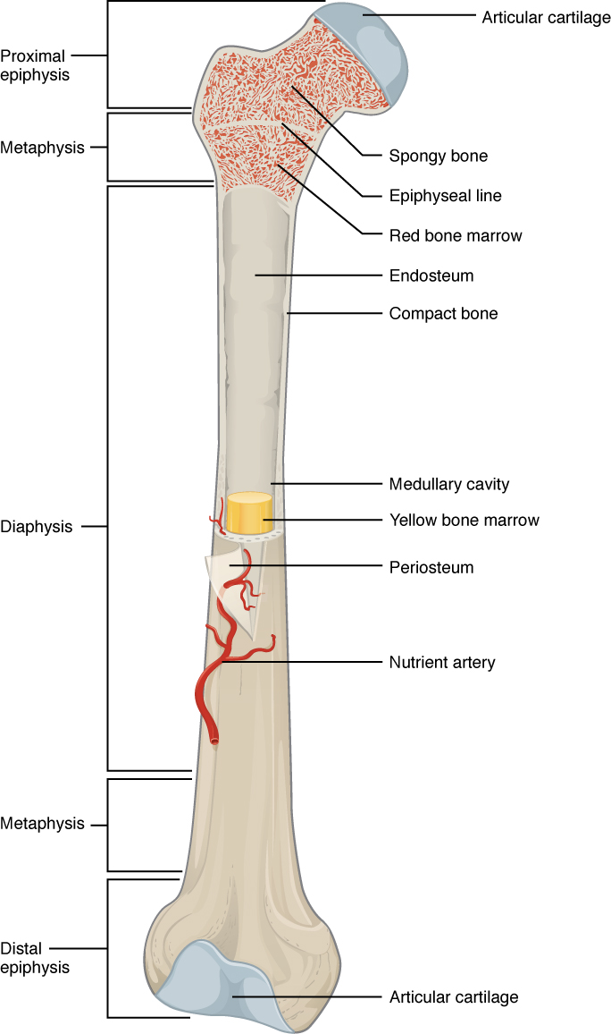
Estructura de un hueso largo, en este caso el fémur, que es el hueso principal del muslo.
Imagen: “A typical long bone shows the gross anatomical characteristics of bone” por OpenStax College. Licencia: CC BY 4.0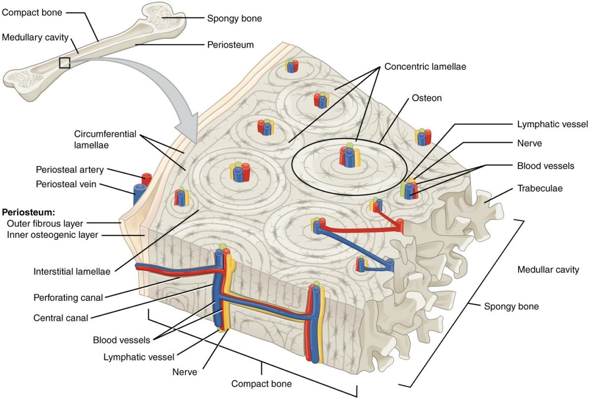
Estructura microscópica del hueso compacto
Imagen: “This cross-sectional view of compact bone shows the basic structural unit, the osteon” por OpenStax College. Licencia: CC BY 4.0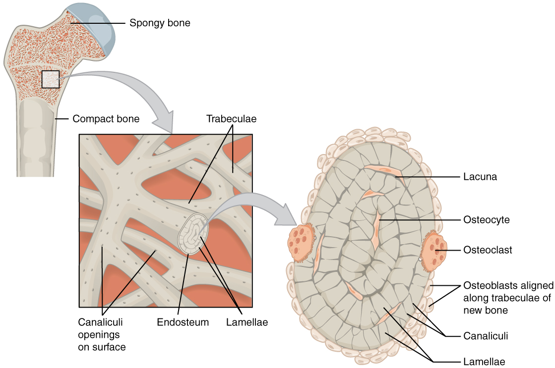
Estructura microscópica del hueso esponjoso
Imagen: “Spongy bone is composed of trabeculae that contain the osteocytes. Red marrow fills the spaces in some bones.” por OpenStax College. Licencia: CC BY 4.0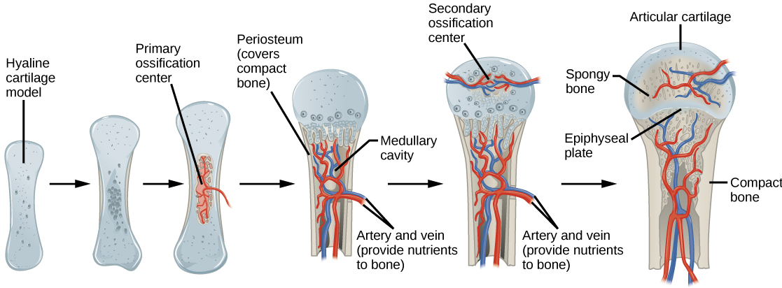
Proceso de osificación endocondral
Imagen: “Process of endochondral ossification” por CNX OpenStax. Licencia: CC BY 4.0La osificación intramembranosa es una conversión directa de células mesenquimales en EN Erythema nodosum is an immune-mediated panniculitis (inflammation of the subcutaneous fat) caused by a type IV (delayed-type) hypersensitivity reaction. It commonly manifests in young women as tender, erythematous nodules on the shins. Erythema Nodosum tejido óseo.
Estos huesos tienen una capa intermedia de hueso esponjoso entre capas de hueso compacto:
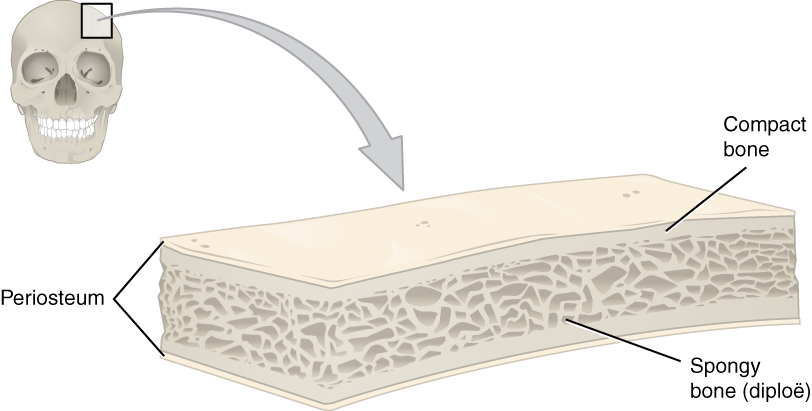
Estructura de un hueso plano
Imagen: “This cross-section of a flat bone shows the spongy bone (diploë) lined on either side by a layer of compact bone” por OpenStax College. Licencia: CC BY 4.0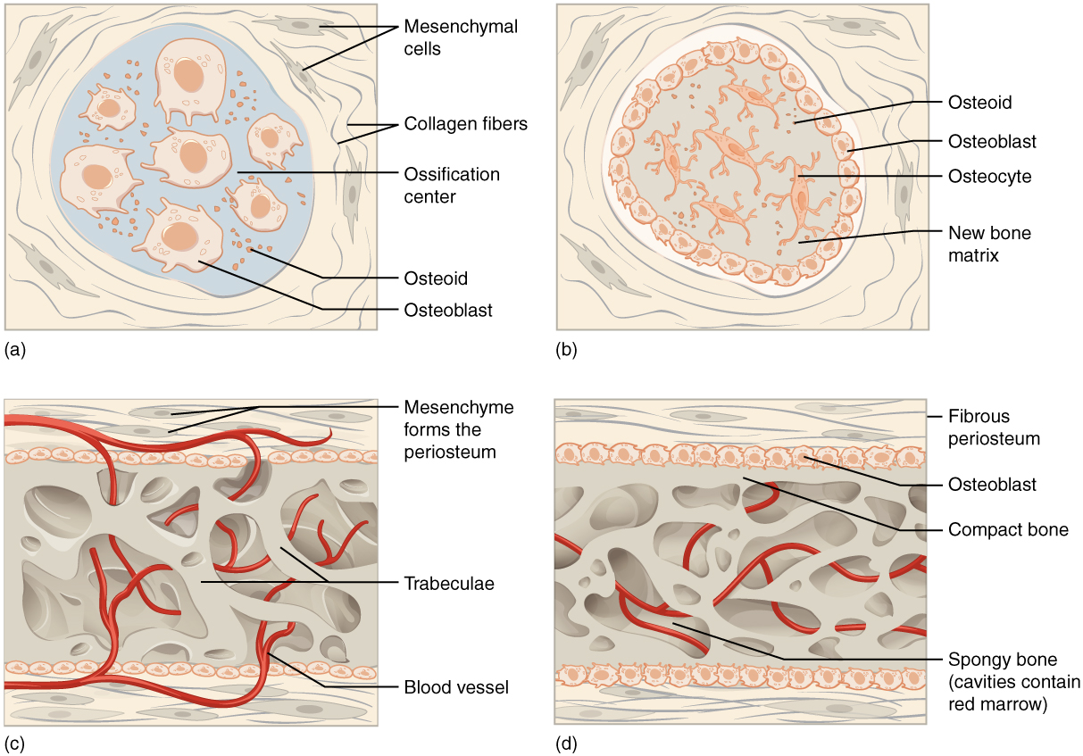
Proceso de osificación intramembranosa
Imagen: “Intramembranous ossification follows four steps. (a) Mesenchymal cells group into clusters, and ossification centers form. (b) Secreted osteoid traps osteoblasts, which then become osteocytes. (c) Trabecular matrix and periosteum form. (d) Compact bone develops superficial to the trabecular bone, and crowded blood vessels condense into red marrow.” por OpenStax College. Licencia: CC BY 4.0Las placas epifisarias se encuentran en EN Erythema nodosum is an immune-mediated panniculitis (inflammation of the subcutaneous fat) caused by a type IV (delayed-type) hypersensitivity reaction. It commonly manifests in young women as tender, erythematous nodules on the shins. Erythema Nodosum la metáfisis de los LOS Neisseria huesos largos, la región de transición entre la diáfisis (eje) y la epífisis (extremos). Existen cinco zonas histológicas distintas:
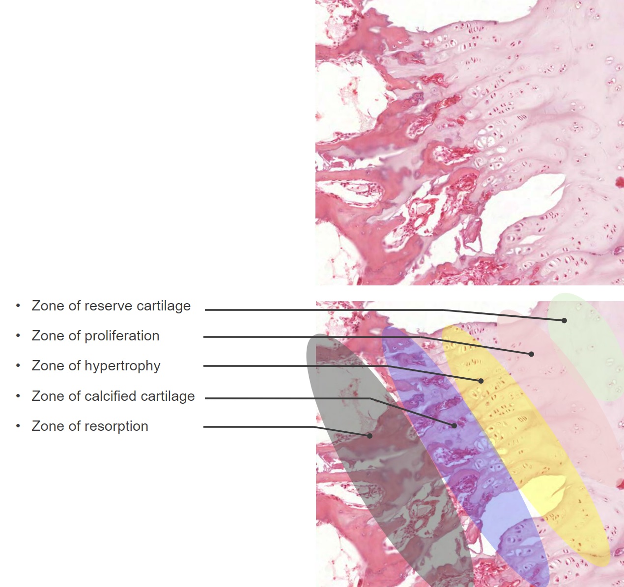
Zonas histológicas de las placas epifisarias
Imagen por Lecturio.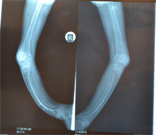
Raquitismo
Imagen: “X-rays of both lower limbs showing severe bowing of the legs and diffuse osteopenia. It also shows dense transverse lines in the tibia suggestive of looser’s zones indicative of rickets” por Al-Sharafi BA et al. Licencia: CC BY 4.0