


O humor Humor Defense Mechanisms vítreo é uma substância transparente e gelatinosa que está presente no espaço entre o cristalino e a retina Retina The ten-layered nervous tissue membrane of the eye. It is continuous with the optic nerve and receives images of external objects and transmits visual impulses to the brain. Its outer surface is in contact with the choroid and the inner surface with the vitreous body. The outermost layer is pigmented, whereas the inner nine layers are transparent. Eye: Anatomy, proporcionando estabilidade estrutural e mantendo a forma do olho. Algumas patologias podem afetar o humor Humor Defense Mechanisms vítreo, nomeadamente o descolamento posterior do vítreo, hemovítreo, sínquise cintilante, hialose asteróide ou persistência da vasculatura fetal. Estas patologias podem ser assintomáticas ou apresentar-se com moscas volantes no campo visual, fotópsias e diminuição da acuidade visual. A fundoscopia e o exame na lâmpada de fenda são frequentemente utilizados no diagnóstico destas doenças. As formas de tratamento dependem da patologia e da gravidade do quadro, e vão desde manter uma atitude expectante até métodos de correção da visão e cirurgia.
Last updated: Nov 12, 2025
O humor Humor Defense Mechanisms vítreo é a substância presente entre o cristalino e a retina Retina The ten-layered nervous tissue membrane of the eye. It is continuous with the optic nerve and receives images of external objects and transmits visual impulses to the brain. Its outer surface is in contact with the choroid and the inner surface with the vitreous body. The outermost layer is pigmented, whereas the inner nine layers are transparent. Eye: Anatomy.
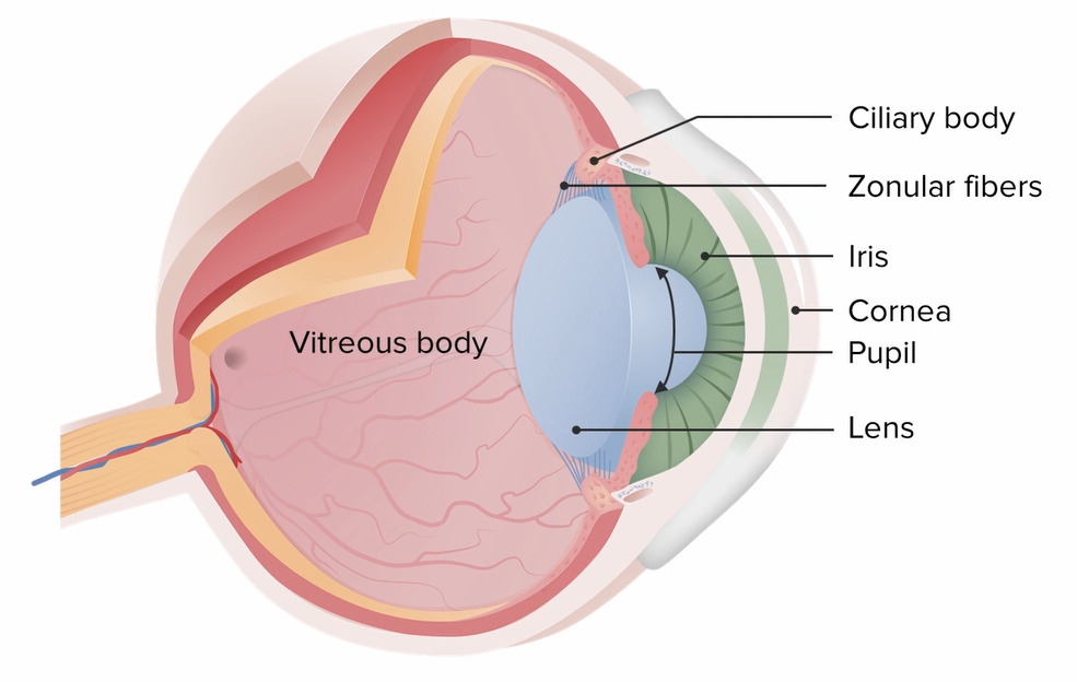
Diagrama da anatomia do olho.
Image by Lecturio.O descolamento posterior do vítreo consiste na separação do humor Humor Defense Mechanisms vítreo da membrana limitante interna da retina Retina The ten-layered nervous tissue membrane of the eye. It is continuous with the optic nerve and receives images of external objects and transmits visual impulses to the brain. Its outer surface is in contact with the choroid and the inner surface with the vitreous body. The outermost layer is pigmented, whereas the inner nine layers are transparent. Eye: Anatomy.
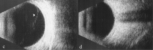
Ecografia Modo-B a revelar um descolamento anterior do vítreo na imagem c.
Descolamento posterior do vítreo na imagem d.
A hemorragia vítrea (ou hemovítreo) corresponde ao extravasamento de sangue para o humor Humor Defense Mechanisms vítreo.
Existem muitas causas para hemovítreo. Algumas causas comuns incluem:
A hemorragia vítrea é geralmente indolor e unilateral. Os sinais e sintomas incluem:
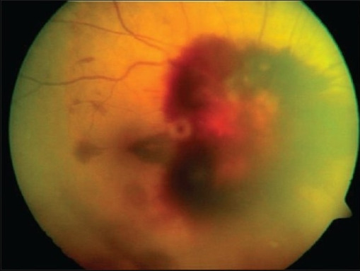
Fotografia de fundo do olho direito com hemorragia vítrea.
Image: “Fundus photographs of right eye showing peripapillary, subhyaloid, vitreous hemorrhage and several flame shaped hemorrhages obscuring the view of the optic disc” by Smt Kanuri Shanthamma Center for Retina Vitreous Diseases, L V Prasad Eye Institute, Kallam Anji Reddy Campus, Banjara Hills, Hyderabad 500 034, India. License: CC BY 2.0, edited by Lecturio.A sínquise cintilante, também conhecida como colesterolosis bulbi, é uma patologia degenerativa definida pela acumulação de cristais de colesterol no humor Humor Defense Mechanisms vítreo liquefeito.
A hialose asteróide é uma patologia na qual complexos de cálcio-lípido (sabão de cálcio) estão ligados à estrutura de colágeno do humor Humor Defense Mechanisms vítreo.
A etiologia da doença é desconhecida.
A hialose asteróide é geralmente assintomática.
O diagnóstico é feito através do exame na lâmpada de fenda, que demonstre:
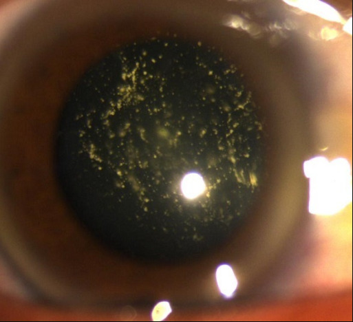
Múltiplas opacidades brilhantes suspensas no vítreo, consistentes com hialose asteróide.
Image: “Asteroid hyalosis: multiple yellow mobile vitreous particles” by University of Mohamed V souissi, hôpital des Spécialités, Ophtalology A Department. License: CC BY 2.0, edited by Lecturio.A persistência da vasculatura fetal, anteriormente conhecida como vítreo primário hiperplásico persistente, é uma patologia na qual os vasos sanguíneos embrionários não regridem.
Esta patologia é geralmente unilateral e pode apresentar-se com:
A persistência da vasculatura fetal geralmente é diagnosticada logo após o nascimento.
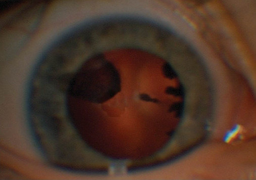
Persistência da vasculatura fetal:
Ao exame com lâmpada de fenda, identifica-se tração dos processos ciliares para o centro da cápsula posterior do cristalino no olho esquerdo e massa retrolenticular.