Domina Conceptos Médicos

Estudia para la escuela de medicina y tus examenes con Lecturio.

Tumores Óseos Benignos

Los LOS Neisseria tumores óseos benignos son un grupo de neoplasias no cancerosas que se forman a partir de hueso o cartílago. Estas lesiones tienen características distintas, como reacciones endósteales con calcificación intraósea y formación de hueso. Hay varios tumores óseos benignos comunes; aquí se comentan el osteoma osteoide, el osteoblastoma Osteoblastoma A benign, painful, tumor of bone characterized by the formation of osteoid tissue, primitive bone and calcified tissue. It occurs frequently in the spine of young persons. Benign Bone Tumors, el osteocondroma, el osteoma, los LOS Neisseria tumores de células gigantes y el encondroma. El diagnóstico se realiza mediante radiografía, que muestra lesiones localizadas en EN Erythema nodosum is an immune-mediated panniculitis (inflammation of the subcutaneous fat) caused by a type IV (delayed-type) hypersensitivity reaction. It commonly manifests in young women as tender, erythematous nodules on the shins. Erythema Nodosum el hueso con márgenes bien delimitados y sin afectación de tejidos blandos. También puede ser necesaria una biopsia. Las lesiones asintomáticas no requieren tratamiento, pero la extirpación quirúrgica o el legrado pueden ser necesarios para las lesiones sintomáticas y/o para prevenir fracturas patológicas.

Last updated: Sep 30, 2026

Editorial responsibility: Stanley Oiseth, Lindsay Jones, Evelin Maza

Descripción General

Los LOS Neisseria osteomas Osteomas A benign tumor composed of bone tissue or a hard tumor of bonelike structure developing on a bone (homoplastic osteoma) or on other structures (heteroplastic osteoma). Familial Adenomatous Polyposis osteoides se presentan con mayor frecuencia en EN Erythema nodosum is an immune-mediated panniculitis (inflammation of the subcutaneous fat) caused by a type IV (delayed-type) hypersensitivity reaction. It commonly manifests in young women as tender, erythematous nodules on the shins. Erythema Nodosum personas jóvenes. Los LOS Neisseria osteoblastomas Osteoblastomas Benign Bone Tumors suelen manifestarse durante la segunda década de vida, aunque pueden aparecer a cualquier edad, mientras que los LOS Neisseria osteomas Osteomas A benign tumor composed of bone tissue or a hard tumor of bonelike structure developing on a bone (homoplastic osteoma) or on other structures (heteroplastic osteoma). Familial Adenomatous Polyposis son más comunes en EN Erythema nodosum is an immune-mediated panniculitis (inflammation of the subcutaneous fat) caused by a type IV (delayed-type) hypersensitivity reaction. It commonly manifests in young women as tender, erythematous nodules on the shins. Erythema Nodosum adultos de mediana edad. Los LOS Neisseria osteocondromas y los LOS Neisseria encondromas son tumores formadores de cartílago. Los LOS Neisseria tumores de células gigantes son osteolíticos. Los LOS Neisseria fibromas y los LOS Neisseria quistes óseos son otras lesiones óseas benignas.
Características de los LOS Neisseria tumores óseos benignos

  • No cancerosos; los LOS Neisseria tumores de células gigantes tienen un bajo potencial metastásico y rara vez pueden metastatizar a los LOS Neisseria pulmones
  • Pueden causar dolor Dolor Inflammation
  • Varían desde lesiones estáticas (permanecen esencialmente sin cambios) hasta lesiones localmente agresivas (se expanden hasta que son tratadas, pero siguen siendo benignas)
  • Se observan hallazgos característicos en EN Erythema nodosum is an immune-mediated panniculitis (inflammation of the subcutaneous fat) caused by a type IV (delayed-type) hypersensitivity reaction. It commonly manifests in young women as tender, erythematous nodules on the shins. Erythema Nodosum las radiografías.
  • A menudo descubiertos incidentalmente
  • Osteoma osteoide:
    • Morfológica y genéticamente similar al AL Amyloidosis osteoblastoma Osteoblastoma A benign, painful, tumor of bone characterized by the formation of osteoid tissue, primitive bone and calcified tissue. It occurs frequently in the spine of young persons. Benign Bone Tumors
    • Pueden ser 2 presentaciones diferentes de la misma entidad genética.

Epidemiología

Estadificación y clasificación

  • Estadificación:
    • Latente: asintomático; descubierto incidentalmente
    • Activo: síntomas leves; crecimiento continuo del tumor Tumor Inflammation
    • Agresivo: crece rápidamente
  • Clasificación:
    • Diafisario
    • Metafisario
    • Epifisario

Osteoma Osteoide (en Niños)

Descripción

  • Lesión hamartomatosa formadora de hueso observada en EN Erythema nodosum is an immune-mediated panniculitis (inflammation of the subcutaneous fat) caused by a type IV (delayed-type) hypersensitivity reaction. It commonly manifests in young women as tender, erythematous nodules on the shins. Erythema Nodosum jóvenes
  • Ovoide: nido típicamente < 1,5 cm
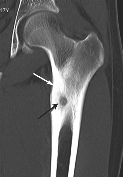
  • El nido es un núcleo altamente vascularizado formado por osteoblastos activos que producen niveles excepcionalmente elevados de prostaglandinas (particularmente PGE2 PGE2 The most common and most biologically active of the mammalian prostaglandins. It exhibits most biological activities characteristic of prostaglandins and has been used extensively as an oxytocic agent. The compound also displays a protective effect on the intestinal mucosa. Fever), las cuales provocan el característico dolor Dolor Inflammation nocturno.
  • Predominantemente localizado en EN Erythema nodosum is an immune-mediated panniculitis (inflammation of the subcutaneous fat) caused by a type IV (delayed-type) hypersensitivity reaction. It commonly manifests in young women as tender, erythematous nodules on the shins. Erythema Nodosum la corteza de la metáfisis y la diáfisis (80%–90%) de los LOS Neisseria huesos largos
  • Localización:
    • Más común:
      • Fémur proximal
    • Otras localizaciones:
      • Tibia Tibia The second longest bone of the skeleton. It is located on the medial side of the lower leg, articulating with the fibula laterally, the talus distally, and the femur proximally. Knee Joint: Anatomy
      • Columna vertebral
      • Otras partes del fémur

Epidemiología

  • Típicamente se presenta durante la segunda década de vida (90 % < 25 años)
  • Afecta a los LOS Neisseria hombres de 2–3 veces más a menudo que a las mujeres

Características clínicas

  • A menudo asintomático y descubierto incidentalmente.
  • Cuando es sintomático, se presenta con:
    • Dolor Dolor Inflammation:
      • Constante, intenso
      • Se exacerba por la noche
      • Se manifiesta antes de que las lesiones sean visibles en EN Erythema nodosum is an immune-mediated panniculitis (inflammation of the subcutaneous fat) caused by a type IV (delayed-type) hypersensitivity reaction. It commonly manifests in young women as tender, erythematous nodules on the shins. Erythema Nodosum la radiografía.
    • Edema Edema Edema is a condition in which excess serous fluid accumulates in the body cavity or interstitial space of connective tissues. Edema is a symptom observed in several medical conditions. It can be categorized into 2 types, namely, peripheral (in the extremities) and internal (in an organ or body cavity). Edema
    • Deformidad
    • Fractura patológica
  • Las lesiones de la columna pueden presentarse con:
    • Cojera
    • Escoliosis
    • Sensibilidad localizada
    • Restricción de movimiento
    • Espasmo de los LOS Neisseria músculos paravertebrales

Diagnóstico

  • Radiografía o TC:
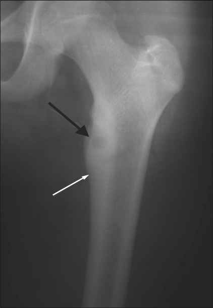
    • Núcleo radiolúcido (nido) rodeado de esclerosis
    • Engrosamiento cortical
    • Esclerosis medular difusa
    • Entre el 25%–40% no se ven en EN Erythema nodosum is an immune-mediated panniculitis (inflammation of the subcutaneous fat) caused by a type IV (delayed-type) hypersensitivity reaction. It commonly manifests in young women as tender, erythematous nodules on the shins. Erythema Nodosum las radiografías y necesitan una TC para su visualización.
    • No se recomienda la RM, ya que muestra edema Edema Edema is a condition in which excess serous fluid accumulates in the body cavity or interstitial space of connective tissues. Edema is a symptom observed in several medical conditions. It can be categorized into 2 types, namely, peripheral (in the extremities) and internal (in an organ or body cavity). Edema de tejidos blandos, que puede oscurecer el nido
  • Gammagrafía: realce intenso; es precisa para localizar el nido
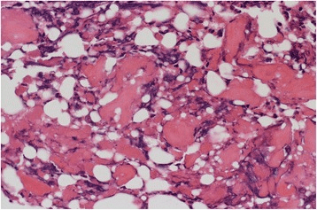
  • Histología: casi idéntica al AL Amyloidosis osteoblastoma Osteoblastoma A benign, painful, tumor of bone characterized by the formation of osteoid tissue, primitive bone and calcified tissue. It occurs frequently in the spine of young persons. Benign Bone Tumors, osteosarcoma Osteosarcoma Osteosarcoma is a primary malignant tumor of the bone characterized by the production of osteoid or immature bone by the tumor cells. The disease is most common in children and young adults and most frequently affects growth plates of the long bones, although it can involve any bone. Osteosarcoma y enostosis

Tratamiento

  • AINE: son eficaces porque el nido produce altos niveles de prostaglandinas.
  • Si el dolor Dolor Inflammation no responde a los LOS Neisseria AINE o los LOS Neisseria síntomas son intolerables: resección quirúrgica, ablación por radiofrecuencia, crioablación o ultrasonido focalizado de alta intensidad guiado por RM.
  • Si los LOS Neisseria síntomas son tolerables o están controlados con AINE, se puede optar por la observación de la lesión mediante exploraciones y radiografías seriadas cada 4 a 6 meses.

Osteoma (en Adultos)

Descripción

  • Se diferencian de los LOS Neisseria osteomas Osteomas A benign tumor composed of bone tissue or a hard tumor of bonelike structure developing on a bone (homoplastic osteoma) or on other structures (heteroplastic osteoma). Familial Adenomatous Polyposis osteoides en EN Erythema nodosum is an immune-mediated panniculitis (inflammation of the subcutaneous fat) caused by a type IV (delayed-type) hypersensitivity reaction. It commonly manifests in young women as tender, erythematous nodules on the shins. Erythema Nodosum que los LOS Neisseria osteomas Osteomas A benign tumor composed of bone tissue or a hard tumor of bonelike structure developing on a bone (homoplastic osteoma) or on other structures (heteroplastic osteoma). Familial Adenomatous Polyposis son crecimientos óseos sólidos (a diferencia de un nido radiolúcido rodeado de esclerosis).
  • Tumores benignos, redondos y formadores de hueso en EN Erythema nodosum is an immune-mediated panniculitis (inflammation of the subcutaneous fat) caused by a type IV (delayed-type) hypersensitivity reaction. It commonly manifests in young women as tender, erythematous nodules on the shins. Erythema Nodosum la superficie cortical de un hueso
  • Bien definidos y solitarios
  • Diámetro generalmente < 1 cm
  • Surgen de los LOS Neisseria osteoblastos:
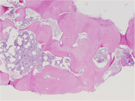
    • Osteomas Osteomas A benign tumor composed of bone tissue or a hard tumor of bonelike structure developing on a bone (homoplastic osteoma) or on other structures (heteroplastic osteoma). Familial Adenomatous Polyposis compactos: compuestos de hueso laminar maduro
    • Osteomas Osteomas A benign tumor composed of bone tissue or a hard tumor of bonelike structure developing on a bone (homoplastic osteoma) or on other structures (heteroplastic osteoma). Familial Adenomatous Polyposis esponjosos: compuestos de hueso trabecular con médula

Epidemiología

  • Pueden desarrollarse a cualquier edad, pero son más comunes en EN Erythema nodosum is an immune-mediated panniculitis (inflammation of the subcutaneous fat) caused by a type IV (delayed-type) hypersensitivity reaction. It commonly manifests in young women as tender, erythematous nodules on the shins. Erythema Nodosum personas de mediana edad
  • Ocurren en EN Erythema nodosum is an immune-mediated panniculitis (inflammation of the subcutaneous fat) caused by a type IV (delayed-type) hypersensitivity reaction. It commonly manifests in young women as tender, erythematous nodules on the shins. Erythema Nodosum hombres un poco más a menudo que en EN Erythema nodosum is an immune-mediated panniculitis (inflammation of the subcutaneous fat) caused by a type IV (delayed-type) hypersensitivity reaction. It commonly manifests in young women as tender, erythematous nodules on the shins. Erythema Nodosum mujeres
  • Asociados al AL Amyloidosis síndrome de Gardner como manifestación extracolónica de poliposis adenomatosa familiar

Presentación Clínica

  • Por lo general, se descubren de forma incidental; a menudo asintomáticos
  • Pueden ser sintomáticos y causar síntomas graves de “efecto de masa” (aunque se consideren benignos)
  • Localización: pueden causar los LOS Neisseria siguientes síntomas:
    • En EN Erythema nodosum is an immune-mediated panniculitis (inflammation of the subcutaneous fat) caused by a type IV (delayed-type) hypersensitivity reaction. It commonly manifests in young women as tender, erythematous nodules on the shins. Erythema Nodosum los LOS Neisseria huesos faciales, pueden provocar:
      • Alteraciones en EN Erythema nodosum is an immune-mediated panniculitis (inflammation of the subcutaneous fat) caused by a type IV (delayed-type) hypersensitivity reaction. It commonly manifests in young women as tender, erythematous nodules on the shins. Erythema Nodosum la visión y la audición
      • Parálisis de los LOS Neisseria nervios craneales/ proptosis Proptosis Retinoblastoma
      • Asimetría facial
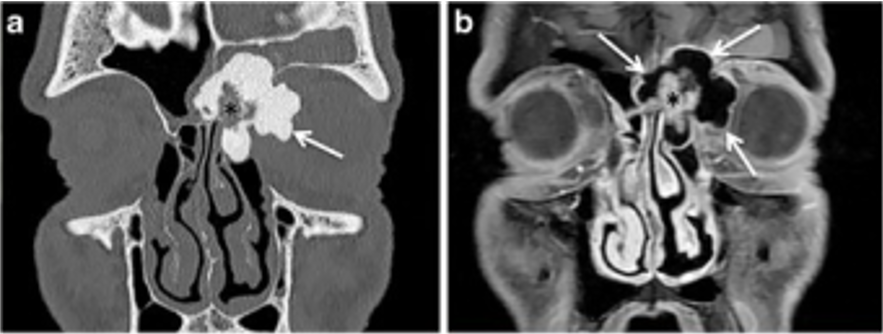
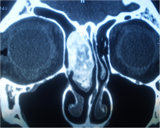
    • En EN Erythema nodosum is an immune-mediated panniculitis (inflammation of the subcutaneous fat) caused by a type IV (delayed-type) hypersensitivity reaction. It commonly manifests in young women as tender, erythematous nodules on the shins. Erythema Nodosum los LOS Neisseria huesos craneales (especialmente los LOS Neisseria senos paranasales), pueden causar:
      • Obstrucción/congestión secundaria de los LOS Neisseria senos nasales
      • Cefalea
    • Columna vertebral: pueden causar compresión de la médula espinal.
  • Hallazgos radiológicos en EN Erythema nodosum is an immune-mediated panniculitis (inflammation of the subcutaneous fat) caused by a type IV (delayed-type) hypersensitivity reaction. It commonly manifests in young women as tender, erythematous nodules on the shins. Erythema Nodosum radiografías o TC:
    • Lesiones radiodensas, bien delimitadas, redondas a ovoides
    • No penetran en EN Erythema nodosum is an immune-mediated panniculitis (inflammation of the subcutaneous fat) caused by a type IV (delayed-type) hypersensitivity reaction. It commonly manifests in young women as tender, erythematous nodules on the shins. Erythema Nodosum los LOS Neisseria tejidos blandos circundantes.

Tratamiento

  • Asintomático → sin tratamiento
  • Crecimiento tumoral sintomático → cirugía

Osteoblastoma

Descripción

  • Lesión formadora de hueso
  • Tumor Tumor Inflammation predominantemente cortical
  • Más grande que el osteoma osteoide (> 2 cm)
  • Predominio de las localizaciones metafisarias sobre las diafisarias
  • Puede ser localmente agresivo

Epidemiología

  • Comprenden aproximadamente el 14% de los LOS Neisseria tumores óseos benignos
  • Por lo general se presenta durante la segunda década de la vida, pero puede ocurrir a cualquier edad.
  • Predominio de hombre a mujer: 2,5 a 1

Características clínicas

  • Localización:
  • Presentación:
    • Dolor Dolor Inflammation insidioso y sordo; se exacerba por la noche
    • Escoliosis dolorosa si hay lesión vertebral
    • Síntomas neurológicos si hay compresión de la médula espinal

Diagnóstico

  • Radiografía:
    • Lesiones predominantemente líticas con borde o esclerosis perifocal
    • Pueden tener una calcificación interna o una apariencia “burbujeante”.
    • La mayoría de las personas presentarán una rápida expansión cortical:
      • A veces, destrucción cortical
      • Aproximadamente el 50% tiene esclerosis o periostitis Periostitis Inflammation of the periosteum. The condition is generally chronic, and is marked by tenderness and swelling of the bone and an aching pain. Acute periostitis is due to infection, is characterized by diffuse suppuration, severe pain, and constitutional symptoms, and usually results in necrosis. Reactive Arthritis circundante.
  • TC:
    • Las lesiones se observan líticas, similares a las radiografías
    • La mineralización de la matriz interna se ve VE Ventilation: Mechanics of Breathing mejor en EN Erythema nodosum is an immune-mediated panniculitis (inflammation of the subcutaneous fat) caused by a type IV (delayed-type) hypersensitivity reaction. It commonly manifests in young women as tender, erythematous nodules on the shins. Erythema Nodosum TC.
  • RM:
    • Las características pueden sobreestimar la lesión.
    • Las áreas de intensidad disminuida corresponden a focos de calcificación.
    • Por lo general, muy vascularizado y realza significativamente con gadolinio.
    • Puede simular una neoplasia maligna, pero rara vez se extiende a los LOS Neisseria tejidos blandos circundantes.

Tratamiento

  • Asintomático → sin tratamiento
  • La mayoría no responde a los LOS Neisseria AINE
  • Cirugía:
    • Resección del nido por síntomas refractarios.
    • Se prefiere el curetaje e injerto óseo
    • Otras opciones incluyen la ablación por radiofrecuencia y la crioablación; la radioterapia se reserva para casos seleccionados irresecables o recurrentes.
  • Radiografías de seguimiento cada 4–5 meses

Osteocondroma

Descripción

  • Aspecto: exostosis Exostosis Benign hypertrophy that projects outward from the surface of bone, often containing a cartilaginous component. Yaws, Bejel, and Pinta ósea (espolón) recubierta por un capuchón cartílago
    • El capuchón de cartílago es gruesa en EN Erythema nodosum is an immune-mediated panniculitis (inflammation of the subcutaneous fat) caused by a type IV (delayed-type) hypersensitivity reaction. It commonly manifests in young women as tender, erythematous nodules on the shins. Erythema Nodosum los LOS Neisseria niños (> 2 cm).
    • Más delgada en EN Erythema nodosum is an immune-mediated panniculitis (inflammation of the subcutaneous fat) caused by a type IV (delayed-type) hypersensitivity reaction. It commonly manifests in young women as tender, erythematous nodules on the shins. Erythema Nodosum adultos (< 1 cm)
  • Localización:
    • Metáfisis de huesos largos adyacentes a las placas de crecimiento.
    • Aproximadamente el 50% se produce alrededor de la rodilla–fémur distal o tibia Tibia The second longest bone of the skeleton. It is located on the medial side of the lower leg, articulating with the fibula laterally, the talus distally, and the femur proximally. Knee Joint: Anatomy proximal.
    • También se observa en EN Erythema nodosum is an immune-mediated panniculitis (inflammation of the subcutaneous fat) caused by a type IV (delayed-type) hypersensitivity reaction. It commonly manifests in young women as tender, erythematous nodules on the shins. Erythema Nodosum el húmero proximal.

Epidemiología

  • Tumor Tumor Inflammation óseo benigno primario más común:
  • Más frecuente en EN Erythema nodosum is an immune-mediated panniculitis (inflammation of the subcutaneous fat) caused by a type IV (delayed-type) hypersensitivity reaction. It commonly manifests in young women as tender, erythematous nodules on the shins. Erythema Nodosum niños y adolescentes, especialmente entre los LOS Neisseria 10 y 15 años
  • Sexo: masculino > femenino
  • Osteocondromas múltiples hereditarios: enfermedad autosómica dominante caracterizada por varias lesiones óseas

Fisiopatología

  • Las mutaciones en EN Erythema nodosum is an immune-mediated panniculitis (inflammation of the subcutaneous fat) caused by a type IV (delayed-type) hypersensitivity reaction. It commonly manifests in young women as tender, erythematous nodules on the shins. Erythema Nodosum los LOS Neisseria genes Genes A category of nucleic acid sequences that function as units of heredity and which code for the basic instructions for the development, reproduction, and maintenance of organisms. DNA Types and Structure supresores de tumores EXT1 o EXT2 alteran la síntesis de heparán sulfato, que normalmente regula la señalización de la placa de crecimiento (incluida la señalización de Indian hedgehog).
  • El cartílago puede herniarse a través de un defecto perióstico y continuar con la osificación endocondral, formando una excrecencia con cubierta cartilaginosa que se continúa con la cortical y la cavidad medular del hueso de origen.

Presentación clínica

  • Por lo general, se presenta como una masa palpable, indolora y de crecimiento lento cerca de los LOS Neisseria extremos de los LOS Neisseria huesos largos.
  • También puede ser asintomático
  • Si es sintomático con dolor Dolor Inflammation, el dolor Dolor Inflammation se debe al AL Amyloidosis efecto de masa directo sobre el tejido blando circundante.
  • El dolor Dolor Inflammation también puede deberse a compresión mecánica o irritación de nervios o vasos sanguíneos adyacentes, fractura a través del pedículo o de la cubierta cartilaginosa, o a un nuevo crecimiento relacionado con una transformación maligna tras la madurez esquelética.

Diagnóstico

  • Macroscópicamente, la lesión presenta un pedículo pedunculado o sésil con una cubierta de cartílago hialino de color blanco azulado y aspecto brillante.
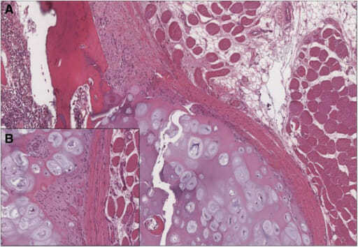
  • Histológicamente, la cubierta se asemeja a una placa de crecimiento desorganizada que experimenta osificación endocondral para transformarse en EN Erythema nodosum is an immune-mediated panniculitis (inflammation of the subcutaneous fat) caused by a type IV (delayed-type) hypersensitivity reaction. It commonly manifests in young women as tender, erythematous nodules on the shins. Erythema Nodosum hueso trabecular maduro en EN Erythema nodosum is an immune-mediated panniculitis (inflammation of the subcutaneous fat) caused by a type IV (delayed-type) hypersensitivity reaction. It commonly manifests in young women as tender, erythematous nodules on the shins. Erythema Nodosum su base.
  • Radiografía:
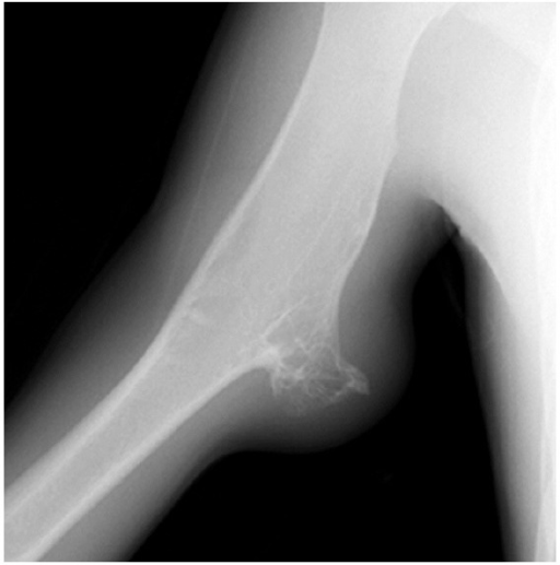
    • Espolón óseo que surge de la superficie de la corteza.
    • La lesión se orienta en EN Erythema nodosum is an immune-mediated panniculitis (inflammation of the subcutaneous fat) caused by a type IV (delayed-type) hypersensitivity reaction. It commonly manifests in young women as tender, erythematous nodules on the shins. Erythema Nodosum dirección opuesta a la articulación, y sus espacios cortical y medular guardan continuidad con los LOS Neisseria del hueso de origen.
    • Puede ser sésil (base > capuchón) o pediculado (capuchón > base)
    • Por lo general, involucra la metáfisis.
  • RM:
    • Se justifica si existe preocupación por pinzamiento del tejido adyacente
    • Indicado si hay posibilidad de resección quirúrgica.

Tratamiento

  • La mayoría de los LOS Neisseria casos no necesitan tratamiento.
  • Casos sintomáticos → escisión
  • Cuando se requiere escisión, esta se realiza en EN Erythema nodosum is an immune-mediated panniculitis (inflammation of the subcutaneous fat) caused by a type IV (delayed-type) hypersensitivity reaction. It commonly manifests in young women as tender, erythematous nodules on the shins. Erythema Nodosum la base del pedículo.
  • Aproximadamente el 1% se transforma en EN Erythema nodosum is an immune-mediated panniculitis (inflammation of the subcutaneous fat) caused by a type IV (delayed-type) hypersensitivity reaction. It commonly manifests in young women as tender, erythematous nodules on the shins. Erythema Nodosum condrosarcoma.

Condroma (Encondroma)

Descripción

  • Tumor Tumor Inflammation benigno compuesto por cartílago hialino maduro.
  • Un condroma situado en EN Erythema nodosum is an immune-mediated panniculitis (inflammation of the subcutaneous fat) caused by a type IV (delayed-type) hypersensitivity reaction. It commonly manifests in young women as tender, erythematous nodules on the shins. Erythema Nodosum la cavidad medular se denomina encondroma; aquel que se encuentra en EN Erythema nodosum is an immune-mediated panniculitis (inflammation of the subcutaneous fat) caused by a type IV (delayed-type) hypersensitivity reaction. It commonly manifests in young women as tender, erythematous nodules on the shins. Erythema Nodosum la superficie ósea recibe el nombre de condroma perióstico.

Epidemiología

  • Por lo general, se presenta entre los LOS Neisseria 20 y los LOS Neisseria 50 años de edad.
  • Afecta por igual a hombres y mujeres.
  • Suele localizarse en EN Erythema nodosum is an immune-mediated panniculitis (inflammation of the subcutaneous fat) caused by a type IV (delayed-type) hypersensitivity reaction. It commonly manifests in young women as tender, erythematous nodules on the shins. Erythema Nodosum los LOS Neisseria huesos pequeños de las manos y los LOS Neisseria pies, particularmente en EN Erythema nodosum is an immune-mediated panniculitis (inflammation of the subcutaneous fat) caused by a type IV (delayed-type) hypersensitivity reaction. It commonly manifests in young women as tender, erythematous nodules on the shins. Erythema Nodosum los LOS Neisseria metacarpianos y las falanges.

Fisiopatología

  • La mayoría de los LOS Neisseria encondromas son solitarios y esporádicos.
  • El cartílago residual de la placa de crecimiento persiste en EN Erythema nodosum is an immune-mediated panniculitis (inflammation of the subcutaneous fat) caused by a type IV (delayed-type) hypersensitivity reaction. It commonly manifests in young women as tender, erythematous nodules on the shins. Erythema Nodosum la cavidad medular en EN Erythema nodosum is an immune-mediated panniculitis (inflammation of the subcutaneous fat) caused by a type IV (delayed-type) hypersensitivity reaction. It commonly manifests in young women as tender, erythematous nodules on the shins. Erythema Nodosum lugar de osificarse normalmente, formando un nódulo de cartílago hialino bien delimitado y de crecimiento lento.
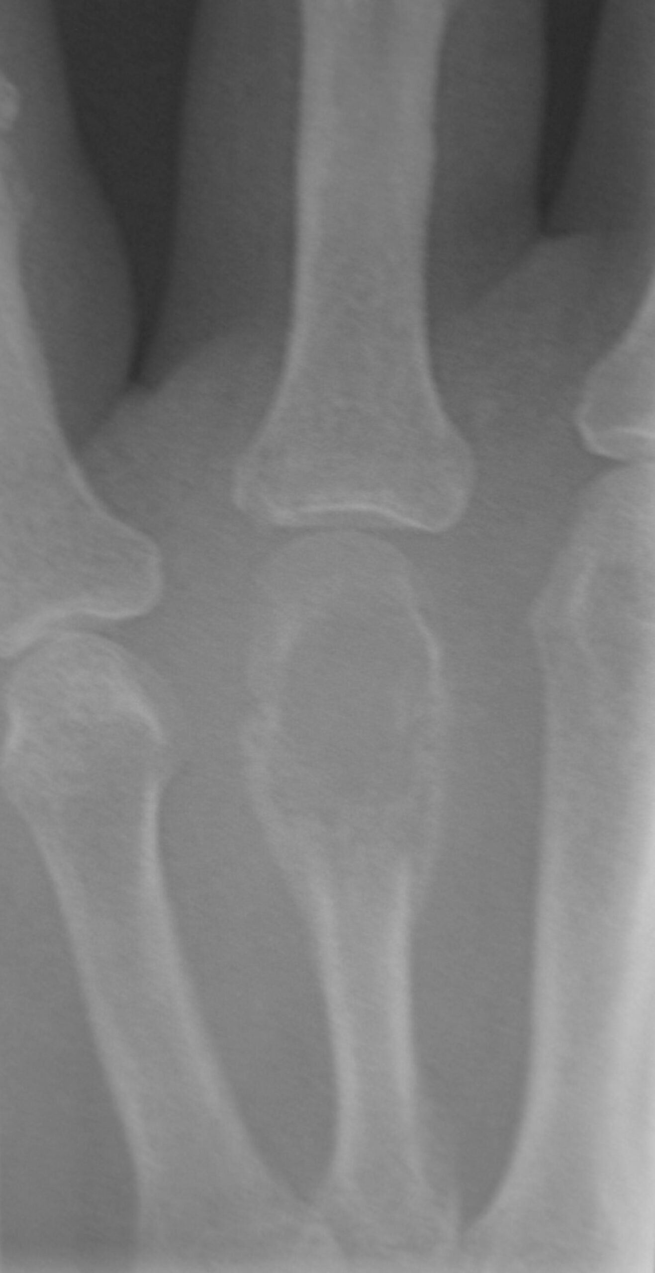
  • La calcificación dentro del nódulo produce la característica imagen en “palomitas de maíz” en EN Erythema nodosum is an immune-mediated panniculitis (inflammation of the subcutaneous fat) caused by a type IV (delayed-type) hypersensitivity reaction. It commonly manifests in young women as tender, erythematous nodules on the shins. Erythema Nodosum los LOS Neisseria estudios radiológicos.
  • El crecimiento de la lesión puede provocar festoneado endóstico y adelgazamiento cortical, lo que debilita el hueso y aumenta el riesgo de fractura patológica.
  • La enfermedad de Ollier y el síndrome de Maffucci se manifiestan con múltiples encondromas y están asociados a mutaciones somáticas en EN Erythema nodosum is an immune-mediated panniculitis (inflammation of the subcutaneous fat) caused by a type IV (delayed-type) hypersensitivity reaction. It commonly manifests in young women as tender, erythematous nodules on the shins. Erythema Nodosum mosaico de los LOS Neisseria genes Genes A category of nucleic acid sequences that function as units of heredity and which code for the basic instructions for the development, reproduction, and maintenance of organisms. DNA Types and Structure IDH1 o IDH2.

Presentación clínica

  • La mayoría son asintomáticos y se descubren de forma incidental.
  • El adelgazamiento cortical puede causar dolor Dolor Inflammation o una fractura patológica, especialmente en EN Erythema nodosum is an immune-mediated panniculitis (inflammation of the subcutaneous fat) caused by a type IV (delayed-type) hypersensitivity reaction. It commonly manifests in young women as tender, erythematous nodules on the shins. Erythema Nodosum los LOS Neisseria dedos.
  • La enfermedad de Ollier se caracteriza por múltiples encondromas, a menudo distribuidos de forma asimétrica o unilateral, y conlleva un mayor riesgo de condrosarcoma.
  • El síndrome de Maffucci se caracteriza por múltiples encondromas y hemangiomas de células fusiformes en EN Erythema nodosum is an immune-mediated panniculitis (inflammation of the subcutaneous fat) caused by a type IV (delayed-type) hypersensitivity reaction. It commonly manifests in young women as tender, erythematous nodules on the shins. Erythema Nodosum tejidos blandos, con un riesgo significativamente mayor de condrosarcoma y otras neoplasias malignas (incluidos tumores ováricos y cerebrales).

Diagnóstico

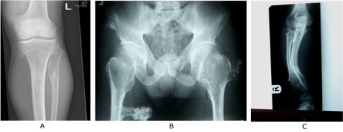
  • Radiografía:
    • Lesión radiolúcida bien delimitada y de localización central en EN Erythema nodosum is an immune-mediated panniculitis (inflammation of the subcutaneous fat) caused by a type IV (delayed-type) hypersensitivity reaction. It commonly manifests in young women as tender, erythematous nodules on the shins. Erythema Nodosum la cavidad medular, frecuentemente en EN Erythema nodosum is an immune-mediated panniculitis (inflammation of the subcutaneous fat) caused by a type IV (delayed-type) hypersensitivity reaction. It commonly manifests in young women as tender, erythematous nodules on the shins. Erythema Nodosum la metáfisis
    • Posibles calcificaciones en EN Erythema nodosum is an immune-mediated panniculitis (inflammation of the subcutaneous fat) caused by a type IV (delayed-type) hypersensitivity reaction. It commonly manifests in young women as tender, erythematous nodules on the shins. Erythema Nodosum «palomitas de maíz» y festoneado endosteal
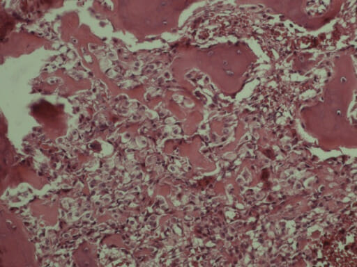
  • Biopsia/histopatología: lobulillos de cartílago hialino maduro y hipocelular rodeados de hueso laminar normal

Tratamiento

  • Lesiones asintomáticas → observación mediante radiografías seriadas para confirmar la estabilidad
  • Lesiones sintomáticas o asociadas a fractura → legrado intralesional seguido de relleno con injerto óseo

Tumor de Células Gigantes

Descripción

  • También conocido como “osteoclastoma”
  • Benigno
  • Localmente agresivo
  • Osteolítico
  • Clasificado como una neoplasia de comportamiento intermedio con bajo potencial metastásico
  • Afecta más comúnmente a la epífisis de los LOS Neisseria huesos largos (~ 50% ocurre alrededor de la rodilla)
  • La porción distal del fémur y la proximal de la tibia Tibia The second longest bone of the skeleton. It is located on the medial side of the lower leg, articulating with the fibula laterally, the talus distally, and the femur proximally. Knee Joint: Anatomy son localizaciones especialmente frecuentes.

Epidemiología

  • Incidencia máxima: edad de 20–40 años
  • Tasa general: 1,3 casos por millón de personas por año
    • 15%–20% de todos los LOS Neisseria tumores óseos benignos
    • Aproximadamente el 4 %–5 % de todos los LOS Neisseria tumores óseos primarios

Presentación clínica

  • Dolor Dolor Inflammation localizado y aumento de volumen en EN Erythema nodosum is an immune-mediated panniculitis (inflammation of the subcutaneous fat) caused by a type IV (delayed-type) hypersensitivity reaction. It commonly manifests in young women as tender, erythematous nodules on the shins. Erythema Nodosum el lugar afectado.
  • Dolor Dolor Inflammation con el movimiento y ↓ amplitud de movimiento de la articulación cercana.
  • ↑ Riesgo de fracturas patológicas.

Diagnóstico

  • Radiografía:
    • Lesión osteolítica multiquística, que da un aspecto de “burbuja de jabón” multiloculada
    • Afecta con mayor frecuencia a la epífisis y puede extenderse a la metáfisis adyacente o al AL Amyloidosis hueso subcondral.
  • Biopsia/histopatología:
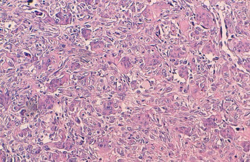
    • No se comprende totalmente la naturaleza exacta del tumor Tumor Inflammation; su componente neoplásico clave es la célula estromal con mutación en EN Erythema nodosum is an immune-mediated panniculitis (inflammation of the subcutaneous fat) caused by a type IV (delayed-type) hypersensitivity reaction. It commonly manifests in young women as tender, erythematous nodules on the shins. Erythema Nodosum H3F3A.
    • Se observarán células gigantes osteoclásticas multinucleadas.
    • Células que expresan el receptor Receptor Receptors are proteins located either on the surface of or within a cell that can bind to signaling molecules known as ligands (e.g., hormones) and cause some type of response within the cell. Receptors activador del factor nuclear kappa-B ( RANKL RANKL A tumor necrosis factor receptor family member that is specific for rank ligand and plays a role in bone homeostasis by regulating osteoclastogenesis. It is also expressed on dendritic cells where it plays a role in regulating dendritic cell survival. Signaling by the activated receptor occurs through its association with tnf receptor-associated factors. Paget Disease of Bone, por sus siglas en EN Erythema nodosum is an immune-mediated panniculitis (inflammation of the subcutaneous fat) caused by a type IV (delayed-type) hypersensitivity reaction. It commonly manifests in young women as tender, erythematous nodules on the shins. Erythema Nodosum inglés)
      • También conocido como “factor de diferenciación de osteoclastos”
      • Se expresa en EN Erythema nodosum is an immune-mediated panniculitis (inflammation of the subcutaneous fat) caused by a type IV (delayed-type) hypersensitivity reaction. It commonly manifests in young women as tender, erythematous nodules on the shins. Erythema Nodosum gran medida por las células del estroma dentro de los LOS Neisseria tumores de células gigantes
      • Expresión de RANKL RANKL A tumor necrosis factor receptor family member that is specific for rank ligand and plays a role in bone homeostasis by regulating osteoclastogenesis. It is also expressed on dendritic cells where it plays a role in regulating dendritic cell survival. Signaling by the activated receptor occurs through its association with tnf receptor-associated factors. Paget Disease of Bone por las células estromales → reclutamiento de células osteoclásticas
      • La detección de la mutación H3F3A, presente en EN Erythema nodosum is an immune-mediated panniculitis (inflammation of the subcutaneous fat) caused by a type IV (delayed-type) hypersensitivity reaction. It commonly manifests in young women as tender, erythematous nodules on the shins. Erythema Nodosum más del 90 % de los LOS Neisseria casos, puede ayudar a confirmar el diagnóstico y a diferenciar este tumor Tumor Inflammation de otras lesiones ricas en EN Erythema nodosum is an immune-mediated panniculitis (inflammation of the subcutaneous fat) caused by a type IV (delayed-type) hypersensitivity reaction. It commonly manifests in young women as tender, erythematous nodules on the shins. Erythema Nodosum células gigantes.

Tratamiento

  • Se necesita un muestreo histológico completo para asegurar el diagnóstico.
  • Cirugía:
    • Curetaje e injerto óseo
    • Resección en EN Erythema nodosum is an immune-mediated panniculitis (inflammation of the subcutaneous fat) caused by a type IV (delayed-type) hypersensitivity reaction. It commonly manifests in young women as tender, erythematous nodules on the shins. Erythema Nodosum bloc para minimizar la tasa de recurrencia
    • Se puede utilizar un adyuvante local, frecuentemente cemento óseo de polimetilmetacrilato.
  • Vigilancia y seguimiento en EN Erythema nodosum is an immune-mediated panniculitis (inflammation of the subcutaneous fat) caused by a type IV (delayed-type) hypersensitivity reaction. It commonly manifests in young women as tender, erythematous nodules on the shins. Erythema Nodosum caso de recurrencia
  • El denosumab Denosumab A humanized monoclonal antibody and an inhibitor of the rank ligand, which regulates osteoclast differentiation and bone remodeling. It is used as a bone density conservation agent in the treatment of osteoporosis. Other Antiresorptive Drugs, un inhibidor de RANKL RANKL A tumor necrosis factor receptor family member that is specific for rank ligand and plays a role in bone homeostasis by regulating osteoclastogenesis. It is also expressed on dendritic cells where it plays a role in regulating dendritic cell survival. Signaling by the activated receptor occurs through its association with tnf receptor-associated factors. Paget Disease of Bone, constituye una opción para la enfermedad irresecable o metastásica, o bien como tratamiento neoadyuvante para reducir el tamaño del tumor Tumor Inflammation antes de la cirugía.

Pronóstico

  • Suele ser benigno; riesgo raro de degeneración maligna ↑ con la edad (aproximadamente 4%)
  • Tendencia a una destrucción ósea significativa y recurrencia local.

Diagnóstico Diferencial

  • Osteosarcoma Osteosarcoma Osteosarcoma is a primary malignant tumor of the bone characterized by the production of osteoid or immature bone by the tumor cells. The disease is most common in children and young adults and most frequently affects growth plates of the long bones, although it can involve any bone. Osteosarcoma: tumor Tumor Inflammation maligno primario del hueso caracterizado por la producción de hueso osteoide o inmaduro por las células tumorales. El osteosarcoma Osteosarcoma Osteosarcoma is a primary malignant tumor of the bone characterized by the production of osteoid or immature bone by the tumor cells. The disease is most common in children and young adults and most frequently affects growth plates of the long bones, although it can involve any bone. Osteosarcoma es más común en EN Erythema nodosum is an immune-mediated panniculitis (inflammation of the subcutaneous fat) caused by a type IV (delayed-type) hypersensitivity reaction. It commonly manifests in young women as tender, erythematous nodules on the shins. Erythema Nodosum niños y adultos jóvenes y se presenta con dolor Dolor Inflammation y aumento de volumen, a veces con una masa palpable o como una fractura patológica. El diagnóstico se establece con imagenología y biopsia. El tratamiento incluye quimioterapia sistémica y resección quirúrgica.
  • Sarcoma de Ewing: neoplasia ósea primaria derivada de células redondas primitivas. El sarcoma de Ewing afecta principalmente a niños y adolescentes y comúnmente se presenta como una masa dolorosa, aumento de volumen y fracturas óseas patológicas. El diagnóstico se establece con imagenología y biopsia. El tratamiento incluye quimioterapia sistémica y control local del tumor Tumor Inflammation con resección quirúrgica o radiación. Con el tratamiento adecuado, la supervivencia a 5 años es superior al AL Amyloidosis 70%.
  • Condrosarcoma: tumor Tumor Inflammation óseo maligno caracterizado por la producción de una matriz cartilaginosa. El condrosarcoma generalmente se presenta con una masa o inflamación que aumenta lentamente y un dolor Dolor Inflammation sordo y doloroso. Dependiendo de la localización, también puede estar asociado con síntomas de compresión nerviosa. El diagnóstico se establece con los LOS Neisseria hallazgos en EN Erythema nodosum is an immune-mediated panniculitis (inflammation of the subcutaneous fat) caused by a type IV (delayed-type) hypersensitivity reaction. It commonly manifests in young women as tender, erythematous nodules on the shins. Erythema Nodosum la imagenología y los LOS Neisseria resultados de la biopsia de tejido. El pilar del tratamiento es la escisión quirúrgica.
  • Mieloma múltiple: afección maligna de las células plasmáticas (linfocitos B activados) que se observa principalmente en EN Erythema nodosum is an immune-mediated panniculitis (inflammation of the subcutaneous fat) caused by a type IV (delayed-type) hypersensitivity reaction. It commonly manifests in young women as tender, erythematous nodules on the shins. Erythema Nodosum ancianos. La proliferación monoclonal de células plasmáticas da como resultado una actividad osteoclástica impulsada por citocinas y una secreción excesiva de anticuerpos IgG IgG The major immunoglobulin isotype class in normal human serum. There are several isotype subclasses of igg, for example, igg1, igg2a, and igg2b. Hypersensitivity Pneumonitis. El diagnóstico se establece mediante electroforesis en EN Erythema nodosum is an immune-mediated panniculitis (inflammation of the subcutaneous fat) caused by a type IV (delayed-type) hypersensitivity reaction. It commonly manifests in young women as tender, erythematous nodules on the shins. Erythema Nodosum plasma Plasma The residual portion of blood that is left after removal of blood cells by centrifugation without prior blood coagulation. Transfusion Products y biopsia de médula ósea. Algunas personas de bajo riesgo pueden permanecer estables durante años sin tratamiento, mientras que otras pueden progresar a pesar del tratamiento. La quimioterapia está indicada para personas que cumplen con criterios de alto riesgo.
  • Craneofaringiomas: tumores epiteliales escamosos raros con una estructura sólida y/o quística que surgen de los LOS Neisseria restos de la bolsa de Rathke a lo largo del tallo hipofisario en EN Erythema nodosum is an immune-mediated panniculitis (inflammation of the subcutaneous fat) caused by a type IV (delayed-type) hypersensitivity reaction. It commonly manifests in young women as tender, erythematous nodules on the shins. Erythema Nodosum la región supraselar. Los LOS Neisseria síntomas de los LOS Neisseria craneofaringiomas incluyen cefalea, náuseas, vómitos, alteraciones visuales, disfunción endocrina y cambios de comportamiento. El diagnóstico se realiza mediante imagenología y biopsia. El tratamiento incluye la escisión quirúrgica y la radioterapia.

Referencias

  1. Librodo, G. (2021). Osteoid osteoma. Emedicine. Retrieved August 30, 2021, from https://emedicine.medscape.com/article/1253443-treatment
  2. Tis, K. (2020). Nonmalignant bone lesions in children and adolescents. UpToDate. Retrieved August 30, 2021, from https://www.uptodate.com/contents/nonmalignant-bone-lesions-in-children-and-adolescents
  3. Ortmann, F. (2020). Osteoblastoma. Emedicine. Retrieved August 11, 2021, from https://emedicine.medscape.com/article/1257927-overview
  4. Dickey, I. (2021). Solitary osteochondroma workup. Emedicine. Retrieved August 11, 2021, from https://emedicine.medscape.com/article/1256477-workup#c6
  5. Lewis, V. (2019). Giant cell tumor of bone treatment & management. Emedicine. Retrieved August 11, 2021, from https://emedicine.medscape.com/article/1255364-treatment
  6. Wei, S. & Stevens, T.M. (2014). Benign tumors and tumor-like conditions of bone. In: McManus, L.M., Mitchell, R.N. (Eds.), Pathobiology of Human Disease. Elsevier. https://doi.org/10.1016/B978-0-12-386456-7.03110-5
  7. Saber, M., Gaillard, F. (2018). Osteoma. Radiopaedia. Retrieved August 30, 2021, from https://radiopaedia.org/articles/osteoma?lang=us
  8. Burt, R.W. (2020). Gardner syndrome. UpToDate. Retrieved August 30, 2021, from https://www.uptodate.com/contents/gardner-syndrome
  9. Becker, M., Stefanelli, S., Rougemont, A.L., Poletti, P.A., Merlini, L. (2017). Non-odontogenic tumors of the facial bones in children and adolescents: role of multiparametric imaging. Neuroradiology 59:327–342. https://pubmed.ncbi.nlm.nih.gov/28289810/
  10. Palmerini, E., Picci, P., Reichardt, P., Downey, G. (2019). Malignancy in giant cell tumor of bone: a review of the literature. Technology in Cancer Research & Treatment 18:1533033819840000. https://pubmed.ncbi.nlm.nih.gov/30935298/
  11. Thomas, D.M., Desai, J., Damron, T.A. (2021). Giant cell tumor of bone. UpToDate. Retrieved August 30, 2021, from https://www.uptodate.com/contents/giant-cell-tumor-of-bone

¡Crea tu cuenta gratis o inicia una sesión para seguir leyendo!

Regístrate ahora y obtén acceso gratuito a Lecturio con páginas de concepto, videos médicos y cuestionarios para tu educación médica.

User Reviews

Details