El tálamo es una gran estructura ovoide en EN Erythema nodosum is an immune-mediated panniculitis (inflammation of the subcutaneous fat) caused by a type IV (delayed-type) hypersensitivity reaction. It commonly manifests in young women as tender, erythematous nodules on the shins. Erythema Nodosum la parte dorsal del diencéfalo que se ubica entre la corteza cerebral y el mesencéfalo, estando constituido de varios núcleos de sustancia gris interconectados, los LOS Neisseria cuales están separados por láminas de sustancia blanca. El tálamo es el principal conductor de la información que pasa entre la corteza cerebral y la periferia, la médula espinal o el tronco encefálico y se divide en EN Erythema nodosum is an immune-mediated panniculitis (inflammation of the subcutaneous fat) caused by a type IV (delayed-type) hypersensitivity reaction. It commonly manifests in young women as tender, erythematous nodules on the shins. Erythema Nodosum una parte anterior, una medial y una lateral. Cada parte contiene grupos de núcleos que funcionan como centros de relevo para los LOS Neisseria impulsos sensoriales y para la modulación de las respuestas motoras a través de interconexiones con los LOS Neisseria ganglios basales.
Last updated: Dec 15, 2025
| Núcleo | Aferencia principal | Eferencia principal | Función |
|---|---|---|---|
| Anterior | Cuerpo mamilar y formación del hipocampo | Circunvolución del cíngulo | Vía límbica |
| Posteromedial ventral | Tracto trigéminotalámico y núcleo solitario rostral | Corteza somatosensorial primaria y corteza gustativa | Tacto, posición, dolor Dolor Inflammation y temperatura de la cara; gusto |
| Posterolateral ventral | Columna dorsal-lemnisco medial y tracto anterolateral | Corteza somatosensorial primaria | Sensación de dolor Dolor Inflammation y temperatura, tacto y posición. |
| Ventral anterior | Ganglios basales | Motor Motor Neurons which send impulses peripherally to activate muscles or secretory cells. Nervous System: Histology, premotor y difuso | Planificación motora |
| Lateral ventral | Cerebelo | Corteza motora y premotora | Planificación motora y control |
| Dorsomedial | Amígdala, corteza olfatoria, ganglios basales límbicos | Corteza frontal Frontal The bone that forms the frontal aspect of the skull. Its flat part forms the forehead, articulating inferiorly with the nasal bone and the cheek bone on each side of the face. Skull: Anatomy | Vía límbica, relevo principal a la corteza frontal Frontal The bone that forms the frontal aspect of the skull. Its flat part forms the forehead, articulating inferiorly with the nasal bone and the cheek bone on each side of the face. Skull: Anatomy |
| Pulvinar Pulvinar Large mass of nuclei forming the most caudal portion of the thalamus and overhanging the geniculate bodies and the dorsolateral surface of the midbrain. It is divided into four parts: the lateral, medial, inferior, and oral pulvinar nuclei. Thalamus: Anatomy | Vías visuales, auditivas y otras sensoriales | Áreas de asociación parietal Parietal One of a pair of irregularly shaped quadrilateral bones situated between the frontal bone and occipital bone, which together form the sides of the cranium. Skull: Anatomy, occipital Occipital Part of the back and base of the cranium that encloses the foramen magnum. Skull: Anatomy y temporal | Integración sensorial y atención visual |
| Geniculado medial | Colículo inferior | Corteza auditiva primaria | Audición |
| Geniculado lateral | Retina Retina The ten-layered nervous tissue membrane of the eye. It is continuous with the optic nerve and receives images of external objects and transmits visual impulses to the brain. Its outer surface is in contact with the choroid and the inner surface with the vitreous body. The outermost layer is pigmented, whereas the inner nine layers are transparent. Eye: Anatomy (tracto óptico) | Corteza visual primaria | Visión |
| Intralaminar | Formación reticular, médula espinal, hipotálamo | Corteza límbica y ganglios basales | Excitación, motivación, afecto, dolor Dolor Inflammation. |
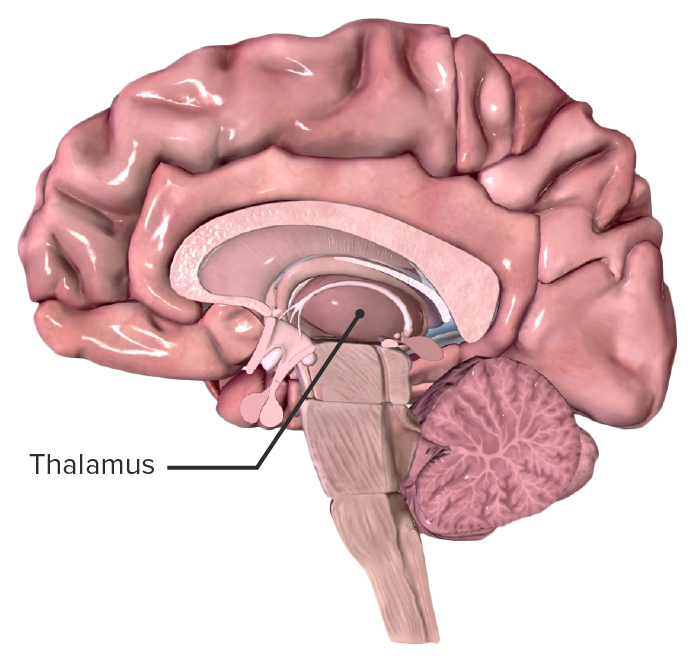
Localización del tálamo en una sección sagital por la línea media del cerebro humano
Imagen por BioDigital, editada por Lecturio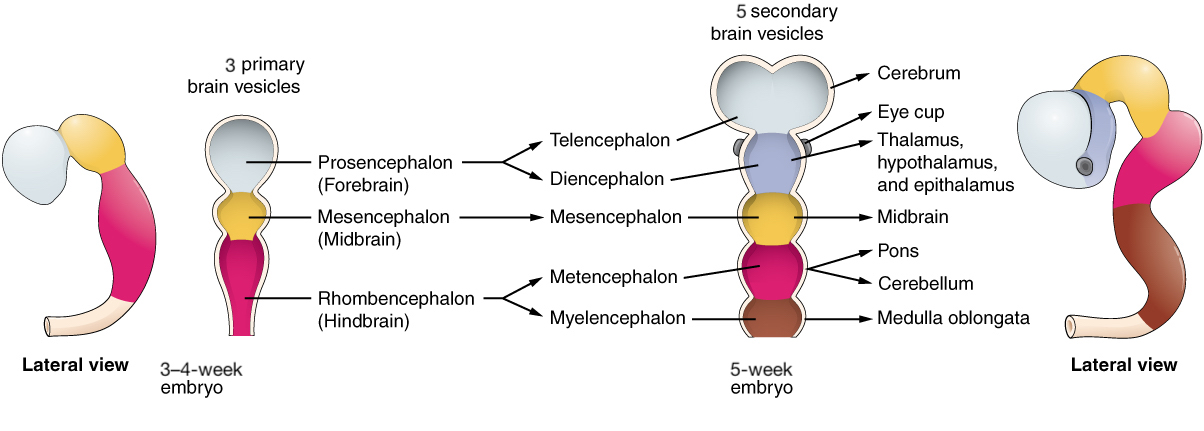
Desarrollo del tálamo a partir del prosencéfalo, donde forma el diencéfalo, que comprende el tálamo, el hipotálamo y el epitálamo.
Imagen: “Primary and Secondary Vesicle Stages of Development” por Phil Schatz. Licencia: CC BY 4.0, editada por Lecturio.Los LOS Neisseria núcleos anteriores se subdividen en EN Erythema nodosum is an immune-mediated panniculitis (inflammation of the subcutaneous fat) caused by a type IV (delayed-type) hypersensitivity reaction. It commonly manifests in young women as tender, erythematous nodules on the shins. Erythema Nodosum 3 secciones con aferencias de los LOS Neisseria cuerpos mamilares y el hipocampo, y eferencias a la circunvolución del cíngulo. Los LOS Neisseria núcleos anteriores están asociados con el aprendizaje, la memoria y las emociones.
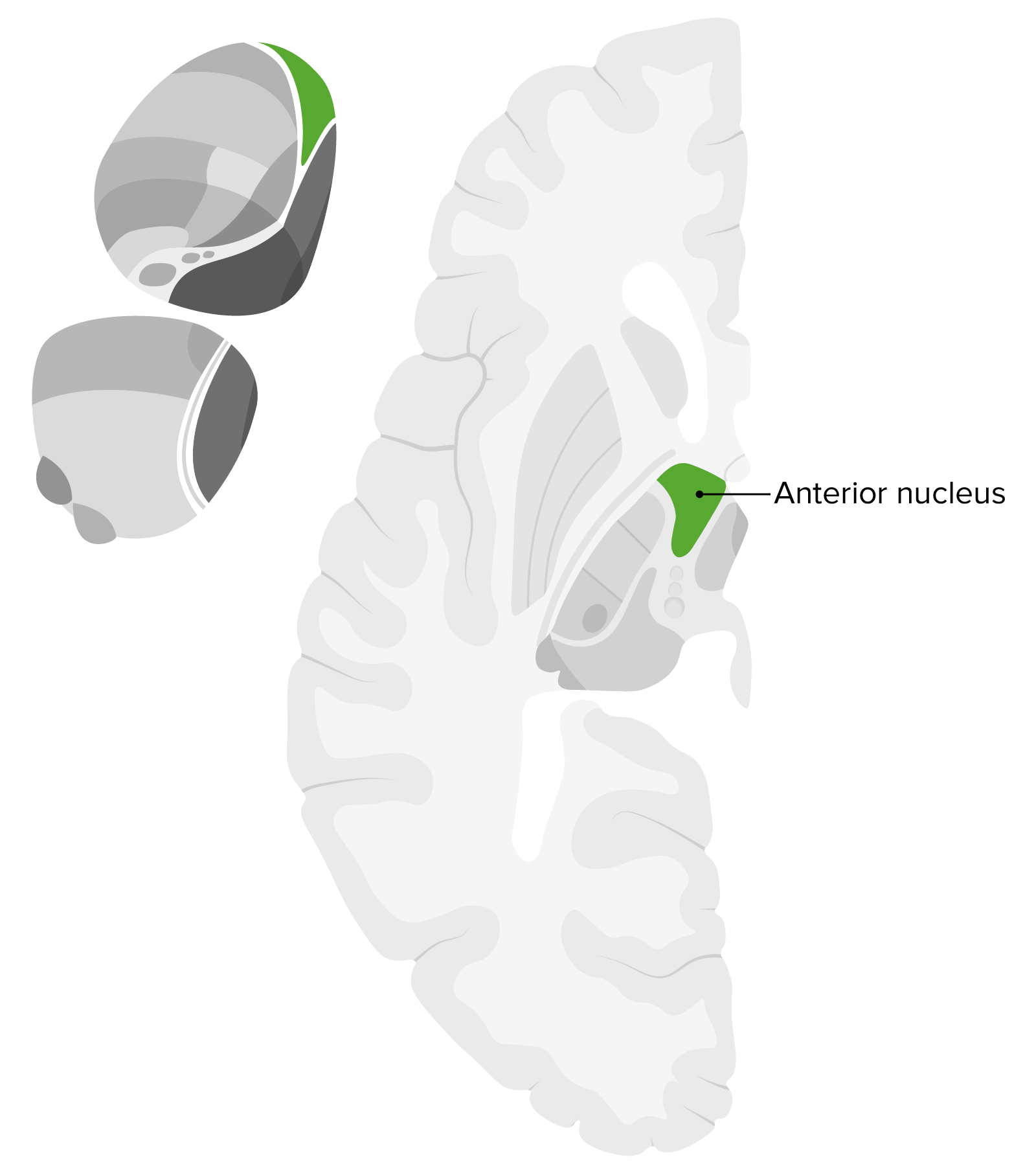
Ubicación del núcleo anterior dentro del tálamo: importante tener en cuenta su ubicación central y anterior dentro de esta hemisección del cerebro.
Imagen por Lecturio.Los LOS Neisseria núcleos ventral posteromedial y ventral posterolateral están asociados con aferencias del núcleo solitario rostral y el tracto trigéminotalámico y eferencias a la corteza somatosensorial primaria y al AL Amyloidosis opérculo/ínsula frontal Frontal The bone that forms the frontal aspect of the skull. Its flat part forms the forehead, articulating inferiorly with the nasal bone and the cheek bone on each side of the face. Skull: Anatomy.
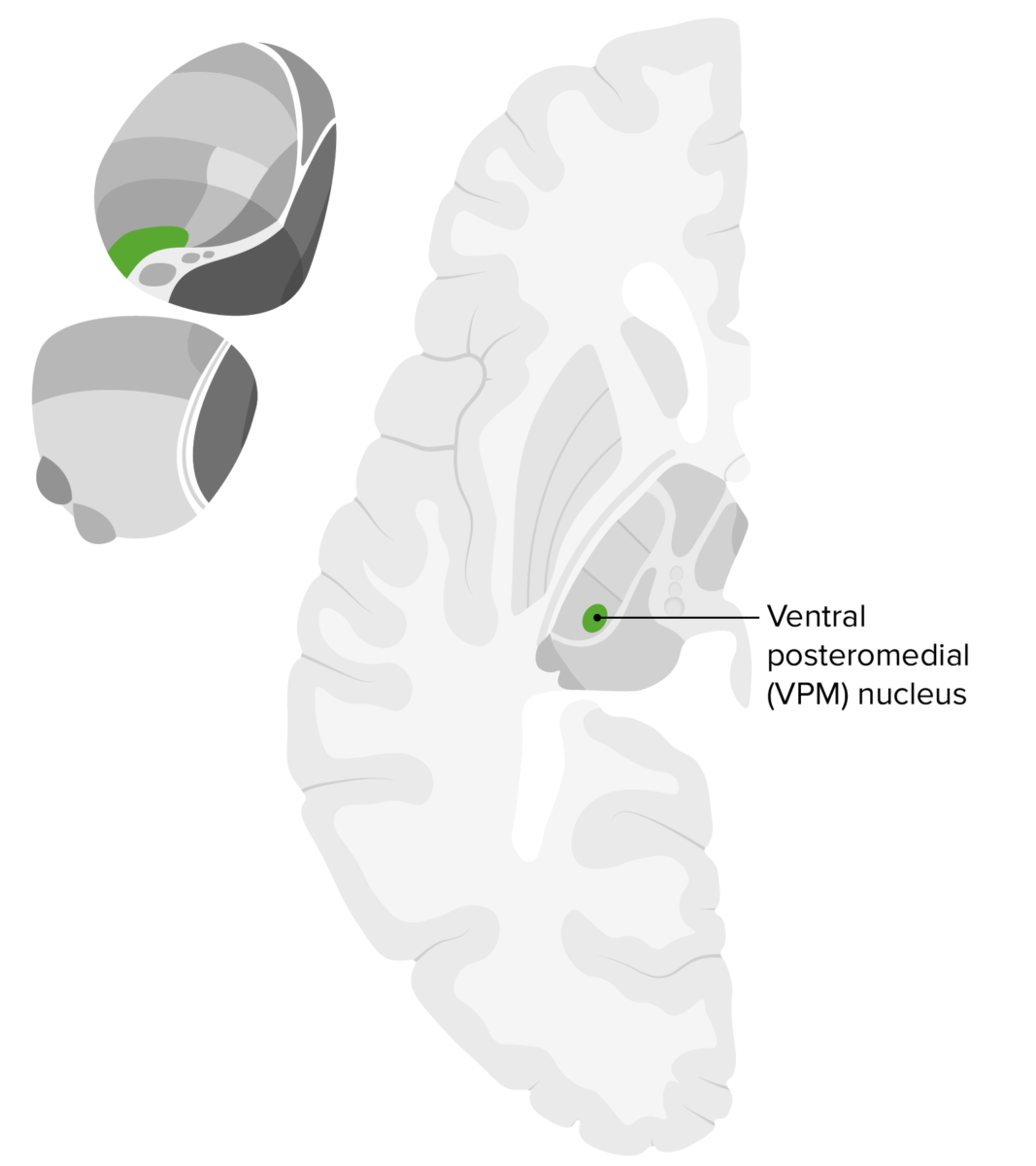
Ubicación del núcleo talámico ventral posteromedial
Imagen por Lecturio.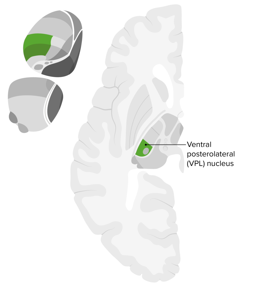
Ubicación del núcleo talámico ventral posterolateral
Imagen por Lecturio.Los LOS Neisseria núcleos ventral anterior y ventral lateral están asociados con aferencias de los LOS Neisseria ganglios basales y el cerebelo y eferencias al AL Amyloidosis lóbulo frontal Frontal The bone that forms the frontal aspect of the skull. Its flat part forms the forehead, articulating inferiorly with the nasal bone and the cheek bone on each side of the face. Skull: Anatomy. Ambos núcleos también están asociados con varias vías motoras.
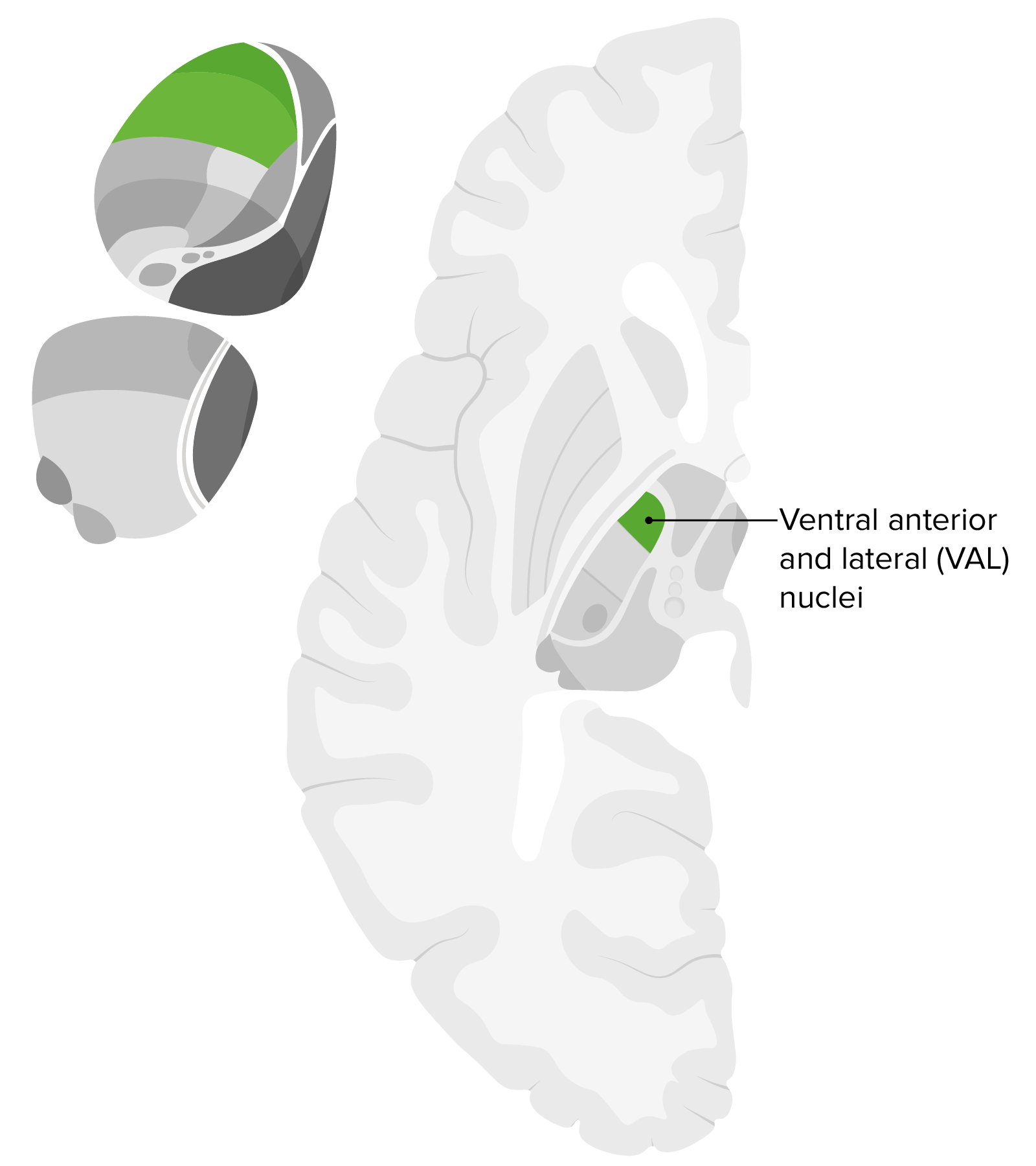
Ubicación de los núcleos ventral anterior y ventral lateral
Imagen por Lecturio.Los LOS Neisseria núcleos dorsomediales están asociados con aferencias de la amígdala, centros olfatorios y ganglios basales y eferencia a la corteza frontal Frontal The bone that forms the frontal aspect of the skull. Its flat part forms the forehead, articulating inferiorly with the nasal bone and the cheek bone on each side of the face. Skull: Anatomy. Los LOS Neisseria núcleos dorsomediales están asociados con diversas vías límbicas.

Núcleos que componen el tálamo y sus respectivas proyecciones: Nótese el núcleo dorsomedial (mediodorsal) ubicado medialmente en la cara dorsal del tálamo con proyecciones hacia la corteza prefrontal.
VA: ventral anterior
VL: ventral lateral
VPL: ventral posterolateral
VPM: ventral posteromedial
LD: lateral dorsal
LP: lateral posterior
El núcleo pulvinar Pulvinar Large mass of nuclei forming the most caudal portion of the thalamus and overhanging the geniculate bodies and the dorsolateral surface of the midbrain. It is divided into four parts: the lateral, medial, inferior, and oral pulvinar nuclei. Thalamus: Anatomy se asocia con aferencias del colículo superior, áreas visuales, complejo auditivo y otras vías sensoriales, y eferencias a las áreas de asociación parietotemporal. El núcleo pulvinar Pulvinar Large mass of nuclei forming the most caudal portion of the thalamus and overhanging the geniculate bodies and the dorsolateral surface of the midbrain. It is divided into four parts: the lateral, medial, inferior, and oral pulvinar nuclei. Thalamus: Anatomy está asociado con varias vías visuales y sensoriales.
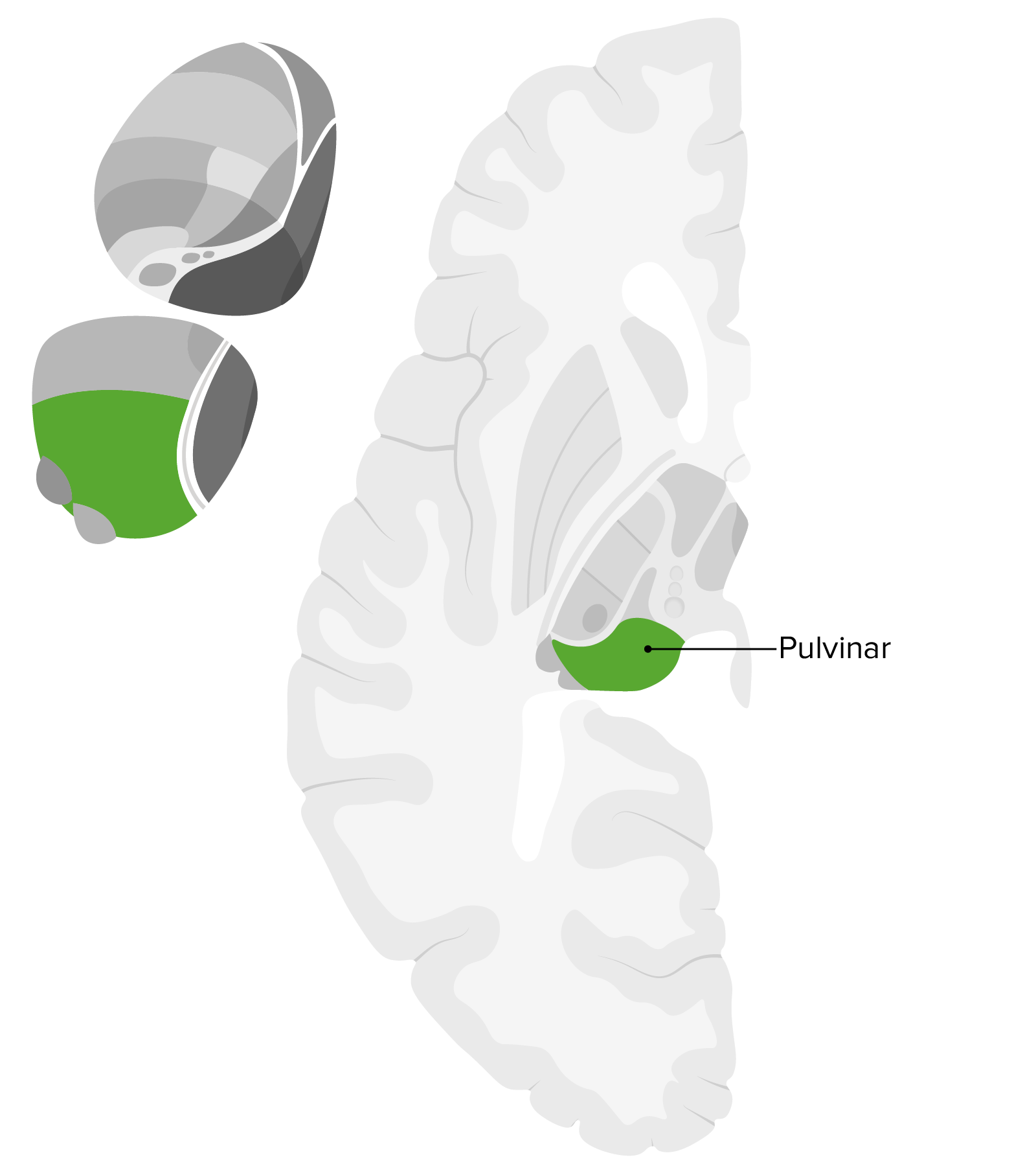
Ubicación del núcleo pulvinar, que es la expansión posterior del tálamo que sobresale del colículo superior
Imagen por Lecturio.Los LOS Neisseria cuerpos geniculados medial y lateral están asociados con las aferencias del colículo inferior y la retina Retina The ten-layered nervous tissue membrane of the eye. It is continuous with the optic nerve and receives images of external objects and transmits visual impulses to the brain. Its outer surface is in contact with the choroid and the inner surface with the vitreous body. The outermost layer is pigmented, whereas the inner nine layers are transparent. Eye: Anatomy y las eferencias del lóbulo temporal y la corteza visual, y participan en EN Erythema nodosum is an immune-mediated panniculitis (inflammation of the subcutaneous fat) caused by a type IV (delayed-type) hypersensitivity reaction. It commonly manifests in young women as tender, erythematous nodules on the shins. Erythema Nodosum varias vías auditivas y visuales.

Ubicación de los cuerpos geniculados lateral y medial del tálamo
Imagen por Lecturio.Los LOS Neisseria núcleos intralaminar y de la línea media están asociados con aferencias de la médula espinal, hipotálamo y formación reticular descendente, y eferencias a los LOS Neisseria ganglios basales y al AL Amyloidosis sistema límbico. Ambos núcleos están asociados con diversas vías emocionales y sensoriales.
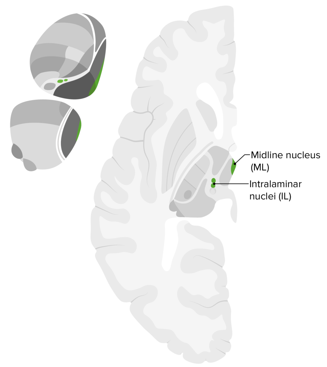
Ubicación de los núcleos intralaminar/de la línea media dentro del tálamo
Imagen por Lecturio.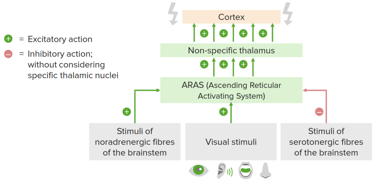
Efectos del sistema activador reticular ascendente: se observan aferencias sensoriales (visuales, auditivas, olfatorias y gustativas) que conducen a la activación del sistema activador reticular ascendente, que tiene su núcleo en el tronco del encéfalo. A su vez, el sistena actuvadir reticular ascendente envía eferencias excitatorias al tejido talámico. El tálamo transmite esta excitación a la corteza, lo que da como resultado la activación.
Imagen por Lecturio.