Domina Conceptos Médicos

Estudia para la escuela de medicina y tus examenes con Lecturio.

Sistema Ventricular: Anatomía

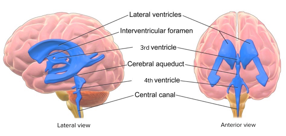
El sistema ventricular es una extensión del espacio subaracnoideo en EN Erythema nodosum is an immune-mediated panniculitis (inflammation of the subcutaneous fat) caused by a type IV (delayed-type) hypersensitivity reaction. It commonly manifests in young women as tender, erythematous nodules on the shins. Erythema Nodosum el cerebro que consiste en EN Erythema nodosum is an immune-mediated panniculitis (inflammation of the subcutaneous fat) caused by a type IV (delayed-type) hypersensitivity reaction. It commonly manifests in young women as tender, erythematous nodules on the shins. Erythema Nodosum una serie de espacios y canales interconectados. Cuatro cámaras están llenas de líquido cefalorraquídeo (LCR): los LOS Neisseria dos ventrículos laterales, el 3er ventrículo y el 4to ventrículo. Las conexiones entre las estructuras se producen a través del agujero interventricular de Monro y el acueducto cerebral (acueducto de Silvio). El agujero de Magendie y los LOS Neisseria agujeros de Luschka, son canales adicionales presentes en EN Erythema nodosum is an immune-mediated panniculitis (inflammation of the subcutaneous fat) caused by a type IV (delayed-type) hypersensitivity reaction. It commonly manifests in young women as tender, erythematous nodules on the shins. Erythema Nodosum el 4to ventrículo.

Last updated: Dec 15, 2025

Editorial responsibility: Stanley Oiseth, Lindsay Jones, Evelin Maza

Topografía y Relaciones del Sistema Ventricular

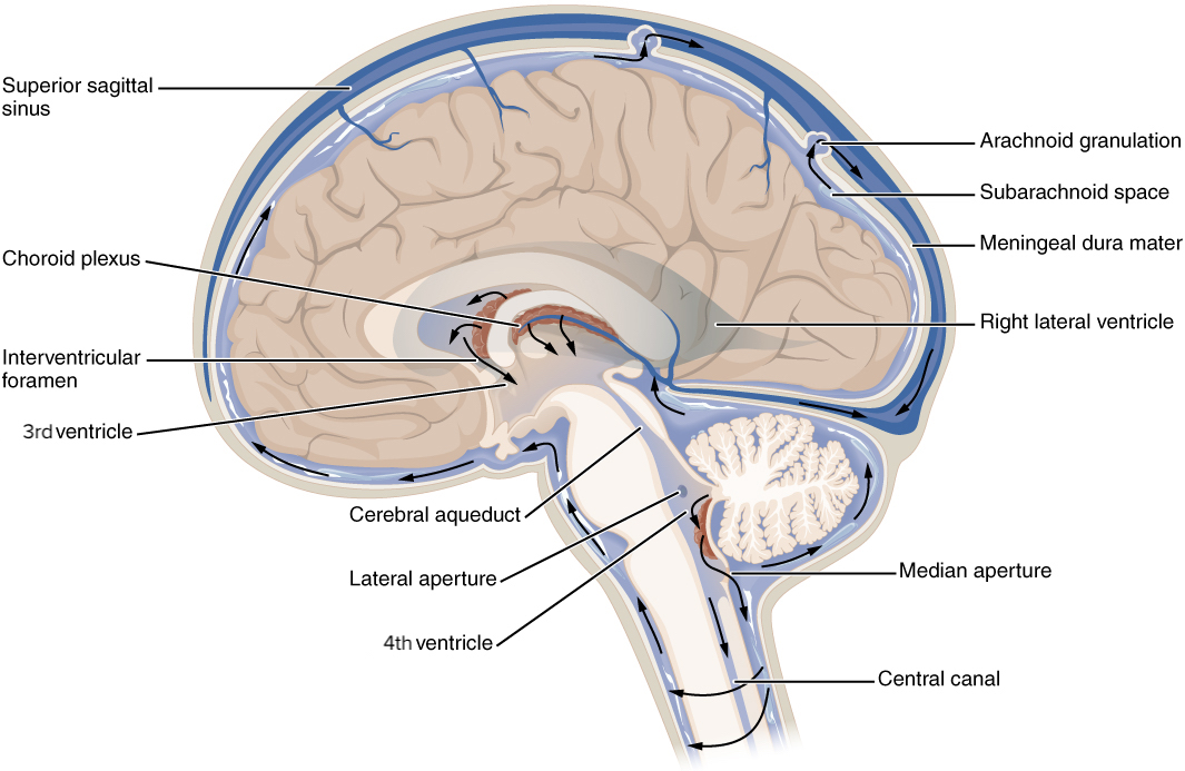
El sistema ventricular consta de cuatro ventrículos con un acueducto que conecta el tercer y el cuarto ventrículo. El líquido cefalorraquídeo (LCR) fluye a través de los LOS Neisseria ventrículos antes de entrar en EN Erythema nodosum is an immune-mediated panniculitis (inflammation of the subcutaneous fat) caused by a type IV (delayed-type) hypersensitivity reaction. It commonly manifests in young women as tender, erythematous nodules on the shins. Erythema Nodosum el espacio subaracnoideo del cerebro y la médula espinal desde el cuarto ventrículo:

  • Ventrículos laterales:
    • Cuerpo:
      • Contenidos en EN Erythema nodosum is an immune-mediated panniculitis (inflammation of the subcutaneous fat) caused by a type IV (delayed-type) hypersensitivity reaction. It commonly manifests in young women as tender, erythematous nodules on the shins. Erythema Nodosum los LOS Neisseria lóbulos frontal Frontal The bone that forms the frontal aspect of the skull. Its flat part forms the forehead, articulating inferiorly with the nasal bone and the cheek bone on each side of the face. Skull: Anatomy y parietal Parietal One of a pair of irregularly shaped quadrilateral bones situated between the frontal bone and occipital bone, which together form the sides of the cranium. Skull: Anatomy
      • Se extienden desde el agujero interventricular de Monro hasta el esplenio del cuerpo calloso
    • Cuerno anterior: en EN Erythema nodosum is an immune-mediated panniculitis (inflammation of the subcutaneous fat) caused by a type IV (delayed-type) hypersensitivity reaction. It commonly manifests in young women as tender, erythematous nodules on the shins. Erythema Nodosum el lóbulo frontal Frontal The bone that forms the frontal aspect of the skull. Its flat part forms the forehead, articulating inferiorly with the nasal bone and the cheek bone on each side of the face. Skull: Anatomy
    • Cuerno posterior: se curva posteromedialmente hacia el lóbulo occipital Occipital Part of the back and base of the cranium that encloses the foramen magnum. Skull: Anatomy
    • Cuerno inferior:
      • Compartimento más grande del ventrículo lateral
      • Se extiende hacia el lóbulo temporal
  • 3er ventrículo:
    • Una cavidad en EN Erythema nodosum is an immune-mediated panniculitis (inflammation of the subcutaneous fat) caused by a type IV (delayed-type) hypersensitivity reaction. It commonly manifests in young women as tender, erythematous nodules on the shins. Erythema Nodosum forma de hendidura en EN Erythema nodosum is an immune-mediated panniculitis (inflammation of the subcutaneous fat) caused by a type IV (delayed-type) hypersensitivity reaction. It commonly manifests in young women as tender, erythematous nodules on the shins. Erythema Nodosum la línea media
    • Derivado de la vesícula primitiva del cerebro anterior
  • Acueducto cerebral (de Silvio):
    • Pequeño tubo que se extiende a lo largo del cuarto dorsal del cerebro medio
    • Rodeado de materia gris periacueductal
    • Conecta el 3er y el 4to ventrículo
  • 4to ventrículo:
    • Entre el tronco cerebral y el cerebelo
    • Los LOS Neisseria agujeros de Luschka (aperturas laterales) y el agujero de Magendie (apertura mediana) permiten que el LCR recién producido entre en EN Erythema nodosum is an immune-mediated panniculitis (inflammation of the subcutaneous fat) caused by a type IV (delayed-type) hypersensitivity reaction. It commonly manifests in young women as tender, erythematous nodules on the shins. Erythema Nodosum el espacio subaracnoideo.
  • Órganos circunventriculares:
    • Estructuras caracterizadas por capilares abundantes y altamente permeables
    • Rodea el 3er y el 4to ventrículo
    • Estas regiones no suelen tener una barrera hematoencefálica, lo que permite la comunicación entre la sangre periférica y el LCR.
Ventrículos del cerebro

Ventrículos del cerebro

Imagen: “Ventricles of the brain” por Bruce Blaus. Licencia: CC BY 3.0, editada por Lecturio.

Plexo Coroideo y Líquido Cefalorraquídeo

Plexo coroideo

  • Piamadre vascular en EN Erythema nodosum is an immune-mediated panniculitis (inflammation of the subcutaneous fat) caused by a type IV (delayed-type) hypersensitivity reaction. It commonly manifests in young women as tender, erythematous nodules on the shins. Erythema Nodosum todos los LOS Neisseria ventrículos
  • Localización:
    • Techo de las astas temporales de los LOS Neisseria ventrículos laterales
    • Suelo del cuerpo de los LOS Neisseria ventrículos laterales
    • Foramen de Monro
    • Techo del tercer ventrículo
    • Velo medular del cuarto ventrículo, se extiende a través de los LOS Neisseria agujeros de Luschka
  • Secreta activamente LCR en EN Erythema nodosum is an immune-mediated panniculitis (inflammation of the subcutaneous fat) caused by a type IV (delayed-type) hypersensitivity reaction. It commonly manifests in young women as tender, erythematous nodules on the shins. Erythema Nodosum los LOS Neisseria ventrículos lateral, 3ro y 4to
Desarrollo del plexo coroideo

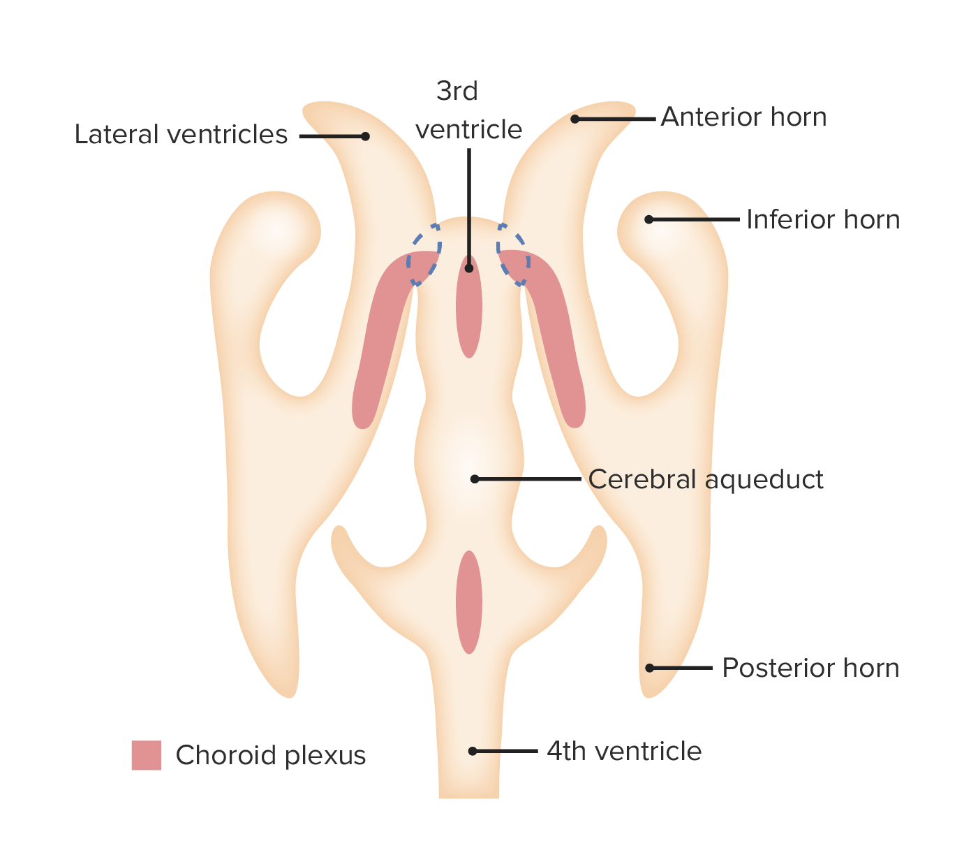
Desarrollo del plexo coroideo: El plexo coroideo se encuentra en todo el sistema ventricular y es responsable de la secreción del líquido cefalorraquídeo.

Imagen por Lecturio.

Líquido cefalorraquídeo

  • Líquido claro e incoloro que contiene una pequeña cantidad de proteínas
  • Proporciona flotabilidad y protección para el cerebro
  • Regula la concentración química de los LOS Neisseria neurotransmisores y los LOS Neisseria residuos metabólicos

Espacio Subaracnoideo y Circulación del Líquido Cefalorraquídeo

Espacio subaracnoideo

  • Entre la aracnoides y la piamadre
  • Contiene el LCR, las arterias y venas más grandes y las porciones intracraneales e intervertebrales de los LOS Neisseria nervios craneales y espinales

Circulación del líquido cefalorraquídeo

  • Tras su producción en EN Erythema nodosum is an immune-mediated panniculitis (inflammation of the subcutaneous fat) caused by a type IV (delayed-type) hypersensitivity reaction. It commonly manifests in young women as tender, erythematous nodules on the shins. Erythema Nodosum los LOS Neisseria ventrículos, el LCR fluye hacia el espacio subaracnoideo.
  • El LCR fluye libremente dentro del espacio subaracnoideo del cerebro y la médula espinal.
  • El LCR es reabsorbido por las granulaciones aracnoideas hacia la circulación venosa.
Circulación

Circulación del líquido cefalorraquídeo (LCR):
Obsérvese la salida del LCR del plexo coroideo hacia los ventrículos. El líquido cefalorraquídeo es reabsorbido por las granulaciones aracnoideas hacia la circulación venosa.

Imagen: “Circulation” por OpenStax. Licencia: CC BY 4.0, editado por Lecturio.

Relevancia Clínica

  • Hidrocefalia: condición potencialmente mortal causada por la acumulación excesiva de LCR dentro del sistema ventricular. La presentación clínica es inespecífica y puede incluir cefalea, cambios de comportamiento, retrasos en el desarrollo o náuseas y vómitos. El diagnóstico se confirma por medio de neuroimagenología (ultrasonido, TC craneal o RM) que muestra ventriculomegalia. El tratamiento es la colocación de una derivación de LCR.
  • Hidrocefalia de presión normal: agrandamiento de los ventrículos sin elevaciones detectables de la presión intracraneal. La hidrocefalia de presión normal se da con mayor frecuencia en adultos > 60 años de edad. Los síntomas incluyen una tríada de incontinencia urinaria, ataxia y disfunción cognitiva y pueden recordarse con la mnemotecnia (en inglés) “wet, wobbly, and wacky” (húmedo, tambaleante y chiflado).
  • Enfermedad isquémica microvascular crónica: enfermedad aterosclerótica crónica que suele provocar hipoperfusión en EN Erythema nodosum is an immune-mediated panniculitis (inflammation of the subcutaneous fat) caused by a type IV (delayed-type) hypersensitivity reaction. It commonly manifests in young women as tender, erythematous nodules on the shins. Erythema Nodosum zonas de la corteza cerebral de manera simétrica y que causa atrofia cerebral u otras malformaciones cerebrales. A diferencia de la hidrocefalia, la presión intracraneal no está elevada.
  • Malformaciones de Chiari: grupo de trastornos definidos por déficits estructurales en EN Erythema nodosum is an immune-mediated panniculitis (inflammation of the subcutaneous fat) caused by a type IV (delayed-type) hypersensitivity reaction. It commonly manifests in young women as tender, erythematous nodules on the shins. Erythema Nodosum el cerebro y la médula espinal que conducen a un espacio limitado en EN Erythema nodosum is an immune-mediated panniculitis (inflammation of the subcutaneous fat) caused by a type IV (delayed-type) hypersensitivity reaction. It commonly manifests in young women as tender, erythematous nodules on the shins. Erythema Nodosum la fosa posterior y obligan a las estructuras cerebelosas a sobresalir a través del foramen magno. Las malformaciones de Chiari tipo II se observan en EN Erythema nodosum is an immune-mediated panniculitis (inflammation of the subcutaneous fat) caused by a type IV (delayed-type) hypersensitivity reaction. It commonly manifests in young women as tender, erythematous nodules on the shins. Erythema Nodosum combinación con la hernia Hernia Protrusion of tissue, structure, or part of an organ through the bone, muscular tissue, or the membrane by which it is normally contained. Hernia may involve tissues such as the abdominal wall or the respiratory diaphragm. Hernias may be internal, external, congenital, or acquired. Abdominal Hernias del vermis cerebeloso, el tronco cerebral y el cuarto ventrículo en EN Erythema nodosum is an immune-mediated panniculitis (inflammation of the subcutaneous fat) caused by a type IV (delayed-type) hypersensitivity reaction. It commonly manifests in young women as tender, erythematous nodules on the shins. Erythema Nodosum el foramen magno. Existe una asociación con el mielomeningocele y múltiples anomalías cerebrales, incluyendo la hidrocefalia y la siringomielia.

Referencias

  1. Kahle, K. T., Kulkarni, A. V., Limbrick, D. D., & Warf, B. C. (2016). Hydrocephalus in children. Lancet, The, 387(10020), 788–799. http://dx.doi.org/10.1016/S0140-6736(15)60694-8
  2. Blázquez, M., & Zarranz, J. J. (2018). Síndrome meníngeo. edema cerebral. hipertensión intracraneal. hidrocefalias. hipotensión intracraneal. In J. J. Zarranz (Ed.), Neurología (pp. 219-233). https://www.clinicalkey.es/#!/content/3-s2.0-B978849113071000012X
  3. Sato, O., Yamguchi, T., Kittaka, M., & Toyama, H. (2001). Hydrocephalus and epilepsy. Child’s nervous system: ChNS: official journal of the International Society for Pediatric Neurosurgery, 17(1-2), 76–86.
  4. Tubbs, R. S., & Oakes, W. J. (2017). Chiari malformations. Neurological surgery. pp. 1531–1540. http://dx.doi.org/10.1016/B978-0-323-28782-1.00190-8
  5. Schijman, E. History, anatomic forms, and pathogenesis of Chiari I malformations. Childs Nerv Syst 20, 323–328 (2004). https://doi.org/10.1007/s00381-003-0878-y
  6. Khoury, C. (2020). Chiari malformations. UpToDate. Retrieved December 1, 2020 from https://www.uptodate.com/contents/chiari-malformations
  7. Abd-El-Barr M. M., Strong C.I., Groff M. W. Chiari malformations: diagnosis, treatments and failures. J Neurosurg Sci. 2014 Dec. 58 (4):215–21.
  8. McClugage, S and Oakes, J. The Chiari I malformation. JNSPG 75th Anniversary Invited Review Article. https://doi.org/10.3171/2019.5.PEDS18382

¡Crea tu cuenta gratis o inicia una sesión para seguir leyendo!

Regístrate ahora y obtén acceso gratuito a Lecturio con páginas de concepto, videos médicos y cuestionarios para tu educación médica.

User Reviews

Que tengas una sesión de estudio alegre y navideña 🎁 Ahorra 50% en todos los planes >>

Details