El molusco contagioso es una infección vírica limitada a la epidermis Epidermis The external, nonvascular layer of the skin. It is made up, from within outward, of five layers of epithelium: (1) basal layer (stratum basale epidermidis); (2) spinous layer (stratum spinosum epidermidis); (3) granular layer (stratum granulosum epidermidis); (4) clear layer (stratum lucidum epidermidis); and (5) horny layer (stratum corneum epidermidis). Skin: Structure and Functions, normalmente sin manifestaciones sistémicas. Esta infección es frecuente en EN Erythema nodosum is an immune-mediated panniculitis (inflammation of the subcutaneous fat) caused by a type IV (delayed-type) hypersensitivity reaction. It commonly manifests in young women as tender, erythematous nodules on the shins. Erythema Nodosum niños menores de 5 años, aunque también puede observarse en EN Erythema nodosum is an immune-mediated panniculitis (inflammation of the subcutaneous fat) caused by a type IV (delayed-type) hypersensitivity reaction. It commonly manifests in young women as tender, erythematous nodules on the shins. Erythema Nodosum adolescentes y adultos sanos, normalmente relacionada con deportes de contacto o, con lesiones en EN Erythema nodosum is an immune-mediated panniculitis (inflammation of the subcutaneous fat) caused by a type IV (delayed-type) hypersensitivity reaction. It commonly manifests in young women as tender, erythematous nodules on the shins. Erythema Nodosum los LOS Neisseria genitales, como infección de transmisión sexual (ITS). Las lesiones aparecen como pápulas agrupadas, de color piel, en EN Erythema nodosum is an immune-mediated panniculitis (inflammation of the subcutaneous fat) caused by a type IV (delayed-type) hypersensitivity reaction. It commonly manifests in young women as tender, erythematous nodules on the shins. Erythema Nodosum forma de cúpula, con umbilicación central. El molusco contagioso es leve en EN Erythema nodosum is an immune-mediated panniculitis (inflammation of the subcutaneous fat) caused by a type IV (delayed-type) hypersensitivity reaction. It commonly manifests in young women as tender, erythematous nodules on the shins. Erythema Nodosum pacientes inmunocompetentes y se resuelve por sí solo en EN Erythema nodosum is an immune-mediated panniculitis (inflammation of the subcutaneous fat) caused by a type IV (delayed-type) hypersensitivity reaction. It commonly manifests in young women as tender, erythematous nodules on the shins. Erythema Nodosum unos meses. Las personas inmunodeprimidas presentan lesiones extensas que requieren tratamiento. El molusco contagioso es altamente transmisible, por lo que la educación del paciente es clave en EN Erythema nodosum is an immune-mediated panniculitis (inflammation of the subcutaneous fat) caused by a type IV (delayed-type) hypersensitivity reaction. It commonly manifests in young women as tender, erythematous nodules on the shins. Erythema Nodosum su tratamiento. La crioterapia con nitrógeno líquido es el tratamiento de primera línea.
Last updated: Dec 15, 2025
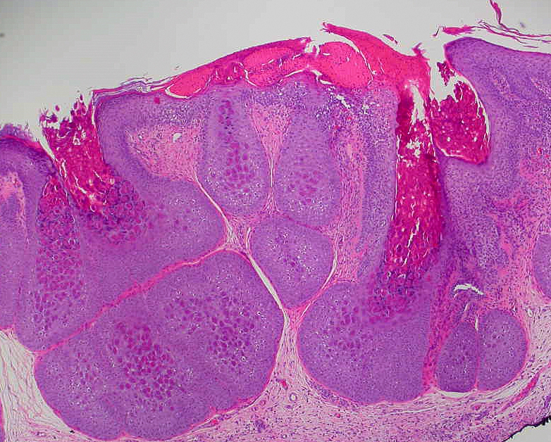
Cuerpos de Henderson-Paterson:
Rasgos histológicos característicos del molusco contagioso. Los cuerpos de Henderson-Paterson son inclusiones visibles en los queratinocitos de las capas basal, espinosa y granular de la epidermis.
Manifestación dermatológica del molusco contagioso:
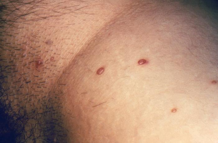
En la región inguinal se encuentran pápulas nacaradas de color carne con umbilicación central, lo que sugiere molusco contagioso.
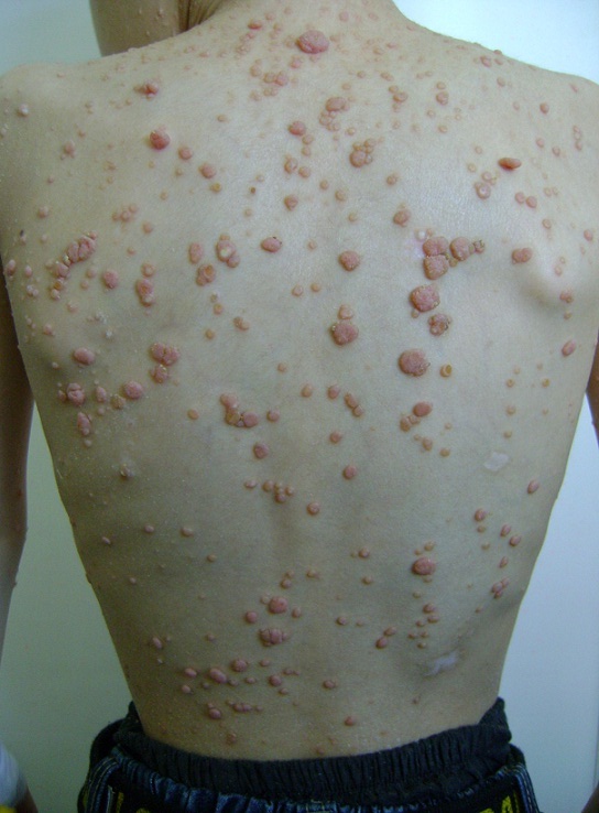
Múltiples lesiones de molusco contagioso en la espalda de un niño:
Pápulas nacaradas color carne con umbilicación central