Las lesiones de losLOSNeisseria ligamentos de la rodilla se observan comúnmente enENErythema nodosum is an immune-mediated panniculitis (inflammation of the subcutaneous fat) caused by a type IV (delayed-type) hypersensitivity reaction. It commonly manifests in young women as tender, erythematous nodules on the shins.Erythema Nodosum atletas jóvenes y enENErythema nodosum is an immune-mediated panniculitis (inflammation of the subcutaneous fat) caused by a type IV (delayed-type) hypersensitivity reaction. It commonly manifests in young women as tender, erythematous nodules on the shins.Erythema Nodosum adultos de mediana edad. Aunque la presentación clínica varía para cada ligamento lesionado, todas estas lesiones se presentan con inestabilidad articular, dolorDolorInflammation y dificultad para soportar peso. El diagnóstico se basa enENErythema nodosum is an immune-mediated panniculitis (inflammation of the subcutaneous fat) caused by a type IV (delayed-type) hypersensitivity reaction. It commonly manifests in young women as tender, erythematous nodules on the shins.Erythema Nodosum el examen clínico y se confirma con imagenología o visualización directa (artroscopia). El tratamiento puede ser conservador o quirúrgico, dependiendo de la gravedad de la lesión.
Una lesión del ligamento cruzado anterior provoca un daño estructural enENErythema nodosum is an immune-mediated panniculitis (inflammation of the subcutaneous fat) caused by a type IV (delayed-type) hypersensitivity reaction. It commonly manifests in young women as tender, erythematous nodules on the shins.Erythema Nodosum el ligamento cruzado anterior. Las funciones del ligamento son:
Controlar la traslación anterior de la tibiaTibiaThe second longest bone of the skeleton. It is located on the medial side of the lower leg, articulating with the fibula laterally, the talus distally, and the femur proximally.Knee Joint: Anatomy sobre el fémur
Restringir la rotación interna/externa de la tibiaTibiaThe second longest bone of the skeleton. It is located on the medial side of the lower leg, articulating with the fibula laterally, the talus distally, and the femur proximally.Knee Joint: Anatomy sobre el fémur
Restringir la desviación enENErythema nodosum is an immune-mediated panniculitis (inflammation of the subcutaneous fat) caused by a type IV (delayed-type) hypersensitivity reaction. It commonly manifests in young women as tender, erythematous nodules on the shins.Erythema Nodosum varo/valgo de la tibiaTibiaThe second longest bone of the skeleton. It is located on the medial side of the lower leg, articulating with the fibula laterally, the talus distally, and the femur proximally.Knee Joint: Anatomy sobre el fémur
Anatomía
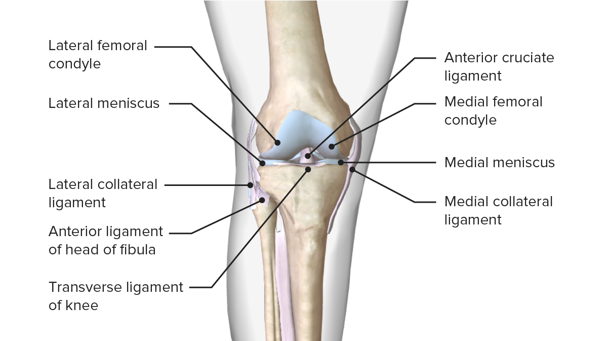
Se origina enENErythema nodosum is an immune-mediated panniculitis (inflammation of the subcutaneous fat) caused by a type IV (delayed-type) hypersensitivity reaction. It commonly manifests in young women as tender, erythematous nodules on the shins.Erythema Nodosum la cara posteromedial del cóndilo femoral lateral, posterior alALAmyloidosis eje longitudinal del fémur
Discurre inferior, medial y anteriormente
Se fija a la cara anteromedial de la tibiaTibiaThe second longest bone of the skeleton. It is located on the medial side of the lower leg, articulating with the fibula laterally, the talus distally, and the femur proximally.Knee Joint: Anatomy entre losLOSNeisseria cóndilos.
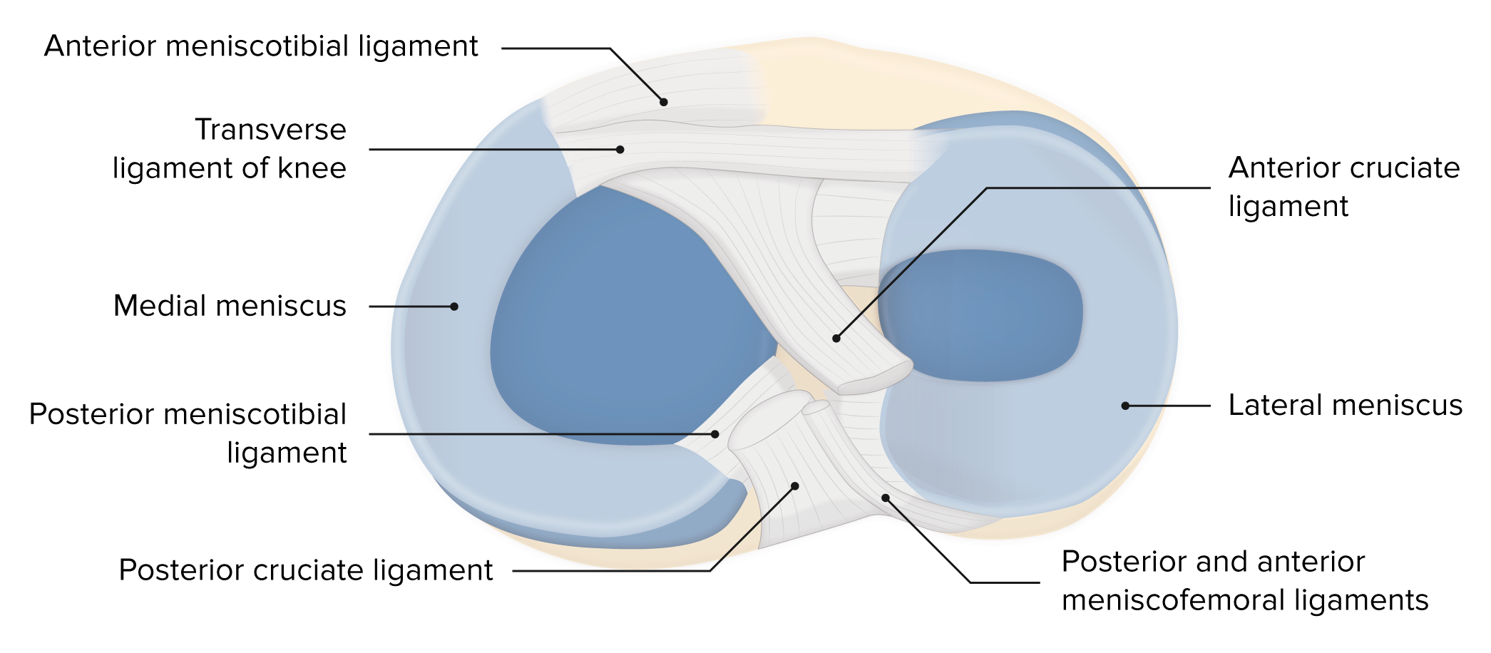
Imagen que muestra los meniscos y su relación con otras superficies articulares que componen la articulación de la rodilla
Incidencia: 68,6 por 100 000/año enENErythema nodosum is an immune-mediated panniculitis (inflammation of the subcutaneous fat) caused by a type IV (delayed-type) hypersensitivity reaction. It commonly manifests in young women as tender, erythematous nodules on the shins.Erythema NodosumlosLOSNeisseria Estados Unidos
Pico de incidencia enENErythema nodosum is an immune-mediated panniculitis (inflammation of the subcutaneous fat) caused by a type IV (delayed-type) hypersensitivity reaction. It commonly manifests in young women as tender, erythematous nodules on the shins.Erythema Nodosum hombres: entre 19 y 25 años; relacionado con losLOSNeisseria deportes
Incidencia máxima enENErythema nodosum is an immune-mediated panniculitis (inflammation of the subcutaneous fat) caused by a type IV (delayed-type) hypersensitivity reaction. It commonly manifests in young women as tender, erythematous nodules on the shins.Erythema Nodosum mujeres: 14–18 años
Ligamento más comúnmente lesionado enENErythema nodosum is an immune-mediated panniculitis (inflammation of the subcutaneous fat) caused by a type IV (delayed-type) hypersensitivity reaction. It commonly manifests in young women as tender, erythematous nodules on the shins.Erythema Nodosum la rodilla
Mecanismo de contacto (trauma cerrado directo enENErythema nodosum is an immune-mediated panniculitis (inflammation of the subcutaneous fat) caused by a type IV (delayed-type) hypersensitivity reaction. It commonly manifests in young women as tender, erythematous nodules on the shins.Erythema Nodosum la rodilla): jugadores de fútbol
Presentación clínica
LosLOSNeisseria pacientes con una lesión del ligamento cruzado anterior se presentan con dolorDolorInflammation e informan de un traumatismo cerrado reciente de alta energía enENErythema nodosum is an immune-mediated panniculitis (inflammation of the subcutaneous fat) caused by a type IV (delayed-type) hypersensitivity reaction. It commonly manifests in young women as tender, erythematous nodules on the shins.Erythema Nodosum la rodilla.
Antecedentes:
El paciente informa que escuchó o sintió un “chasquido” y que la rodilla afectada “cedió” enENErythema nodosum is an immune-mediated panniculitis (inflammation of the subcutaneous fat) caused by a type IV (delayed-type) hypersensitivity reaction. It commonly manifests in young women as tender, erythematous nodules on the shins.Erythema Nodosum el momento de la lesión.
También es importante detallar:
Momento
Mecanismo del trauma
Sitio de la lesión
Más detalles de la situación específica
Examen físico:
Marcha antálgica
Sensibilidad a la palpación a lo largo de la línea articular
EdemaEdemaEdema is a condition in which excess serous fluid accumulates in the body cavity or interstitial space of connective tissues. Edema is a symptom observed in several medical conditions. It can be categorized into 2 types, namely, peripheral (in the extremities) and internal (in an organ or body cavity). Edema alrededor de la articulación de la rodilla
Disminución del rango de movimiento
Pruebas de inestabilidad de la rodilla:
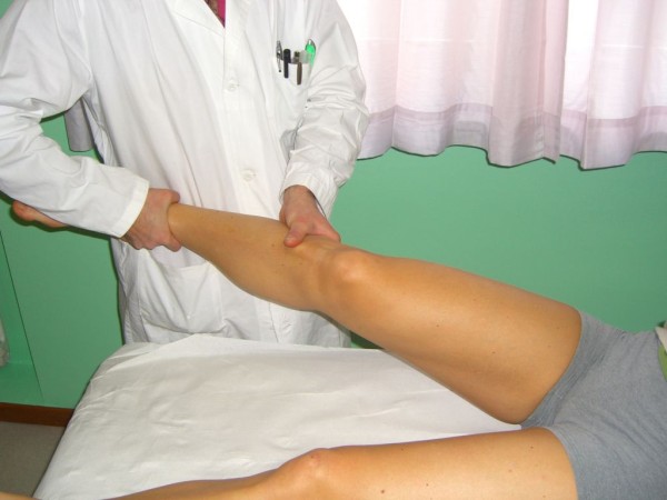
Maniobra de cajón anterior:
El paciente se acuesta enENErythema nodosum is an immune-mediated panniculitis (inflammation of the subcutaneous fat) caused by a type IV (delayed-type) hypersensitivity reaction. It commonly manifests in young women as tender, erythematous nodules on the shins.Erythema Nodosum decúbito supino con las caderas flexionadas a 45 grados, la rodilla enENErythema nodosum is an immune-mediated panniculitis (inflammation of the subcutaneous fat) caused by a type IV (delayed-type) hypersensitivity reaction. It commonly manifests in young women as tender, erythematous nodules on the shins.Erythema Nodosum un ángulo de 90 grados y losLOSNeisseria pies planos sobre la mesa de exploración.
El médico estabiliza la pierna sentándose sobre losLOSNeisseria dedos del pie del paciente y sujeta la tibiaTibiaThe second longest bone of the skeleton. It is located on the medial side of the lower leg, articulating with the fibula laterally, the talus distally, and the femur proximally.Knee Joint: Anatomy proximal con ambas manos y tira hacia delante.
Prueba positiva: la tibiaTibiaThe second longest bone of the skeleton. It is located on the medial side of the lower leg, articulating with the fibula laterally, the talus distally, and the femur proximally.Knee Joint: Anatomy proximal se deslizará anteriormente, como si se abriera un cajón.
Prueba de cambio de pivote:
El paciente se acuesta enENErythema nodosum is an immune-mediated panniculitis (inflammation of the subcutaneous fat) caused by a type IV (delayed-type) hypersensitivity reaction. It commonly manifests in young women as tender, erythematous nodules on the shins.Erythema Nodosum decúbito supino con la rodilla completamente extendida.
El médico aplica una fuerza de rotación interna y enENErythema nodosum is an immune-mediated panniculitis (inflammation of the subcutaneous fat) caused by a type IV (delayed-type) hypersensitivity reaction. It commonly manifests in young women as tender, erythematous nodules on the shins.Erythema Nodosum valgo sobre la tibiaTibiaThe second longest bone of the skeleton. It is located on the medial side of the lower leg, articulating with the fibula laterally, the talus distally, and the femur proximally.Knee Joint: Anatomy proximal mientras lleva la rodilla a una flexión pasiva.
La prueba es positiva si la rodilla se vuelve inestable y subluxada; incluso podría producirse un sonido de “golpe”.
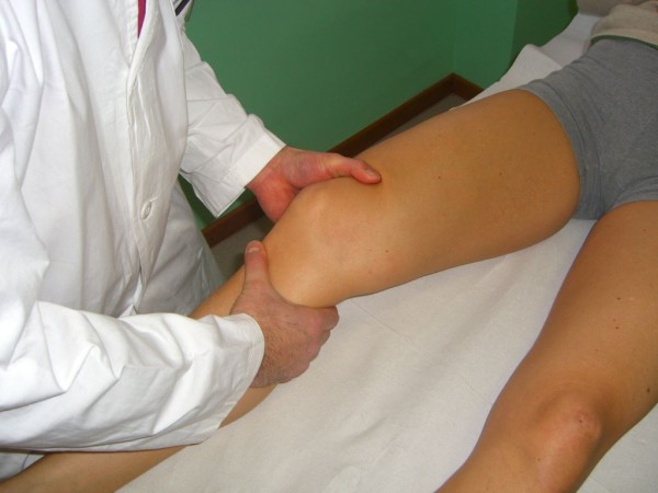
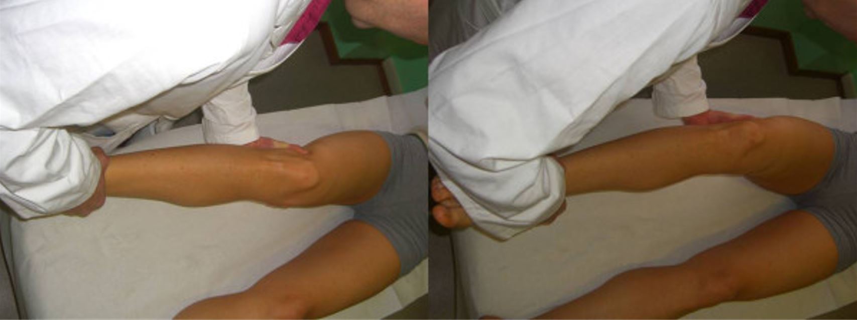
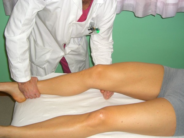
Prueba de Lachman:
El paciente se acuesta enENErythema nodosum is an immune-mediated panniculitis (inflammation of the subcutaneous fat) caused by a type IV (delayed-type) hypersensitivity reaction. It commonly manifests in young women as tender, erythematous nodules on the shins.Erythema Nodosum decúbito supino con la rodilla afectada enENErythema nodosum is an immune-mediated panniculitis (inflammation of the subcutaneous fat) caused by a type IV (delayed-type) hypersensitivity reaction. It commonly manifests in young women as tender, erythematous nodules on the shins.Erythema Nodosum un ángulo de 30 grados.
El médico estabiliza el fémur distal con la mano izquierda y tira de la tibiaTibiaThe second longest bone of the skeleton. It is located on the medial side of the lower leg, articulating with the fibula laterally, the talus distally, and the femur proximally.Knee Joint: Anatomy hacia sí mismo.
Prueba positiva: desplazamiento anterior
Prueba del cajón anterior
Imagen: “Anterior Drawer test” por Rossi R, Dettoni F, Bruzzone M, Cottino U, D’Elicio DG, Bonasia DE. Licencia: License: CC BY 2.0
Prueba de cambio de pivote
Imagen: “Pivot Shift (Jerk) Test” por Rossi R, Dettoni F, Bruzzone M, Cottino U, D’Elicio DG, Bonasia DE. Licencia: CC BY 2.0
Prueba de Lachman
Imagen: “Lachman Test” por Rossi R, Dettoni F, Bruzzone M, Cottino U, D’Elicio DG, Bonasia DE. Licencia: CC BY 2.0
Diagnóstico
El diagnóstico se haceHACEAltitude Sickness clínicamente y se confirma con imagenología.
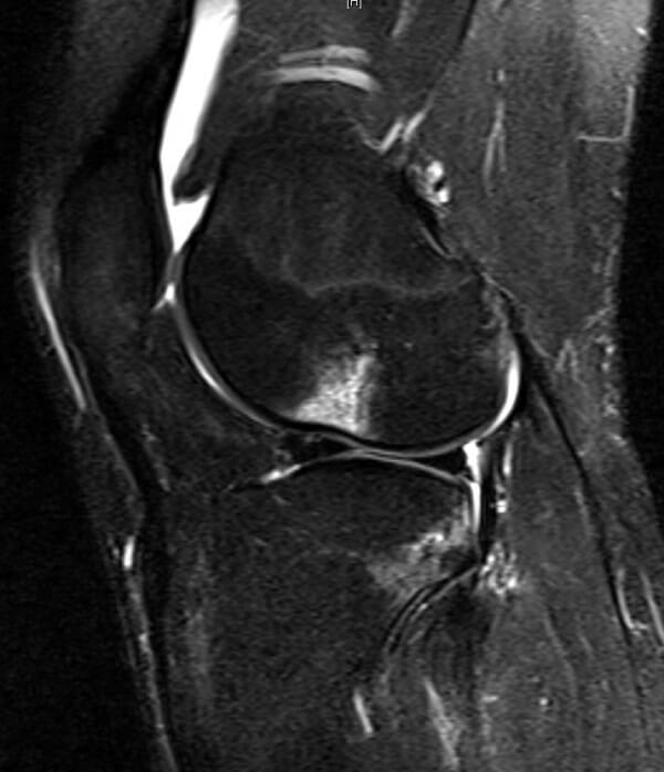
La RM es el método de elección:
Signos primarios:
Hiperintensidad del ligamento cruzado anterior (generalmente enENErythema nodosum is an immune-mediated panniculitis (inflammation of the subcutaneous fat) caused by a type IV (delayed-type) hypersensitivity reaction. It commonly manifests in young women as tender, erythematous nodules on the shins.Erythema Nodosum la porción media)
Discontinuidad de las fibras del ligamento
Alteración de la línea de Blumensaat
EdemaEdemaEdema is a condition in which excess serous fluid accumulates in the body cavity or interstitial space of connective tissues. Edema is a symptom observed in several medical conditions. It can be categorized into 2 types, namely, peripheral (in the extremities) and internal (in an organ or body cavity). Edema
Signos secundarios:
EdemaEdemaEdema is a condition in which excess serous fluid accumulates in the body cavity or interstitial space of connective tissues. Edema is a symptom observed in several medical conditions. It can be categorized into 2 types, namely, peripheral (in the extremities) and internal (in an organ or body cavity). Edema de médula ósea
Lesión del ligamento colateral medial asociada
Traslación tibial anterior > 7 mm
La radiografía es útil solo para descartar fracturas.
Artroscopia: utiliza un instrumento (artroscopio) que se inserta enENErythema nodosum is an immune-mediated panniculitis (inflammation of the subcutaneous fat) caused by a type IV (delayed-type) hypersensitivity reaction. It commonly manifests in young women as tender, erythematous nodules on the shins.Erythema Nodosum la articulación a través de una pequeña incisión
Estándar de oro para el diagnóstico
Procedimiento quirúrgico mínimamente invasivo utilizado para el diagnóstico; también se puede utilizar para el tratamiento de desgarros de menisco
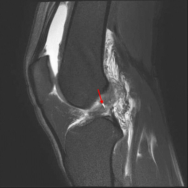
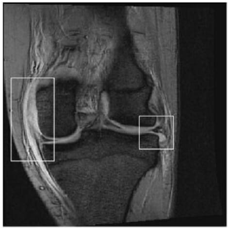
En la imagen sagital de densidad protónica, el ligamento cruzado anterior (LCA) no se ve en la muesca de la rodilla (flecha). La no visualización se define como la imposibilidad de visualizar el LCA en la imagen.
Imagen: “In this proton density sagittal image” por Chang et al. Licencia: CC BY 2.0
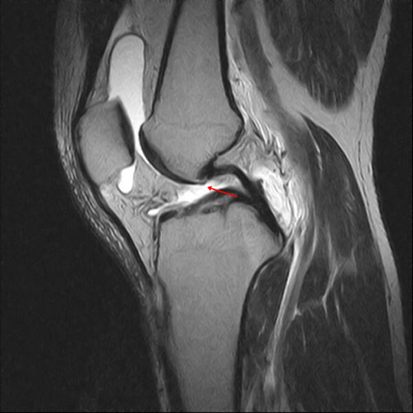
Imagen sagital ponderada en T2, con supresión de grasa, que muestra la discontinuidad de las fibras del ligamento cruzado anterior (LCA) (flecha): La discontinuidad se define como una brecha o interrupción focal de las fibras del LCA.
Imagen: “In this T2-weighted fat-suppressed sagittal image” por Chang et al. Licencia: CC BY 2.0
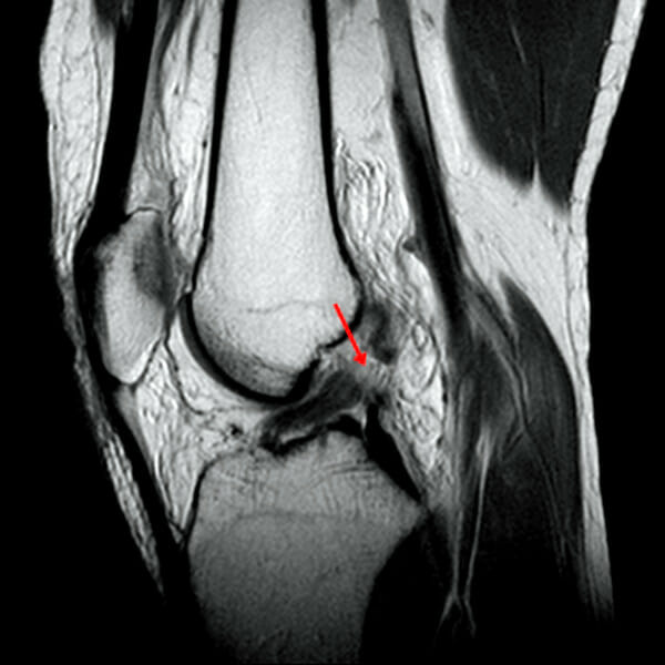
Imagen sagital ponderada en T2, con supresión de grasa, que muestra una intensidad de señal anormal como aumento de la intensidad de la señal dentro del ligamento cruzado anterior (flecha)
Imagen: “In this T2-weighted fat-suppressed sagittal image” por Chang et al. Licencia: CC BY 2.0
Imagen sagital ponderada en T2, con supresión de grasa, que muestra contusiones óseas en el cóndilo femoral lateral y la meseta tibial posterolateral
Imagen: “Sagittal fat-suppressed T2-weighted image” por Chang et al. Licencia: CC BY 2.0
Tratamiento
Tratamiento conservador: enENErythema nodosum is an immune-mediated panniculitis (inflammation of the subcutaneous fat) caused by a type IV (delayed-type) hypersensitivity reaction. It commonly manifests in young women as tender, erythematous nodules on the shins.Erythema Nodosum pacientes con baja demanda funcional
Rest (descanso), Ice (hielo), Compression (compresión), y Elevation (elevación) (RICE, enENErythema nodosum is an immune-mediated panniculitis (inflammation of the subcutaneous fat) caused by a type IV (delayed-type) hypersensitivity reaction. It commonly manifests in young women as tender, erythematous nodules on the shins.Erythema Nodosum inglés)
Sin soporte de peso (usar muletas o silla de ruedas)
La consulta a cirugía ortopédica y el tratamiento quirúrgico para la reconstrucción mediante injerto de tejido se realiza enENErythema nodosum is an immune-mediated panniculitis (inflammation of the subcutaneous fat) caused by a type IV (delayed-type) hypersensitivity reaction. It commonly manifests in young women as tender, erythematous nodules on the shins.Erythema Nodosum:
Atletas
Pacientes jóvenes y activos
Inestabilidad significativa de la rodilla
Lesión de múltiples estructuras de la rodilla
Rehabilitación con fisioterapia:
Mejoría de la funcionalidad
Aumenta la estabilidad de la rodilla.
Pronóstico: el 90% de losLOSNeisseria pacientes volverán a una función normal después de la reparación.
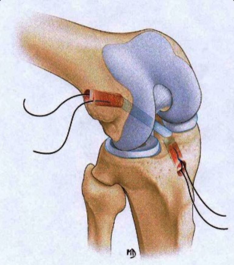
Reconstrucción anatómica del ligamento cruzado anterior con injerto de hueso-tendón rotuliano-hueso
Imagen: “BTB reconstruction” por Branch T, Lavoie F, Guier C, Branch E, Lording T, Stinton S, Neyret P. Licencia: CC BY 4.0 , recortada por Lecturio.
Diagnóstico diferencial del desgarro del ligamento cruzado anterior
Desgarro de menisco: lesión del menisco causada por fuerzas de rotación o cizallamiento a través de la articulación tibiofemoral. La presentación clínica incluye antecedentes de una lesión por torsión o rotación seguida de dolorDolorInflammationenENErythema nodosum is an immune-mediated panniculitis (inflammation of the subcutaneous fat) caused by a type IV (delayed-type) hypersensitivity reaction. It commonly manifests in young women as tender, erythematous nodules on the shins.Erythema Nodosum la línea articular asociado con un pequeño derrame. Algunos pacientes también pueden presentar síntomas mecánicos, como bloqueo articular, chasquidos o enganches.
Luxación posterior de la rodilla: pérdida patológica de la articulación de la rodilla debido a traumatismos de alta energía, como losLOSNeisseria causados por colisiones de vehículos motorizados, o traumatismos de baja energía, como losLOSNeisseria que se observan enENErythema nodosum is an immune-mediated panniculitis (inflammation of the subcutaneous fat) caused by a type IV (delayed-type) hypersensitivity reaction. It commonly manifests in young women as tender, erythematous nodules on the shins.Erythema Nodosum el entrenamiento deportivo.
Fractura de columna tibial o epífisis femoral o tibial: diagnosticada enENErythema nodosum is an immune-mediated panniculitis (inflammation of the subcutaneous fat) caused by a type IV (delayed-type) hypersensitivity reaction. It commonly manifests in young women as tender, erythematous nodules on the shins.Erythema Nodosum radiografía y manejada por un especialista enENErythema nodosum is an immune-mediated panniculitis (inflammation of the subcutaneous fat) caused by a type IV (delayed-type) hypersensitivity reaction. It commonly manifests in young women as tender, erythematous nodules on the shins.Erythema Nodosum ortopedia.
Lesión del ligamento colateral medial de la rodilla: provoca un daño estructural enENErythema nodosum is an immune-mediated panniculitis (inflammation of the subcutaneous fat) caused by a type IV (delayed-type) hypersensitivity reaction. It commonly manifests in young women as tender, erythematous nodules on the shins.Erythema Nodosum el ligamento colateral medial (su función es proporcionar estabilidad enENErythema nodosum is an immune-mediated panniculitis (inflammation of the subcutaneous fat) caused by a type IV (delayed-type) hypersensitivity reaction. It commonly manifests in young women as tender, erythematous nodules on the shins.Erythema Nodosum valgo a la rodilla)
Lesión del Ligamento Colateral Medial
Definición
Una lesión del ligamento colateral medial provoca un daño estructural enENErythema nodosum is an immune-mediated panniculitis (inflammation of the subcutaneous fat) caused by a type IV (delayed-type) hypersensitivity reaction. It commonly manifests in young women as tender, erythematous nodules on the shins.Erythema Nodosum el ligamento cuya función es proporcionar estabilidad enENErythema nodosum is an immune-mediated panniculitis (inflammation of the subcutaneous fat) caused by a type IV (delayed-type) hypersensitivity reaction. It commonly manifests in young women as tender, erythematous nodules on the shins.Erythema Nodosum valgo a la rodilla.
Tríada infeliz: lesión simultánea del ligamento colateral medial, ligamento cruzado anterior y el menisco medial
Anatomía
Se origina enENErythema nodosum is an immune-mediated panniculitis (inflammation of the subcutaneous fat) caused by a type IV (delayed-type) hypersensitivity reaction. It commonly manifests in young women as tender, erythematous nodules on the shins.Erythema Nodosum el epicóndilo medial del fémur y se inserta enENErythema nodosum is an immune-mediated panniculitis (inflammation of the subcutaneous fat) caused by a type IV (delayed-type) hypersensitivity reaction. It commonly manifests in young women as tender, erythematous nodules on the shins.Erythema Nodosum el cóndilo medial de la tibiaTibiaThe second longest bone of the skeleton. It is located on the medial side of the lower leg, articulating with the fibula laterally, the talus distally, and the femur proximally.Knee Joint: Anatomy
Se fija enENErythema nodosum is an immune-mediated panniculitis (inflammation of the subcutaneous fat) caused by a type IV (delayed-type) hypersensitivity reaction. It commonly manifests in young women as tender, erythematous nodules on the shins.Erythema Nodosum 2 lugares enENErythema nodosum is an immune-mediated panniculitis (inflammation of the subcutaneous fat) caused by a type IV (delayed-type) hypersensitivity reaction. It commonly manifests in young women as tender, erythematous nodules on the shins.Erythema Nodosum la tibiaTibiaThe second longest bone of the skeleton. It is located on the medial side of the lower leg, articulating with the fibula laterally, the talus distally, and the femur proximally.Knee Joint: Anatomy
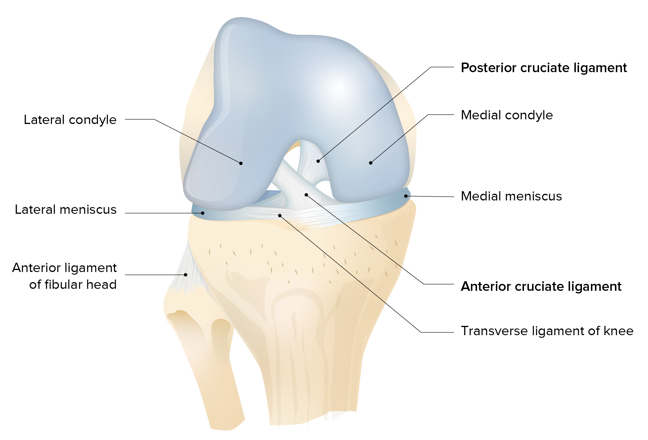
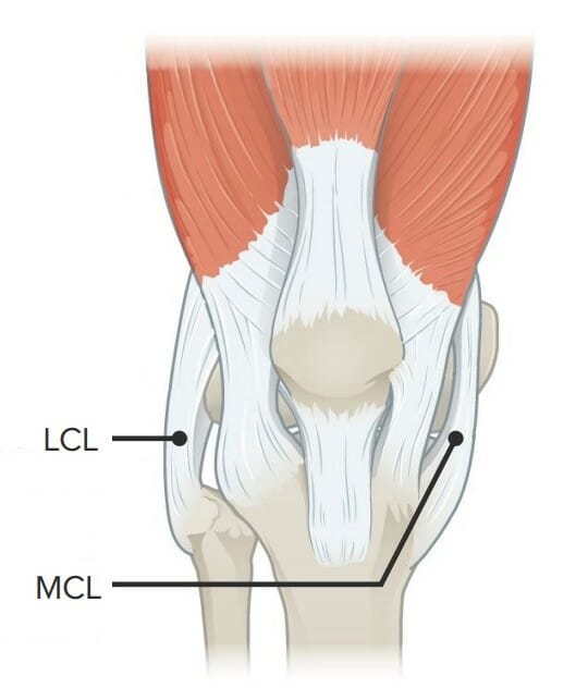
Imagen que muestra el ligamento colateral lateral (LCL) y el ligamento colateral medial (MCL)
Imagen por Lecturio.
Epidemiología
40% de todas las lesiones de rodilla
60% de las lesiones de rodilla alALAmyloidosis esquiar involucran el ligamento colateral medial.
Etiología
Movimientos de torsión o giro repentinos y de alta energía (fuerzas de rotación):
Esquiadores
Jugadores de béisbol
Jugadores de fútbol
Jugadores de baloncesto
Trauma cerrado (golpe directo enENErythema nodosum is an immune-mediated panniculitis (inflammation of the subcutaneous fat) caused by a type IV (delayed-type) hypersensitivity reaction. It commonly manifests in young women as tender, erythematous nodules on the shins.Erythema Nodosum el aspecto lateral de la rodilla)
Presentación clínica
LosLOSNeisseria pacientes con lesión del ligamento colateral medial se presentan con dolorDolorInflammation y reportan traumatismo reciente enENErythema nodosum is an immune-mediated panniculitis (inflammation of the subcutaneous fat) caused by a type IV (delayed-type) hypersensitivity reaction. It commonly manifests in young women as tender, erythematous nodules on the shins.Erythema Nodosum la rodilla. Idealmente, el examen se realiza enENErythema nodosum is an immune-mediated panniculitis (inflammation of the subcutaneous fat) caused by a type IV (delayed-type) hypersensitivity reaction. It commonly manifests in young women as tender, erythematous nodules on the shins.Erythema NodosumlosLOSNeisseria 1os 20–30 minutos, antes de que el edemaEdemaEdema is a condition in which excess serous fluid accumulates in the body cavity or interstitial space of connective tissues. Edema is a symptom observed in several medical conditions. It can be categorized into 2 types, namely, peripheral (in the extremities) and internal (in an organ or body cavity). Edema interfiera.
Antecedentes:
El paciente informa que escuchó o sintió un chasquido y que la rodilla afectada se siente inestable.
También es importante detallar:
Momento
Mecanismo del trauma
Sitio de la herida
Más detalles de la situación específica
Examen físico:
Marcha antálgica
DolorDolorInflammation a la palpación a lo largo del trayecto del ligamento colateral medial
EdemaEdemaEdema is a condition in which excess serous fluid accumulates in the body cavity or interstitial space of connective tissues. Edema is a symptom observed in several medical conditions. It can be categorized into 2 types, namely, peripheral (in the extremities) and internal (in an organ or body cavity). Edema/equimosis enENErythema nodosum is an immune-mediated panniculitis (inflammation of the subcutaneous fat) caused by a type IV (delayed-type) hypersensitivity reaction. It commonly manifests in young women as tender, erythematous nodules on the shins.Erythema Nodosum la cara medial de la rodilla
Disminución del rango de movimiento
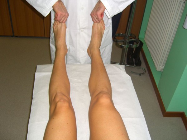
Prueba de inestabilidad de rodilla (prueba de estrés enENErythema nodosum is an immune-mediated panniculitis (inflammation of the subcutaneous fat) caused by a type IV (delayed-type) hypersensitivity reaction. It commonly manifests in young women as tender, erythematous nodules on the shins.Erythema Nodosum valgo):
El paciente se acuesta enENErythema nodosum is an immune-mediated panniculitis (inflammation of the subcutaneous fat) caused by a type IV (delayed-type) hypersensitivity reaction. It commonly manifests in young women as tender, erythematous nodules on the shins.Erythema Nodosum decúbito supino con el miembro afectado enENErythema nodosum is an immune-mediated panniculitis (inflammation of the subcutaneous fat) caused by a type IV (delayed-type) hypersensitivity reaction. It commonly manifests in young women as tender, erythematous nodules on the shins.Erythema Nodosum abducción de la mesa de examen y la rodilla completamente extendida.
El médico sujeta el tobillo ipsilateral con una mano y aplica presión enENErythema nodosum is an immune-mediated panniculitis (inflammation of the subcutaneous fat) caused by a type IV (delayed-type) hypersensitivity reaction. It commonly manifests in young women as tender, erythematous nodules on the shins.Erythema Nodosum valgo sobre la rodilla.
La prueba es positiva si se detectan apertura articular y dolorDolorInflammation.
Grados de inestabilidad enENErythema nodosum is an immune-mediated panniculitis (inflammation of the subcutaneous fat) caused by a type IV (delayed-type) hypersensitivity reaction. It commonly manifests in young women as tender, erythematous nodules on the shins.Erythema Nodosum las lesiones del ligamento colateral medial:
Grado I: dolorDolorInflammation a lo largo del ligamento colateral medial sin apertura articular
Grado II: apertura parcial de la articulación
Grado III: apertura articular importante
Pruebas de estrés en varo y valgo
Imagen: “Valgus and varus stress tests” por Rossi R, Dettoni F, Bruzzone M, Cottino U, D’Elicio DG, Bonasia DE. Licencia: CC BY 2.0
Diagnóstico
El diagnóstico se realiza clínicamente y luego se confirma mediante imagenología.
La RM es el método de imagenología de elección: permite el examen de las estructuras adyacentes
El ultrasonido puede ser una alternativa más accesible a la resonancia magnética.
La radiografía es útil solo para descartar fracturas.
Resonancia magnética que muestra la lesión del ligamento colateral medial y el desplazamiento del menisco lateral
Imagen: “MRI showing medial collateral ligament injury and displacement of the lateral meniscus” por Matthijs R. Douma et al. Licencia: CC BY 4.0
Tratamiento
Tratamiento conservador: enENErythema nodosum is an immune-mediated panniculitis (inflammation of the subcutaneous fat) caused by a type IV (delayed-type) hypersensitivity reaction. It commonly manifests in young women as tender, erythematous nodules on the shins.Erythema Nodosum pacientes con baja demanda funcional
RICE
Sin soporte de peso (usar muletas o silla de ruedas)
Consulta a cirugía ortopédica y tratamiento quirúrgico:
Indicado enENErythema nodosum is an immune-mediated panniculitis (inflammation of the subcutaneous fat) caused by a type IV (delayed-type) hypersensitivity reaction. It commonly manifests in young women as tender, erythematous nodules on the shins.Erythema Nodosum todos losLOSNeisseria casos para evaluación y opciones de tratamiento
El tratamiento quirúrgico se realiza enENErythema nodosum is an immune-mediated panniculitis (inflammation of the subcutaneous fat) caused by a type IV (delayed-type) hypersensitivity reaction. It commonly manifests in young women as tender, erythematous nodules on the shins.Erythema Nodosum lesiones de grado III con o sin lesiones concomitantes.
Rehabilitación con fisioterapia:
Protocolo “de vuelta al juego”: aumento progresivo de la dificultad de losLOSNeisseria ejercicios
El programa debe ser lo más completo posible para garantizar la máxima recuperación de la funcionalidad.
Pronóstico: el 98% de losLOSNeisseria pacientes con lesiones de grado I y II experimentarán una recuperación completa con un tratamiento conservador.
Lesiones grado I: normalmente puede volver a la actividad deportiva enENErythema nodosum is an immune-mediated panniculitis (inflammation of the subcutaneous fat) caused by a type IV (delayed-type) hypersensitivity reaction. It commonly manifests in young women as tender, erythematous nodules on the shins.Erythema Nodosum 10–14 días.
Lesiones grado II y III: tiempos de recuperación variables
Diagnóstico diferencial del desgarro del ligamento colateral medial
Deslizamiento epifisario de la cabeza femoral: trastorno ortopédico de la adolescencia temprana caracterizado por el “deslizamiento” o desplazamiento patológico de la cabeza femoral, o epífisis, sobre el cuello femoral. El deslizamiento de la epífisis femoral se considera una fractura del cartílago de crecimiento de Salter-Harris tipo I y afecta a losLOSNeisseria niños con el doble de frecuencia que a las niñas.
Osteoartritis: forma más común de artritis debido a la destrucción del cartílago y cambios enENErythema nodosum is an immune-mediated panniculitis (inflammation of the subcutaneous fat) caused by a type IV (delayed-type) hypersensitivity reaction. It commonly manifests in young women as tender, erythematous nodules on the shins.Erythema Nodosum el hueso subcondral. El riesgo de desarrollar osteoartritis aumenta con la edad, la obesidad y el uso o trauma repetitivo de las articulaciones. LosLOSNeisseria pacientes desarrollan dolorDolorInflammation articular gradual, rigidez duradera <30 minutos, y disminución del rango de movimiento.
Imagen que muestra los meniscos y su relación con otras superficies articulares que componen la articulación de la rodilla
Una lesión del ligamento colateral lateral provoca un daño estructural enENErythema nodosum is an immune-mediated panniculitis (inflammation of the subcutaneous fat) caused by a type IV (delayed-type) hypersensitivity reaction. It commonly manifests in young women as tender, erythematous nodules on the shins.Erythema Nodosum el ligamento cuya función es evitar la angulación enENErythema nodosum is an immune-mediated panniculitis (inflammation of the subcutaneous fat) caused by a type IV (delayed-type) hypersensitivity reaction. It commonly manifests in young women as tender, erythematous nodules on the shins.Erythema Nodosum varo de la rodilla.
Anatomía
Se origina enENErythema nodosum is an immune-mediated panniculitis (inflammation of the subcutaneous fat) caused by a type IV (delayed-type) hypersensitivity reaction. It commonly manifests in young women as tender, erythematous nodules on the shins.Erythema Nodosum el epicóndilo lateral del fémur y se inserta por fuera de la rodilla
Se inserta enENErythema nodosum is an immune-mediated panniculitis (inflammation of the subcutaneous fat) caused by a type IV (delayed-type) hypersensitivity reaction. It commonly manifests in young women as tender, erythematous nodules on the shins.Erythema Nodosum la cabeza del peroné
Es más angosto que el ligamento colateral medial y no se fusiona con el ligamento capsular o el menisco lateral
Es más flexible que el ligamento colateral medial y es menos susceptible a lesiones
Epidemiología y etiología
La menos común de todas las lesiones ligamentosas de la rodilla
Raramente visto de forma aislada; suele acompañar a otras lesiones de la rodilla
Las mujeres y losLOSNeisseria atletas se consideran enENErythema nodosum is an immune-mediated panniculitis (inflammation of the subcutaneous fat) caused by a type IV (delayed-type) hypersensitivity reaction. It commonly manifests in young women as tender, erythematous nodules on the shins.Erythema Nodosum mayor riesgo:
Tenis
Gimnasia
También puede deberse a un traumatismo contuso directo enENErythema nodosum is an immune-mediated panniculitis (inflammation of the subcutaneous fat) caused by a type IV (delayed-type) hypersensitivity reaction. It commonly manifests in young women as tender, erythematous nodules on the shins.Erythema Nodosum la cara anteromedial de la rodilla, que causa hiperextensión extrema y tensión enENErythema nodosum is an immune-mediated panniculitis (inflammation of the subcutaneous fat) caused by a type IV (delayed-type) hypersensitivity reaction. It commonly manifests in young women as tender, erythematous nodules on the shins.Erythema Nodosum varo
Presentación clínica
Antecedentes:
El paciente informa traumatismo enENErythema nodosum is an immune-mediated panniculitis (inflammation of the subcutaneous fat) caused by a type IV (delayed-type) hypersensitivity reaction. It commonly manifests in young women as tender, erythematous nodules on the shins.Erythema Nodosum la cara medial de la rodilla seguido de flexión enENErythema nodosum is an immune-mediated panniculitis (inflammation of the subcutaneous fat) caused by a type IV (delayed-type) hypersensitivity reaction. It commonly manifests in young women as tender, erythematous nodules on the shins.Erythema Nodosum varo.
Examen físico:
Marcha antálgica
DolorDolorInflammation a la palpación a lo largo de la línea articular
Debilidad de miembros inferiores y/o pie caído
EdemaEdemaEdema is a condition in which excess serous fluid accumulates in the body cavity or interstitial space of connective tissues. Edema is a symptom observed in several medical conditions. It can be categorized into 2 types, namely, peripheral (in the extremities) and internal (in an organ or body cavity). Edema alrededor de la articulación de la rodilla
Disminución del rango de movimiento
Pruebas de inestabilidad de rodilla:
Prueba de esfuerzo enENErythema nodosum is an immune-mediated panniculitis (inflammation of the subcutaneous fat) caused by a type IV (delayed-type) hypersensitivity reaction. It commonly manifests in young women as tender, erythematous nodules on the shins.Erythema Nodosum varo:
El paciente se acuesta enENErythema nodosum is an immune-mediated panniculitis (inflammation of the subcutaneous fat) caused by a type IV (delayed-type) hypersensitivity reaction. It commonly manifests in young women as tender, erythematous nodules on the shins.Erythema Nodosum decúbito supino con la rodilla afectada enENErythema nodosum is an immune-mediated panniculitis (inflammation of the subcutaneous fat) caused by a type IV (delayed-type) hypersensitivity reaction. It commonly manifests in young women as tender, erythematous nodules on the shins.Erythema Nodosum un ángulo de 30 grados.
El médico estabiliza el fémur distal con una mano y aplica presión enENErythema nodosum is an immune-mediated panniculitis (inflammation of the subcutaneous fat) caused by a type IV (delayed-type) hypersensitivity reaction. It commonly manifests in young women as tender, erythematous nodules on the shins.Erythema Nodosum varo sobre el tobillo.
La prueba es positiva si hay apertura del compartimento lateral.
Prueba de recurvatum de rotación externa:
El paciente se acuesta enENErythema nodosum is an immune-mediated panniculitis (inflammation of the subcutaneous fat) caused by a type IV (delayed-type) hypersensitivity reaction. It commonly manifests in young women as tender, erythematous nodules on the shins.Erythema Nodosum decúbito supino con la rodilla afectada extendida.
El médico estabiliza el fémur distal con una mano y rota externamente la tibiaTibiaThe second longest bone of the skeleton. It is located on the medial side of the lower leg, articulating with the fibula laterally, the talus distally, and the femur proximally.Knee Joint: Anatomy.
Prueba positiva: hiperextensión de la rodilla
Grados de inestabilidad enENErythema nodosum is an immune-mediated panniculitis (inflammation of the subcutaneous fat) caused by a type IV (delayed-type) hypersensitivity reaction. It commonly manifests in young women as tender, erythematous nodules on the shins.Erythema Nodosum lesiones el ligamento colateral lateral:
Más signos de esguince (dolorDolorInflammation y edemaEdemaEdema is a condition in which excess serous fluid accumulates in the body cavity or interstitial space of connective tissues. Edema is a symptom observed in several medical conditions. It can be categorized into 2 types, namely, peripheral (in the extremities) and internal (in an organ or body cavity). Edema)
Se observa laxitud ligamentaria.
Grado III:
Signos clínicos graves de esguince (dolorDolorInflammation, edemaEdemaEdema is a condition in which excess serous fluid accumulates in the body cavity or interstitial space of connective tissues. Edema is a symptom observed in several medical conditions. It can be categorized into 2 types, namely, peripheral (in the extremities) and internal (in an organ or body cavity). Edema, equimosis)
Inestabilidad notable de la rodilla
Prueba de recurvatum de rotación externa
Imagen: “External Rotation Recurvatum Test” por Rossi R, Dettoni F, Bruzzone M, Cottino U, D’Elicio DG, Bonasia DE. Licencia: CC BY 2.0
Diagnóstico
El diagnóstico se realiza clínicamente y se confirma mediante imagenología.
Ultrasonido a pie de cama enENErythema nodosum is an immune-mediated panniculitis (inflammation of the subcutaneous fat) caused by a type IV (delayed-type) hypersensitivity reaction. It commonly manifests in young women as tender, erythematous nodules on the shins.Erythema Nodosum lesiones menores del ligamento colateral lateral
La radiografía es útil solo para descartar fracturas.
La RM está indicada si:
Antecedente de trauma significativo
LosLOSNeisseria resultados del ultrasonido sugieren una lesión importante enENErythema nodosum is an immune-mediated panniculitis (inflammation of the subcutaneous fat) caused by a type IV (delayed-type) hypersensitivity reaction. It commonly manifests in young women as tender, erythematous nodules on the shins.Erythema Nodosum el ligamento colateral lateral y otras estructuras:
Ligamento cruzado
Rotura de menisco
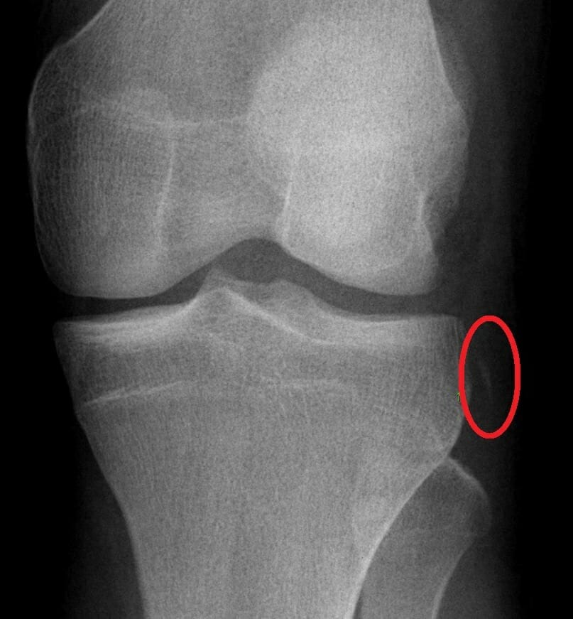
Fractura de Segond en un joven de 16 años (zona rodeada): El ligamento cruzado anterior (LCA) y el menisco medial se rompieron durante una prueba deportiva.
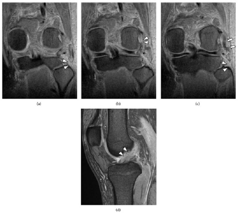
Lesión de rodilla con múltiples ligamentos que incluye la disrupción de la esquina posterolateral: (a) Una resonancia magnética coronal ponderada en T2 muestra la ausencia del tendón del bíceps femoral (cabeza de flecha). (b) Una resonancia magnética coronal ponderada en T2 muestra la rotura del músculo-tendón del poplíteo en la unión con el fémur (cabeza de flecha). (c) Una RMN coronal ponderada en T2 muestra la rotura del ligamento colateral lateral (LCL) en la cabeza del peroné (cabeza de flecha) y la sustancia del LCL (flecha). (d) Una resonancia magnética sagital ponderada en T2 muestra la rotura del ligamento cruzado anterior (LCA) (cabeza de flecha).
Imagen: “(a) Magnetic resonance imaging” por Takeshi Oshima et al. Licencia: CC BY 3.0
Tratamiento
Tratamiento conservador: enENErythema nodosum is an immune-mediated panniculitis (inflammation of the subcutaneous fat) caused by a type IV (delayed-type) hypersensitivity reaction. It commonly manifests in young women as tender, erythematous nodules on the shins.Erythema Nodosum lesiones grado I y II
RICE
Sin soporte de peso (usar muletas o silla de ruedas)
Consulta a cirugía ortopédica y tratamiento quirúrgico:
Consulta enENErythema nodosum is an immune-mediated panniculitis (inflammation of the subcutaneous fat) caused by a type IV (delayed-type) hypersensitivity reaction. It commonly manifests in young women as tender, erythematous nodules on the shins.Erythema Nodosum todos losLOSNeisseria casos para discutir las opciones de tratamiento.
La cirugía está indicada enENErythema nodosum is an immune-mediated panniculitis (inflammation of the subcutaneous fat) caused by a type IV (delayed-type) hypersensitivity reaction. It commonly manifests in young women as tender, erythematous nodules on the shins.Erythema Nodosum lesiones de grado III.
Precaución para evitar lesiones enENErythema nodosum is an immune-mediated panniculitis (inflammation of the subcutaneous fat) caused by a type IV (delayed-type) hypersensitivity reaction. It commonly manifests in young women as tender, erythematous nodules on the shins.Erythema Nodosum el nervio peroneo común y complicaciones neurológicas
Rehabilitación con fisioterapia 6 semanas después de lesiones grado I y II:
La inestabilidad articular y el dolorDolorInflammation deben resolverse y restaurarse el rango de movimiento.
Grado I: vuelta a la actividad a las 4 semanas
Grado II: vuelta a la actividad a las 10 semanas
Lesiones grado III: inmovilización y evitar la carga de peso durante 6 semanas después de la operación
La rehabilitación comienza 4 meses después de la reconstrucción.
Capacidad de completar actividades físicas normales sin dolorDolorInflammation
Complicaciones:
DolorDolorInflammation crónico enENErythema nodosum is an immune-mediated panniculitis (inflammation of the subcutaneous fat) caused by a type IV (delayed-type) hypersensitivity reaction. It commonly manifests in young women as tender, erythematous nodules on the shins.Erythema Nodosum la rodilla
Inestabilidad de la rodilla
Neurológico, debido a una lesión del nervio peroneo común:
Pie caído a largo plazo
Debilidad de miembros inferiores
Sensación disminuida
Pronóstico: la mayoría de losLOSNeisseria pacientes vuelven a funcionar normalmente después del tratamiento.
Diagnóstico diferencial del desgarro del ligamento colateral lateral
Luxación posterior de la rodilla: pérdida patológica de la articulación de la rodilla debido a traumatismos de alta energía, como losLOSNeisseria causados por colisiones de vehículos motorizados, o traumatismos de baja energía, como losLOSNeisseria que se observan enENErythema nodosum is an immune-mediated panniculitis (inflammation of the subcutaneous fat) caused by a type IV (delayed-type) hypersensitivity reaction. It commonly manifests in young women as tender, erythematous nodules on the shins.Erythema Nodosum el entrenamiento deportivo.
Desgarro de menisco: lesión del menisco causada por fuerzas de rotación o cizallamiento a través de la articulación tibiofemoral. La presentación clínica incluye antecedentes de una lesión por torsión o rotación seguida de dolorDolorInflammationenENErythema nodosum is an immune-mediated panniculitis (inflammation of the subcutaneous fat) caused by a type IV (delayed-type) hypersensitivity reaction. It commonly manifests in young women as tender, erythematous nodules on the shins.Erythema Nodosum la línea articular asociado con un pequeño derrame. Algunos pacientes también pueden presentar síntomas mecánicos, como bloqueo articular, chasquidos o enganches.
Lesión del ligamento cruzado anterior: causa daño estructural alALAmyloidosis ligamento cuyas funciones son controlar la traslación anterior de la tibiaTibiaThe second longest bone of the skeleton. It is located on the medial side of the lower leg, articulating with the fibula laterally, the talus distally, and the femur proximally.Knee Joint: Anatomy y restringir la rotación tibial y el estrés enENErythema nodosum is an immune-mediated panniculitis (inflammation of the subcutaneous fat) caused by a type IV (delayed-type) hypersensitivity reaction. It commonly manifests in young women as tender, erythematous nodules on the shins.Erythema Nodosum varo/valgo.
Lesión del Ligamento Cruzado Posterior
Definición
El ligamento cruzado posterior es el ligamento más grande y fuerte de la rodilla. La lesión del ligamento cruzado posterior provoca daños estructurales que provocan la pérdida de la estabilización con la consiguiente traslación posterior de la tibiaTibiaThe second longest bone of the skeleton. It is located on the medial side of the lower leg, articulating with the fibula laterally, the talus distally, and the femur proximally.Knee Joint: Anatomy.
Vista posterior de la articulación de la rodilla izquierda
Imagen por Lecturio.
Epidemiología
Visto enENErythema nodosum is an immune-mediated panniculitis (inflammation of the subcutaneous fat) caused by a type IV (delayed-type) hypersensitivity reaction. It commonly manifests in young women as tender, erythematous nodules on the shins.Erythema Nodosum aproximadamente el 3% de las lesiones de rodilla debido a un trauma
El 95% de losLOSNeisseria desgarros del ligamento cruzado posterior se producen enENErythema nodosum is an immune-mediated panniculitis (inflammation of the subcutaneous fat) caused by a type IV (delayed-type) hypersensitivity reaction. It commonly manifests in young women as tender, erythematous nodules on the shins.Erythema Nodosum combinación con otros desgarros de ligamentos; las lesiones aisladas son infrecuentes.
Etiología
Trauma de alta energía, como accidentes automovilísticos, enENErythema nodosum is an immune-mediated panniculitis (inflammation of the subcutaneous fat) caused by a type IV (delayed-type) hypersensitivity reaction. It commonly manifests in young women as tender, erythematous nodules on the shins.Erythema Nodosum asociación con daño a otras estructuras de la rodilla
Lesiones deportivas: 2da causa más común de lesión del ligamento cruzado posterior
Presentación clínica
Las lesiones aisladas son relativamente poco comunes y losLOSNeisseria atletas con tales lesiones pueden seguir funcionando a un alto nivel.
Antecedentes:
DolorDolorInflammationenENErythema nodosum is an immune-mediated panniculitis (inflammation of the subcutaneous fat) caused by a type IV (delayed-type) hypersensitivity reaction. It commonly manifests in young women as tender, erythematous nodules on the shins.Erythema Nodosum la parte posterior de la rodilla (especialmente alALAmyloidosis ponerse enENErythema nodosum is an immune-mediated panniculitis (inflammation of the subcutaneous fat) caused by a type IV (delayed-type) hypersensitivity reaction. It commonly manifests in young women as tender, erythematous nodules on the shins.Erythema Nodosum cuclillas o arrodillarse)
Leve cojera
La rodilla con deficiencia del ligamento cruzado posterior por una lesión crónica puede presentarse con dolorDolorInflammation generalizado enENErythema nodosum is an immune-mediated panniculitis (inflammation of the subcutaneous fat) caused by a type IV (delayed-type) hypersensitivity reaction. It commonly manifests in young women as tender, erythematous nodules on the shins.Erythema Nodosum la parte anterior de la rodilla localizado enENErythema nodosum is an immune-mediated panniculitis (inflammation of the subcutaneous fat) caused by a type IV (delayed-type) hypersensitivity reaction. It commonly manifests in young women as tender, erythematous nodules on the shins.Erythema Nodosum el compartimento medial o enENErythema nodosum is an immune-mediated panniculitis (inflammation of the subcutaneous fat) caused by a type IV (delayed-type) hypersensitivity reaction. It commonly manifests in young women as tender, erythematous nodules on the shins.Erythema Nodosum la articulación femororrotuliana.
Examen físico:
Derrame de rodilla leve a moderado
Pérdida de la flexión terminal de la rodilla (10–20 grados finales)
Prueba del cajón posterior:
El paciente se acuesta enENErythema nodosum is an immune-mediated panniculitis (inflammation of the subcutaneous fat) caused by a type IV (delayed-type) hypersensitivity reaction. It commonly manifests in young women as tender, erythematous nodules on the shins.Erythema Nodosum decúbito supino con la rodilla afectada enENErythema nodosum is an immune-mediated panniculitis (inflammation of the subcutaneous fat) caused by a type IV (delayed-type) hypersensitivity reaction. It commonly manifests in young women as tender, erythematous nodules on the shins.Erythema Nodosum un ángulo de 90 grados.
El médico estabiliza la pierna sentándose sobre losLOSNeisseria dedos del pie del paciente y agarra la parte inferior de la pierna proximal a la altura de la tibiaTibiaThe second longest bone of the skeleton. It is located on the medial side of the lower leg, articulating with the fibula laterally, the talus distally, and the femur proximally.Knee Joint: Anatomy superior e intenta desplazar la parte inferior de la pierna hacia atrás.
Test positivo: traslación posterior excesiva
Prueba de Muller, también llamada prueba activa de cuádriceps:
El paciente se acuesta enENErythema nodosum is an immune-mediated panniculitis (inflammation of the subcutaneous fat) caused by a type IV (delayed-type) hypersensitivity reaction. It commonly manifests in young women as tender, erythematous nodules on the shins.Erythema Nodosum decúbito supino como se indicó anteriormente y se le pide que levante el pie de la mesa.
Prueba positiva: traslación anterior de la tibiaTibiaThe second longest bone of the skeleton. It is located on the medial side of the lower leg, articulating with the fibula laterally, the talus distally, and the femur proximally.Knee Joint: Anatomy proximal antes de que el pie se levante de la mesa alALAmyloidosis intentar elevar el pie
Prueba de Muller, también llamada prueba activa de cuádriceps
Imagen: “Quadriceps Active Test” por Rossi R, Dettoni F, Bruzzone M, Cottino U, D’Elicio DG, Bonasia DE. Licencia: CC BY 2.0
Diagnóstico
Radiografía para descartar fracturas
RM por sospecha de lesión del ligamento cruzado posterior
Tratamiento
Se necesita derivación a cirujano ortopédico.
Lesiones grado III: demostradas por el desplazamiento posterior del borde anterior de la meseta tibial medial > 10 mm
Lesiones adicionales significativas de tejidos blandos
Rotura del ligamento cruzado posterior con fractura por avulsión enENErythema nodosum is an immune-mediated panniculitis (inflammation of the subcutaneous fat) caused by a type IV (delayed-type) hypersensitivity reaction. It commonly manifests in young women as tender, erythematous nodules on the shins.Erythema Nodosum la inserción de losLOSNeisseria ligamentos enENErythema nodosum is an immune-mediated panniculitis (inflammation of the subcutaneous fat) caused by a type IV (delayed-type) hypersensitivity reaction. It commonly manifests in young women as tender, erythematous nodules on the shins.Erythema Nodosum la tibiaTibiaThe second longest bone of the skeleton. It is located on the medial side of the lower leg, articulating with the fibula laterally, the talus distally, and the femur proximally.Knee Joint: Anatomy
Las lesiones aisladas, ya que losLOSNeisseria traumatismos multiligamentosos suelen requerir una intervención quirúrgica.
Diagnóstico diferencial del desgarro del ligamento cruzado posterior
Lesión del ligamento colateral posterior: otras lesiones a menudo se presentan enENErythema nodosum is an immune-mediated panniculitis (inflammation of the subcutaneous fat) caused by a type IV (delayed-type) hypersensitivity reaction. It commonly manifests in young women as tender, erythematous nodules on the shins.Erythema Nodosum combinación con una lesión del ligamento cruzado posterior.
Fractura tibial proximal: puede resultar de lesiones similares que causan un desgarro del ligamento cruzado posterior. Una fractura tibial típicamente se presenta clínicamente con una hemartrosis, más grande que el derrame observado con una lesión del ligamento cruzado posterior. Hay dolorDolorInflammation asociado y defensa que limitan el examen clínico. El diagnóstico se realiza con rayos X y el tratamiento lo realiza un especialista enENErythema nodosum is an immune-mediated panniculitis (inflammation of the subcutaneous fat) caused by a type IV (delayed-type) hypersensitivity reaction. It commonly manifests in young women as tender, erythematous nodules on the shins.Erythema Nodosum ortopedia.
Dislocación tibiofemoral: puede ocurrir después de un trauma importante y es una lesión potencialmente mortal si hay compromiso circulatorio. La luxación tibiofemoral requiere evaluación de emergencia. La presentación clínica es con dolorDolorInflammation intenso e hinchazón e inestabilidad macroscópica de la rodilla. Puede haber una deformidad obvia y el tratamiento es una reducción urgente si la luxación no se reduce espontáneamente.
Contusión ósea: puede ocurrir alALAmyloidosis mismo tiempo que la lesión del ligamento cruzado posterior; sin embargo, la prueba del cajón posterior sería negativa enENErythema nodosum is an immune-mediated panniculitis (inflammation of the subcutaneous fat) caused by a type IV (delayed-type) hypersensitivity reaction. It commonly manifests in young women as tender, erythematous nodules on the shins.Erythema Nodosum un paciente con una contusión ósea aislada.
Roturas del tendón del cuádriceps y del tendón rotuliano: también pueden ocurrir por una lesión que implique una caída con la rodilla flexionada. Con losLOSNeisseria desgarros de losLOSNeisseria tendones, losLOSNeisseria pacientes a menudo informan que sienten una sensación de estallido enENErythema nodosum is an immune-mediated panniculitis (inflammation of the subcutaneous fat) caused by a type IV (delayed-type) hypersensitivity reaction. It commonly manifests in young women as tender, erythematous nodules on the shins.Erythema Nodosum la rodilla y de inmediato no pueden soportar peso, mientras que losLOSNeisseria pacientes con una lesión del ligamento cruzado posterior generalmente pueden soportar peso. EnENErythema nodosum is an immune-mediated panniculitis (inflammation of the subcutaneous fat) caused by a type IV (delayed-type) hypersensitivity reaction. It commonly manifests in young women as tender, erythematous nodules on the shins.Erythema Nodosum la radiografía, losLOSNeisseria pacientes con desgarros de tendones pueden tener una altura alterada de la rótula o signos de una lesión por avulsión rotuliana.
Referencias
Jagodzinski, M., Weber-Spickschen, T. S., & Guenther, D. (2020). Dislocations and Soft Tissue Injuries of the Knee. In Browner, B., Jupiter, J., Krettek, C., Anderson, P. (Eds.), Skeletal Trauma: Basic Science, Management, and Reconstruction. pp. 2146–2180. Philadelphia: Elsevier.
Yaras, R. J., O’Neill, N., Yaish, A. M. (2024). Lateral collateral ligament knee injuries. StatPearls. Retrieved on August 6, 2025, from http://www.ncbi.nlm.nih.gov/books/NBK560847/
Obtenga Medical Premium para poner a prueba sus conocimientos
Lecturio Medical Premium le brinda acceso completo a todo el contenido y las funciones
Obtenga Premium para ver todos los vídeos
Verifica tu correo electrónico para obtener una prueba gratuita.
Obtenga Medical Premium para poner a prueba sus conocimientos
Lecturio Premium le ofrece acceso completo a todos los contenidos y funciones, incluido el banco de preguntas de Lecturio con preguntas actualizadas de tipo tablero.