Domina Conceptos Médicos

Estudia para la escuela de medicina y tus examenes con Lecturio.

Fracturas de Le Fort

Las fracturas de Le Fort son un grupo de patrones de fracturas de la región facial medialque se clasifican en EN Erythema nodosum is an immune-mediated panniculitis (inflammation of the subcutaneous fat) caused by a type IV (delayed-type) hypersensitivity reaction. It commonly manifests in young women as tender, erythematous nodules on the shins. Erythema Nodosum 3 tipos: Le Fort I Le Fort I Le Fort Fractures, II y III. Las fracturas de Le Fort representan entre el 10%–20% de todas las fracturas faciales y pueden ser causadas por cualquier traumatismo contuso significativo en EN Erythema nodosum is an immune-mediated panniculitis (inflammation of the subcutaneous fat) caused by a type IV (delayed-type) hypersensitivity reaction. It commonly manifests in young women as tender, erythematous nodules on the shins. Erythema Nodosum la cara, más comúnmente por accidentes automovilísticos. La presentación clínica incluye sangrado facial severo, edema Edema Edema is a condition in which excess serous fluid accumulates in the body cavity or interstitial space of connective tissues. Edema is a symptom observed in several medical conditions. It can be categorized into 2 types, namely, peripheral (in the extremities) and internal (in an organ or body cavity). Edema y movilidad de diferentes segmentos óseos, según el tipo de fractura del que se trate. El diagnóstico es clínico y se apoya en EN Erythema nodosum is an immune-mediated panniculitis (inflammation of the subcutaneous fat) caused by a type IV (delayed-type) hypersensitivity reaction. It commonly manifests in young women as tender, erythematous nodules on the shins. Erythema Nodosum las técnicas de imagenología. El tratamiento inicial se centra en EN Erythema nodosum is an immune-mediated panniculitis (inflammation of the subcutaneous fat) caused by a type IV (delayed-type) hypersensitivity reaction. It commonly manifests in young women as tender, erythematous nodules on the shins. Erythema Nodosum la estabilización del paciente y el control del sangrado. El tratamiento definitivo es quirúrgico. El tratamiento a largo plazo es la preservación y rehabilitación de la función facial.

Last updated: Dec 15, 2025

Editorial responsibility: Stanley Oiseth, Lindsay Jones, Evelin Maza

Descripción General

Definición

  • Un grupo de fracturas de la región facial medial típicamente provocadas por un traumatismo
  • Descritas por el Dr. Rene Le Fort en EN Erythema nodosum is an immune-mediated panniculitis (inflammation of the subcutaneous fat) caused by a type IV (delayed-type) hypersensitivity reaction. It commonly manifests in young women as tender, erythematous nodules on the shins. Erythema Nodosum 1901

Clasificación

  • Le Fort I Le Fort I Le Fort Fractures (más inferior):
    • Fractura transmaxilar (también conocida como fractura de Guerin)
    • La disrupción ósea implica:
      • Superior al AL Amyloidosis proceso alveolar maxilar
      • Se extiende por el cuerpo del tabique nasal.
      • Puede involucrar a las placas pterigoideas del hueso esfenoides
  • Le Fort II Le Fort II Le Fort Fractures:
    • Fractura piramidal
    • La disrupción ósea implica:
      • Porciones posterolaterales de los LOS Neisseria senos maxilares
      • Suele atravesar el orificio infraorbitario
      • Pueden estar afectados los LOS Neisseria huesos lagrimales y/o etmoidales
  • Le Fort III Le Fort III Le Fort Fractures (más superior):
    • Disyunción craneofacial: la cara está separada del cráneo
    • También conocida como fractura horizontal
    • La disrupción ósea implica:
      • Fisuras orbitarias superiores
      • Huesos lagrimales
      • Huesos etmoidales
      • Huesos nasales
      • Alas ALAS An enzyme of the transferase class that catalyzes condensation of the succinyl group from succinyl coenzyme a with glycine to form delta-aminolevulinate. It is a pyridoxal phosphate protein and the reaction occurs in mitochondria as the first step of the heme biosynthetic pathway. The enzyme is a key regulatory enzyme in heme biosynthesis. In liver feedback is inhibited by heme. Heme Metabolism mayores del hueso esfenoides
      • Suturas frontocigomáticas
    • Los LOS Neisseria nervios ópticos ayudan a mantener a los LOS Neisseria ojos en EN Erythema nodosum is an immune-mediated panniculitis (inflammation of the subcutaneous fat) caused by a type IV (delayed-type) hypersensitivity reaction. It commonly manifests in young women as tender, erythematous nodules on the shins. Erythema Nodosum su lugar.
  • Le Fort IV Le Fort IV Le Fort Fractures:
    • Características del tipo III + involucramiento del hueso frontal Frontal The bone that forms the frontal aspect of the skull. Its flat part forms the forehead, articulating inferiorly with the nasal bone and the cheek bone on each side of the face. Skull: Anatomy

Epidemiología

  • 10%–20% de todas las fracturas faciales
  • Habitualmente se observan en EN Erythema nodosum is an immune-mediated panniculitis (inflammation of the subcutaneous fat) caused by a type IV (delayed-type) hypersensitivity reaction. It commonly manifests in young women as tender, erythematous nodules on the shins. Erythema Nodosum el contexto de politraumatismos
  • Asociado con el uso de sustancias tóxicas
  • Disminución de la incidencia relacionada con choques de vehículos de motor Motor Neurons which send impulses peripherally to activate muscles or secretory cells. Nervous System: Histology debido al AL Amyloidosis mejoramiento de los LOS Neisseria sistemas de seguridad

Etiología

  • Cualquier traumatismo facial que transfiera una significativa cantidad de energía cinética:
    • Accidentes de vehículos de motor Motor Neurons which send impulses peripherally to activate muscles or secretory cells. Nervous System: Histology
    • Lesiones deportivas
    • Agresión física (e.g., abuso infantil, violencia doméstica, abuso en EN Erythema nodosum is an immune-mediated panniculitis (inflammation of the subcutaneous fat) caused by a type IV (delayed-type) hypersensitivity reaction. It commonly manifests in young women as tender, erythematous nodules on the shins. Erythema Nodosum ancianos)
    • Caídas
  • La velocidad del trauma está asociada con la gravedad de las fracturas de Le Fort.
Tipos de fracturas de Le Fort

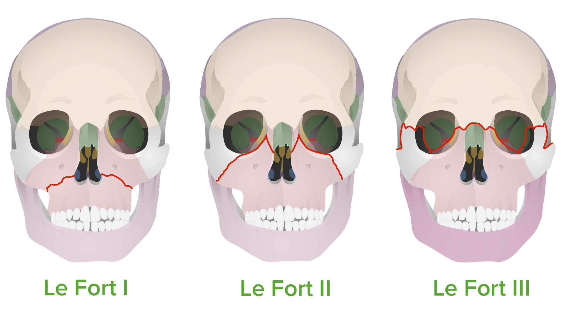
Tipos de fracturas de Le Fort:
Estas fracturas siguen las “líneas de debilidad” propuestas por Rene Le Fort en 1901.

Imagen por Lecturio.

Fisiopatología

  • Traumatismo cerrado: una cantidad significativa de energía cinética se transfiere directamente a la región facial medial.
    • Accidentes de vehículos de motor Motor Neurons which send impulses peripherally to activate muscles or secretory cells. Nervous System: Histology: la cara de un paciente sin cinturón de seguridad que es golpeada contra el tablero.
    • Deportes (e.g., fútbol, béisbol y hockey): lesiones por contacto o proyectiles (e.g., pelota o disco) que golpean la cara
    • Agresión física: individuos severamente maltratados
    • Caídas desde alturas significativas
    • También pueden incluir:
      • Lesiones penetrantes
      • Lesiones lacerantes
  • Le Fort I Le Fort I Le Fort Fractures: La fuerza se dirige inferiormente hacia el borde maxilar.
  • Le Fort II Le Fort II Le Fort Fractures: la fuerza se dirige inferior o medialmente al AL Amyloidosis maxilar
  • Le Fort III Le Fort III Le Fort Fractures: La fuerza impacta el puente nasal o superiormente del maxilar

Presentación Clínica

Antecedentes

  • Recuerda: debido a la naturaleza de la fractura, lo más probable es que la persona no pueda articular ningún detalle sobre el incidente.
  • Los LOS Neisseria socorristas o acompañantes le informarán sobre un trauma contundente reciente de alta energía.
    • Accidente en EN Erythema nodosum is an immune-mediated panniculitis (inflammation of the subcutaneous fat) caused by a type IV (delayed-type) hypersensitivity reaction. It commonly manifests in young women as tender, erythematous nodules on the shins. Erythema Nodosum vehículo a motor Motor Neurons which send impulses peripherally to activate muscles or secretory cells. Nervous System: Histology
    • Caída desde una gran altura
    • Ataque con un objeto contundente
  • El médico debe determinar:
    • El mecanismo del traumatismo
    • Más detalles de ser posible

Examen físico

  • Vía aérea:
    • Evaluar la permeabilidad.
    • Determinar la necesidad de una vía aérea de emergencia.
  • Evaluación de la cavidad oral:
    • Determinar la presencia de una fractura del paladar duro
    • Para determinar la presencia/fuente de una hemorragia orofaríngea
  • Evaluación de la cavidad nasal:
    • Para determinar la presencia de fracturas nasales
    • Para determinar la presencia/fuente de una hemorragia nasofaríngea
    • Evaluar si existe fuga de LCR.
  • Evaluación de la columna cervical/cuello:
    • Mantener la columna cervical inmovilizada durante la examinación física.
    • Siempre se debe sospechar una fractura de la columna cervical hasta que se demuestre lo contrario.
  • Evaluación facial:
    • Es probable que se presenten hemorragias y edema Edema Edema is a condition in which excess serous fluid accumulates in the body cavity or interstitial space of connective tissues. Edema is a symptom observed in several medical conditions. It can be categorized into 2 types, namely, peripheral (in the extremities) and internal (in an organ or body cavity). Edema facial importante.
    • Posible desarrollo de equimosis temprana
    • Deformidad en EN Erythema nodosum is an immune-mediated panniculitis (inflammation of the subcutaneous fat) caused by a type IV (delayed-type) hypersensitivity reaction. It commonly manifests in young women as tender, erythematous nodules on the shins. Erythema Nodosum forma de plato: aplanamiento de la cara (tipo III)
  • Determinar los LOS Neisseria segmentos óseos móviles (con la frente estabilizada):
  • Evaluación orbital/ocular:
    • Equimosis orbital bilateral (también conocida como ojos de mapache)
    • Ampliación del espacio intercantal
    • La afectación de las órbitas suelen ocurrir con las fracturas de los LOS Neisseria tipos II y III (requieren de una consulta oftalmológica urgente).
  • Evaluación neurológica:
    • Determinar el estado mental y el nivel de conciencia.
    • Si el nivel de conciencia está alterado, determinar la puntuación inicial de la Escala de coma Coma Coma is defined as a deep state of unarousable unresponsiveness, characterized by a score of 3 points on the GCS. A comatose state can be caused by a multitude of conditions, making the precise epidemiology and prognosis of coma difficult to determine. Coma de Glasgow.
    • Valorar la función de los LOS Neisseria pares craneales y la presencia de una lesión neurológica focal.
    • Evaluar los LOS Neisseria síntomas/signos de aumento de la presión intracraneal.
    • Evaluar los LOS Neisseria síntomas/signos de hemorragia intracraneal / lesión cerebral traumática.
Fractura de Le Fort III

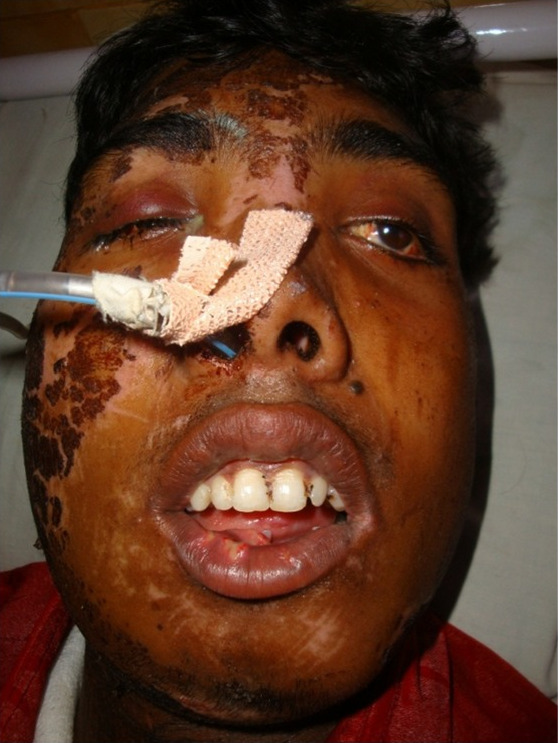
Paciente con una fractura de Le Fort III:
Nótese el ensanchamiento del espacio intercantal y el profuso edema facial. Los médicos de urgencias optaron por una vía aérea segura a través de la nariz.

Imagen: “Preoperative” por Santosh Kumar Yadav et al. Licencia: CC0 1.0

Diagnóstico

Diagnóstico clínico

  • Antes de evaluar los LOS Neisseria hallazgos específicos de las fracturas de Le Fort, se debe evaluar, diagnosticar y clasificar adecuadamente lo siguiente:
    • Permeabilidad de la vía aérea
    • Estabilidad de la columna cervical
    • Traumatismo potencialmente mortal
    • Hemorragia potencialmente mortal
    • Estado hemodinámico
  • El diagnóstico de una fractura de Le Fort debe considerarse y descartarse en EN Erythema nodosum is an immune-mediated panniculitis (inflammation of the subcutaneous fat) caused by a type IV (delayed-type) hypersensitivity reaction. It commonly manifests in young women as tender, erythematous nodules on the shins. Erythema Nodosum cualquier individuo con lo siguiente:
    • Antecedentes de traumatismo craneal, facial o cervical significativo
    • Hemorragia facial
    • Edema Edema Edema is a condition in which excess serous fluid accumulates in the body cavity or interstitial space of connective tissues. Edema is a symptom observed in several medical conditions. It can be categorized into 2 types, namely, peripheral (in the extremities) and internal (in an organ or body cavity). Edema facial
    • Equimosis facial
  • La anamnesis y el examen físico deberían ser suficientes para la clasificación inicial si el individuo está estable y la sospecha clínica de fractura de Le Fort es baja.

Imagenología

  • Debe obtenerse solamente después de que el paciente haya sido estabilizado.
  • Radiografías simples de la cara/columna cervical:
    • Pueden ser útiles en EN Erythema nodosum is an immune-mediated panniculitis (inflammation of the subcutaneous fat) caused by a type IV (delayed-type) hypersensitivity reaction. It commonly manifests in young women as tender, erythematous nodules on the shins. Erythema Nodosum la ausencia de la tomografía computarizada (TC) para identificar fracturas superficiales
    • Utilidad limitada para identificar fracturas más profundas y lesiones asociadas de tejidos blandos/vasculares
    • Puede ser útil en EN Erythema nodosum is an immune-mediated panniculitis (inflammation of the subcutaneous fat) caused by a type IV (delayed-type) hypersensitivity reaction. It commonly manifests in young women as tender, erythematous nodules on the shins. Erythema Nodosum ausencia de TC para evaluar la estabilidad de la columna cervical y/o la presencia de fractura
  • TC:
    • Útil para la identificación de fracturas faciales superficiales y profundas
    • Útil para la identificación de lesiones vasculares/de tejidos blandos asociadas
    • Útil en EN Erythema nodosum is an immune-mediated panniculitis (inflammation of the subcutaneous fat) caused by a type IV (delayed-type) hypersensitivity reaction. It commonly manifests in young women as tender, erythematous nodules on the shins. Erythema Nodosum la evaluación de lesiones penetrantes asociadas
    • Útil en EN Erythema nodosum is an immune-mediated panniculitis (inflammation of the subcutaneous fat) caused by a type IV (delayed-type) hypersensitivity reaction. It commonly manifests in young women as tender, erythematous nodules on the shins. Erythema Nodosum la evaluación de lesiones cerebrales traumáticas y/o hemorragias intracraneales asociadas
Imagen de una TC de una fractura de Le Fort III

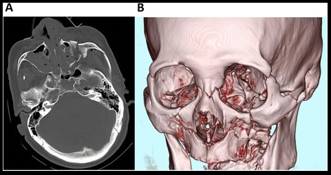
TC craneofacial (A) y reconstrucción tridimensional (B) de un paciente con una fractura de Le Fort III

Imagen: “Craniofacial computed tomography scan obtained on arrival.” por Momoko Mishima, Tetsuya Yumoto, Hiroaki Hashimoto et al. Licencia: CC BY 4.0

Tratamiento

Objetivos del tratamiento

  • Restaurar la integridad nasal y orbital, junto con la altura y la proyección de la región facial medial
  • Recuperar la oclusión adecuada de la mandíbula
  • Evaluar y tratar adecuadamente las lesiones asociadas:
    • Traumatismo craneoencefálico/lesión cerebral traumática
    • Hemorragia intracraneal
    • Aumento de la presión intracraneal
    • Fuga de LCR
    • Lesión vascular/de tejidos blandos asociada
    • Fractura de la columna cervical/lesión de la médula espinal
    • Lesiones oculares/pérdida de visión
    • Lesiones dentales

Triaje inicial

  • Asegurar la vía aérea
    • No se recomienda la técnica de intubación nasotraqueal a ciegas
    • Se prefiere la intubación endotraqueal con visualización directa.
    • La epistaxis Epistaxis Bleeding from the nose. Granulomatosis with Polyangiitis/hemorragia orofacial puede complicar la permeabilidad de la vía aérea y/o la visualización para la intubación.
  • Se deben seguir los LOS Neisseria protocolos de soporte vital cardíaco avanzado/soporte vital avanzado de trauma para el tratamiento de:
    • Inestabilidad hemodinámica
    • Insuficiencia respiratoria
    • Traumatismo no facial asociado
    • Hemorragia

Tratamiento inicial

  • En EN Erythema nodosum is an immune-mediated panniculitis (inflammation of the subcutaneous fat) caused by a type IV (delayed-type) hypersensitivity reaction. It commonly manifests in young women as tender, erythematous nodules on the shins. Erythema Nodosum paralelo con la evaluación de la vía aérea, la respiración y la circulación (ABC, por sus siglas en EN Erythema nodosum is an immune-mediated panniculitis (inflammation of the subcutaneous fat) caused by a type IV (delayed-type) hypersensitivity reaction. It commonly manifests in young women as tender, erythematous nodules on the shins. Erythema Nodosum inglés):
  • Protección de la vía aérea:
    • Intubación orotraqueal o vía aérea quirúrgica
    • Soporte ventilatorio solamente si está indicado
  • Resucitación con fluidos
  • Estabilización de la columna cervical (e.g., collarín cervical)
  • Taponamiento nasal

Tratamiento quirúrgico

  • Se realiza después de la estabilización y el tratamiento de las lesiones potencialmente mortales
  • Cirugía Maxilofacial:
    • Reducción de fracturas con osteosíntesis de placa y tornillos
    • La oclusión apropiada asegura una reparación adecuada.
  • Consulta de neurocirugía: en EN Erythema nodosum is an immune-mediated panniculitis (inflammation of the subcutaneous fat) caused by a type IV (delayed-type) hypersensitivity reaction. It commonly manifests in young women as tender, erythematous nodules on the shins. Erythema Nodosum casos con fugas de LCR

Complicaciones

  • Exanguinación potencialmente mortal
  • Hemorragia intracraneal
  • Lesión cerebral traumática
  • Fuga de LCR
  • Epistaxis Epistaxis Bleeding from the nose. Granulomatosis with Polyangiitis severa
  • Desfiguración permanente

Pronóstico

  • Depende del mecanismo, la gravedad y la ubicación de la lesión
  • Potencialmente mortal si no se brinda un tratamiento oportuno
  • Las morbilidades comunes después de una lesión incluyen:
    • Problemas visuales
    • Dificultades para masticar
    • Parálisis de los LOS Neisseria pares craneales
    • Anormalidades en EN Erythema nodosum is an immune-mediated panniculitis (inflammation of the subcutaneous fat) caused by a type IV (delayed-type) hypersensitivity reaction. It commonly manifests in young women as tender, erythematous nodules on the shins. Erythema Nodosum el gusto/olfato

Diagnóstico Diferencial

  • Fractura frontal Frontal The bone that forms the frontal aspect of the skull. Its flat part forms the forehead, articulating inferiorly with the nasal bone and the cheek bone on each side of the face. Skull: Anatomy: ruptura en EN Erythema nodosum is an immune-mediated panniculitis (inflammation of the subcutaneous fat) caused by a type IV (delayed-type) hypersensitivity reaction. It commonly manifests in young women as tender, erythematous nodules on the shins. Erythema Nodosum la corteza del hueso frontal Frontal The bone that forms the frontal aspect of the skull. Its flat part forms the forehead, articulating inferiorly with the nasal bone and the cheek bone on each side of the face. Skull: Anatomy, generalmente debido a un traumatismo directo o penetrante en EN Erythema nodosum is an immune-mediated panniculitis (inflammation of the subcutaneous fat) caused by a type IV (delayed-type) hypersensitivity reaction. It commonly manifests in young women as tender, erythematous nodules on the shins. Erythema Nodosum la frente. La presentación clínica incluye sangrado activo y edema Edema Edema is a condition in which excess serous fluid accumulates in the body cavity or interstitial space of connective tissues. Edema is a symptom observed in several medical conditions. It can be categorized into 2 types, namely, peripheral (in the extremities) and internal (in an organ or body cavity). Edema, crepitación en EN Erythema nodosum is an immune-mediated panniculitis (inflammation of the subcutaneous fat) caused by a type IV (delayed-type) hypersensitivity reaction. It commonly manifests in young women as tender, erythematous nodules on the shins. Erythema Nodosum el área de la fractura y/o movilidad de los LOS Neisseria segmentos óseos y estado mental alterado asociado con una lesión cerebral traumática acompañante. El diagnóstico es clínico, respaldado por imagenología y el tratamiento definitivo puede ser conservador o quirúrgico.
  • Luxación mandibular: describe el desacoplamiento unilateral o bilateral de los LOS Neisseria componentes de la articulación temporomandibular. Las causas pueden ser traumáticas o atraumáticas, como una sobreapertura forzada de la mandíbula. El diagnóstico es clínico. El tratamiento consiste en EN Erythema nodosum is an immune-mediated panniculitis (inflammation of the subcutaneous fat) caused by a type IV (delayed-type) hypersensitivity reaction. It commonly manifests in young women as tender, erythematous nodules on the shins. Erythema Nodosum una reducción bimanual.
  • Fractura de la mandíbula: ruptura de la corteza de la mandíbula, generalmente debido a un traumatismo contuso observado en EN Erythema nodosum is an immune-mediated panniculitis (inflammation of the subcutaneous fat) caused by a type IV (delayed-type) hypersensitivity reaction. It commonly manifests in young women as tender, erythematous nodules on the shins. Erythema Nodosum accidentes de tráfico o lesiones en EN Erythema nodosum is an immune-mediated panniculitis (inflammation of the subcutaneous fat) caused by a type IV (delayed-type) hypersensitivity reaction. It commonly manifests in young women as tender, erythematous nodules on the shins. Erythema Nodosum el lugar de trabajo. La presentación clínica incluye deformidad maxilofacial, edema Edema Edema is a condition in which excess serous fluid accumulates in the body cavity or interstitial space of connective tissues. Edema is a symptom observed in several medical conditions. It can be categorized into 2 types, namely, peripheral (in the extremities) and internal (in an organ or body cavity). Edema y equimosis en EN Erythema nodosum is an immune-mediated panniculitis (inflammation of the subcutaneous fat) caused by a type IV (delayed-type) hypersensitivity reaction. It commonly manifests in young women as tender, erythematous nodules on the shins. Erythema Nodosum el sitio de la lesión. El diagnóstico es clínico, respaldado por imagenología y el tratamiento definitivo es quirúrgico.

Referencias

  1. Phillips, B. J., Turco, L. M. (2017). Le Fort fractures: a collective review. Bulletin of Emergency & Trauma, 5:221.
  2. Rodriguez, E. D., Dorafshar, A. H., Manson, P. N. (2018). Facial injuries. In: Rodriguez, E. D., Losee, J. E., P. C. Neligan, P. C. (Eds.), Plastic Surgery. Volume 3: Craniofacial, Head and Neck Surgery and Pediatric Plastic Surgery, 4th ed. pp. 47–81. Elsevier.
  3. Hedayati, T., Amin, D. P. (2020). Trauma to the face. Chapter 259 of Tintinalli, J. E., Ma, O. J., Yealy, D. M., Meckler, G. D., Stapczynski, J. S., Cline, D. M., Thomas, S. H. (Eds.), Tintinalli’s Emergency Medicine: A Comprehensive Study Guide, 9th ed. McGraw-Hill Education. http://accessmedicine.mhmedical.com/content.aspx?aid=1167028185
  4. Hohman, M. H., Patel, B. C., & Waseem, M. (2024). Le Fort fractures. In StatPearls [Internet]. StatPearls Publishing. Retrieved June 15, 2025, from https://www.ncbi.nlm.nih.gov/books/NBK526

¡Crea tu cuenta gratis o inicia una sesión para seguir leyendo!

Regístrate ahora y obtén acceso gratuito a Lecturio con páginas de concepto, videos médicos y cuestionarios para tu educación médica.

User Reviews

Details