Domina Conceptos Médicos

Estudia para la escuela de medicina y tus examenes con Lecturio.

Enfermedades del Cuerpo Vítreo

El cuerpo vítreo es una sustancia transparente y gelatinosa que está presente en EN Erythema nodosum is an immune-mediated panniculitis (inflammation of the subcutaneous fat) caused by a type IV (delayed-type) hypersensitivity reaction. It commonly manifests in young women as tender, erythematous nodules on the shins. Erythema Nodosum el espacio entre el cristalino y la retina Retina The ten-layered nervous tissue membrane of the eye. It is continuous with the optic nerve and receives images of external objects and transmits visual impulses to the brain. Its outer surface is in contact with the choroid and the inner surface with the vitreous body. The outermost layer is pigmented, whereas the inner nine layers are transparent. Eye: Anatomy, proporcionando estabilidad estructural y manteniendo la forma del ojo. Algunas afecciones que pueden alterar al AL Amyloidosis cuerpo vítreo son el desprendimiento vítreo posterior, hemorragia vítrea, sínquisis centelleante, hialosis asteroidea y la vasculatura fetal persistente. Las afecciones pueden ser asintomáticas o presentar miodesopsias en EN Erythema nodosum is an immune-mediated panniculitis (inflammation of the subcutaneous fat) caused by a type IV (delayed-type) hypersensitivity reaction. It commonly manifests in young women as tender, erythematous nodules on the shins. Erythema Nodosum el campo visual, fotopsia y disminución de la agudeza visual. La fundoscopia y la microscopía con lámpara de hendidura se utilizan habitualmente en EN Erythema nodosum is an immune-mediated panniculitis (inflammation of the subcutaneous fat) caused by a type IV (delayed-type) hypersensitivity reaction. It commonly manifests in young women as tender, erythematous nodules on the shins. Erythema Nodosum el diagnóstico de estas enfermedades. Los LOS Neisseria métodos de tratamiento dependen de la afección y la gravedad, pero pueden incluir la observación, corrección de la visión y cirugía.

Last updated: Nov 12, 2025

Editorial responsibility: Stanley Oiseth, Lindsay Jones, Evelin Maza

Descripción General

El cuerpo vítreo es la sustancia presente entre el cristalino y la retina Retina The ten-layered nervous tissue membrane of the eye. It is continuous with the optic nerve and receives images of external objects and transmits visual impulses to the brain. Its outer surface is in contact with the choroid and the inner surface with the vitreous body. The outermost layer is pigmented, whereas the inner nine layers are transparent. Eye: Anatomy.

  • Material similar al AL Amyloidosis gel que proporciona:
    • Un medio óptico claro
    • Integridad estructural al AL Amyloidosis ojo
  • Consiste en EN Erythema nodosum is an immune-mediated panniculitis (inflammation of the subcutaneous fat) caused by a type IV (delayed-type) hypersensitivity reaction. It commonly manifests in young women as tender, erythematous nodules on the shins. Erythema Nodosum gran parte de:
    • Agua (99%)
    • Red de fibrillas de colágeno
    • Ácido hialurónico
    • Células periféricas (hialocitos)
Anatomía del ojo

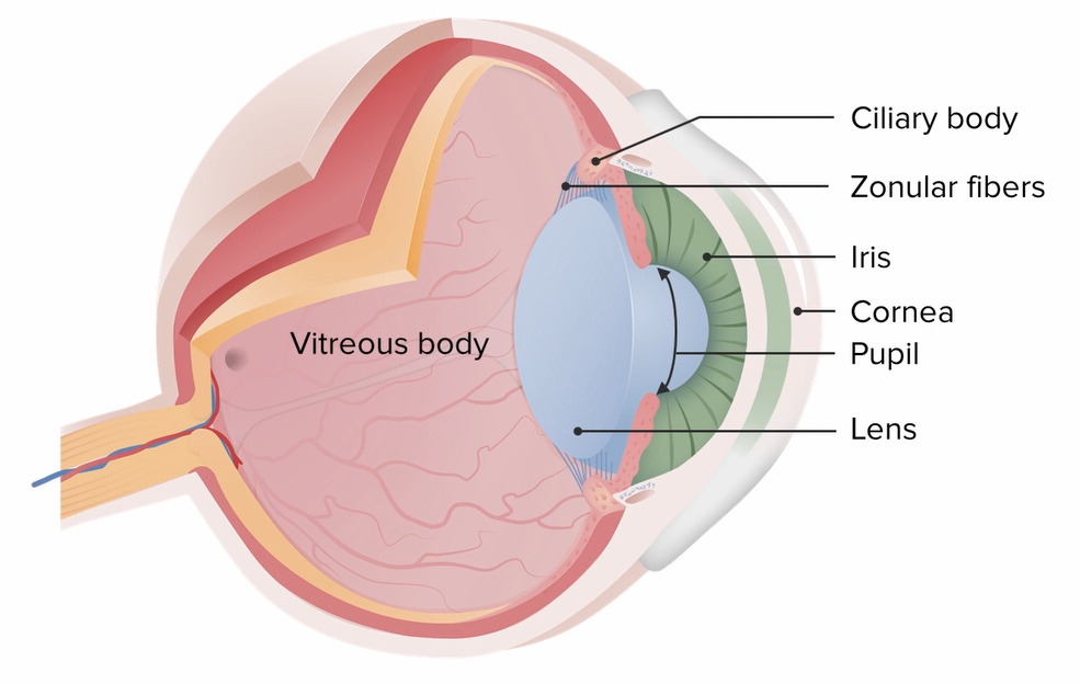
Diagrama que muestra la anatomía del ojo.

Imagen por Lecturio.

Desprendimiento Vítreo Posterior

Definición

El desprendimiento vítreo posterior es la separación del cuerpo vítreo de la membrana limitante interna de la retina Retina The ten-layered nervous tissue membrane of the eye. It is continuous with the optic nerve and receives images of external objects and transmits visual impulses to the brain. Its outer surface is in contact with the choroid and the inner surface with the vitreous body. The outermost layer is pigmented, whereas the inner nine layers are transparent. Eye: Anatomy.

Epidemiología

  • Más común en EN Erythema nodosum is an immune-mediated panniculitis (inflammation of the subcutaneous fat) caused by a type IV (delayed-type) hypersensitivity reaction. It commonly manifests in young women as tender, erythematous nodules on the shins. Erythema Nodosum las personas mayores:
  • Hombres y mujeres igualmente afectados

Etiología

  • Degeneración vítrea relacionada con la edad (causa más común):
    • Con la edad, el humor Humor Defense Mechanisms vítreo pasa de ser un gel espeso a una sustancia líquida fina.
    • El vítreo comienza a encogerse → puede provocar su desprendimiento de la retina Retina The ten-layered nervous tissue membrane of the eye. It is continuous with the optic nerve and receives images of external objects and transmits visual impulses to the brain. Its outer surface is in contact with the choroid and the inner surface with the vitreous body. The outermost layer is pigmented, whereas the inner nine layers are transparent. Eye: Anatomy
  • Traumatismo ocular
  • Cirugía ocular (cataratas)
  • Enfermedad ocular inflamatoria (uveítis)
  • Las personas con miopía tienen un mayor riesgo debido a la elongación del globo ocular.

Presentación clínica

  • La mayoría de los LOS Neisseria casos son asintomáticos.
  • Miodesopsia (cuerpos flotantes):
    • Acumulación de depósitos en EN Erythema nodosum is an immune-mediated panniculitis (inflammation of the subcutaneous fat) caused by a type IV (delayed-type) hypersensitivity reaction. It commonly manifests in young women as tender, erythematous nodules on the shins. Erythema Nodosum el cuerpo vítreo
    • Las miodesopsias son más visibles sobre un fondo claro y aparecen en EN Erythema nodosum is an immune-mediated panniculitis (inflammation of the subcutaneous fat) caused by a type IV (delayed-type) hypersensitivity reaction. It commonly manifests in young women as tender, erythematous nodules on the shins. Erythema Nodosum diferentes formas y tamaños.
  • Fotopsia:
    • Destellos de luz repentinos y breves
    • Suelen ser unilaterales y se dan en EN Erythema nodosum is an immune-mediated panniculitis (inflammation of the subcutaneous fat) caused by a type IV (delayed-type) hypersensitivity reaction. It commonly manifests in young women as tender, erythematous nodules on the shins. Erythema Nodosum zonas oscuras
    • Inducidos por el movimiento de la cabeza o de los LOS Neisseria ojos
    • Causados por la tracción vitreoretinal en EN Erythema nodosum is an immune-mediated panniculitis (inflammation of the subcutaneous fat) caused by a type IV (delayed-type) hypersensitivity reaction. It commonly manifests in young women as tender, erythematous nodules on the shins. Erythema Nodosum la retina Retina The ten-layered nervous tissue membrane of the eye. It is continuous with the optic nerve and receives images of external objects and transmits visual impulses to the brain. Its outer surface is in contact with the choroid and the inner surface with the vitreous body. The outermost layer is pigmented, whereas the inner nine layers are transparent. Eye: Anatomy
  • Visión borrosa

Diagnóstico

  • Microscopía con lámpara de hendidura: el anillo de Weiss es una membrana característica y móvil dentro de la cavidad vítrea que es brillante y arrugada.
  • Ultrasonido B-scan:
    • Se utiliza para estudiar el estado del gel vítreo
    • También ayuda a determinar la extensión del desprendimiento vítreo posterior
Ultrasonido B-scan que muestra la extensión del desprendimiento vítreo.

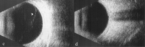
Ultrasonido B-scan que muestra un desprendimiento de vítreo anterior en la imagen c.
El desprendimiento vítreo posterior se ve en la imagen d.

Imagen: “B-scan ultrasonography showing extent of vitreous detachment” por Ophthalmology Unit, DAI Head/Neck, Azienda Policlinico Umberto I, University of Rome “Sapienza”, viale del Policlinico 155, Rome, 00161, Italy. Licencia: CC BY 4.0., editado por Lecturio.

Tratamiento

  • No es necesario ningún tratamiento en EN Erythema nodosum is an immune-mediated panniculitis (inflammation of the subcutaneous fat) caused by a type IV (delayed-type) hypersensitivity reaction. It commonly manifests in young women as tender, erythematous nodules on the shins. Erythema Nodosum los LOS Neisseria pacientes sin lesión de la retina Retina The ten-layered nervous tissue membrane of the eye. It is continuous with the optic nerve and receives images of external objects and transmits visual impulses to the brain. Its outer surface is in contact with the choroid and the inner surface with the vitreous body. The outermost layer is pigmented, whereas the inner nine layers are transparent. Eye: Anatomy.
  • La adaptación a los LOS Neisseria síntomas visuales se desarrollará con el tiempo, y las miodesopsias pueden resolverse.
  • La vitrectomía se considera para los LOS Neisseria síntomas persistentes.

Complicaciones

  • Desprendimiento de retina Retina The ten-layered nervous tissue membrane of the eye. It is continuous with the optic nerve and receives images of external objects and transmits visual impulses to the brain. Its outer surface is in contact with the choroid and the inner surface with the vitreous body. The outermost layer is pigmented, whereas the inner nine layers are transparent. Eye: Anatomy
  • Desgarro de la retina Retina The ten-layered nervous tissue membrane of the eye. It is continuous with the optic nerve and receives images of external objects and transmits visual impulses to the brain. Its outer surface is in contact with the choroid and the inner surface with the vitreous body. The outermost layer is pigmented, whereas the inner nine layers are transparent. Eye: Anatomy
  • Hemorragia vítrea

Related videos

Hemorragia Vítrea

Definición

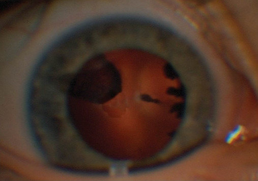
La hemorragia vítrea es la extravasación de sangre en EN Erythema nodosum is an immune-mediated panniculitis (inflammation of the subcutaneous fat) caused by a type IV (delayed-type) hypersensitivity reaction. It commonly manifests in young women as tender, erythematous nodules on the shins. Erythema Nodosum el humor Humor Defense Mechanisms vítreo.

Etiología

Hay muchas causas de hemorragia vítrea. Algunas causas comunes son:

  • Retinopatía diabética proliferativa
  • Desprendimiento vítreo posterior
  • Traumatismo ocular
  • Desgarro espontáneo de la retina Retina The ten-layered nervous tissue membrane of the eye. It is continuous with the optic nerve and receives images of external objects and transmits visual impulses to the brain. Its outer surface is in contact with the choroid and the inner surface with the vitreous body. The outermost layer is pigmented, whereas the inner nine layers are transparent. Eye: Anatomy
  • Degeneración macular
  • Retinopatía falciforme

Presentación clínica

La hemorragia vítrea suele ser indolora y unilateral. Los LOS Neisseria signos y síntomas incluyen:

  • Visión borrosa
  • Miodesopsias
  • Fotopsia
  • Fotofobia
  • Percepción de sombras o telarañas
  • La agudeza visual puede verse afectada, dependiendo de la cantidad de sangre presente.

Diagnóstico

  • El examen fundoscópico puede utilizarse para visualizar la hemorragia.
  • Microscopía con lámpara de hendidura: Se utiliza para examinar el vítreo en EN Erythema nodosum is an immune-mediated panniculitis (inflammation of the subcutaneous fat) caused by a type IV (delayed-type) hypersensitivity reaction. It commonly manifests in young women as tender, erythematous nodules on the shins. Erythema Nodosum busca de glóbulos rojos.
  • El ultrasonido ocular puede utilizarse en EN Erythema nodosum is an immune-mediated panniculitis (inflammation of the subcutaneous fat) caused by a type IV (delayed-type) hypersensitivity reaction. It commonly manifests in young women as tender, erythematous nodules on the shins. Erythema Nodosum los LOS Neisseria casos en EN Erythema nodosum is an immune-mediated panniculitis (inflammation of the subcutaneous fat) caused by a type IV (delayed-type) hypersensitivity reaction. It commonly manifests in young women as tender, erythematous nodules on the shins. Erythema Nodosum que el segmento posterior no es visible debido a una hemorragia excesiva.
  • Puede ser necesaria una tomografía computarizada/resonancia magnética (TC/RM) para evaluar las estructuras óseas y descartar cuerpos extraños.
Fotografías de fondo de ojo de hemorragia vítrea

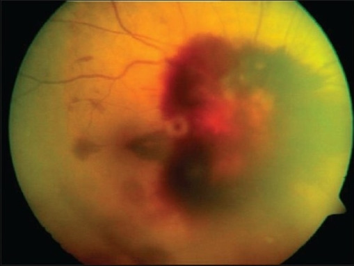
Fotografía del fondo del ojo derecho que muestra una hemorragia vítrea.

Imagen: “Fundus photographs of right eye showing peripapillary, subhyaloid, vitreous hemorrhage and several flame shaped hemorrhages obscuring the view of the optic disc” por Smt Kanuri Shanthamma Center for Retina Vitreous Diseases, L V Prasad Eye Institute, Kallam Anji Reddy Campus, Banjara Hills, Hyderabad 500 034, India. Licencia: CC BY 2.0, editado por Lecturio.

Tratamiento

  • La hemorragia vítrea puede resolverse por sí sola.
  • El tratamiento se dirige a la causa subyacente.
  • La vitrectomía se realiza en EN Erythema nodosum is an immune-mediated panniculitis (inflammation of the subcutaneous fat) caused by a type IV (delayed-type) hypersensitivity reaction. It commonly manifests in young women as tender, erythematous nodules on the shins. Erythema Nodosum casos de enfermedades graves y casos de desprendimiento de retina Retina The ten-layered nervous tissue membrane of the eye. It is continuous with the optic nerve and receives images of external objects and transmits visual impulses to the brain. Its outer surface is in contact with the choroid and the inner surface with the vitreous body. The outermost layer is pigmented, whereas the inner nine layers are transparent. Eye: Anatomy.
  • La fotocoagulación con láser está prevista para las retinopatías proliferativas o el desgarro de retina Retina The ten-layered nervous tissue membrane of the eye. It is continuous with the optic nerve and receives images of external objects and transmits visual impulses to the brain. Its outer surface is in contact with the choroid and the inner surface with the vitreous body. The outermost layer is pigmented, whereas the inner nine layers are transparent. Eye: Anatomy.

Sínquisis Centelleante

Definición

La sínquisis centelleante, también conocida como colesterolosis bulbi, es una afección degenerativa definida por la acumulación de cristales de colesterol en EN Erythema nodosum is an immune-mediated panniculitis (inflammation of the subcutaneous fat) caused by a type IV (delayed-type) hypersensitivity reaction. It commonly manifests in young women as tender, erythematous nodules on the shins. Erythema Nodosum un humor Humor Defense Mechanisms vítreo licuado.

Epidemiología

  • Una afección muy rara
  • Generalmente, ocurre en EN Erythema nodosum is an immune-mediated panniculitis (inflammation of the subcutaneous fat) caused by a type IV (delayed-type) hypersensitivity reaction. It commonly manifests in young women as tender, erythematous nodules on the shins. Erythema Nodosum la 3ra década de la vida
  • No hay predominio de sexo o raza

Factores de riesgo

  • Hemorragia vítrea crónica o recurrente (más común)
  • Retinopatía diabética
  • Uveítis crónica
  • Desprendimiento de retina Retina The ten-layered nervous tissue membrane of the eye. It is continuous with the optic nerve and receives images of external objects and transmits visual impulses to the brain. Its outer surface is in contact with the choroid and the inner surface with the vitreous body. The outermost layer is pigmented, whereas the inner nine layers are transparent. Eye: Anatomy
  • Traumatismo ocular

Presentación clínica

  • Generalmente asintomático
  • Puede haber miodesopsias

Diagnóstico

  • Microscopía con lámpara de hendidura:
    • Pueden verse miodesopsias bilaterales en EN Erythema nodosum is an immune-mediated panniculitis (inflammation of the subcutaneous fat) caused by a type IV (delayed-type) hypersensitivity reaction. It commonly manifests in young women as tender, erythematous nodules on the shins. Erythema Nodosum la cámara posterior de los LOS Neisseria ojos
      • Con refracción alta
      • Planas
      • Doradas/blancas
    • Los LOS Neisseria cristales flotan libremente (efecto “bola de nieve”)
  • Biopsia vítrea: puede realizarse como prueba de confirmación para identificar los LOS Neisseria cristales de colesterol.

Tratamiento

  • En EN Erythema nodosum is an immune-mediated panniculitis (inflammation of the subcutaneous fat) caused by a type IV (delayed-type) hypersensitivity reaction. It commonly manifests in young women as tender, erythematous nodules on the shins. Erythema Nodosum general, no se requiere ningún tratamiento.
  • Es necesario el tratamiento de las enfermedades subyacentes.

Hialosis Asteroidea

Definición

La hialosis asteroidea es una afección en EN Erythema nodosum is an immune-mediated panniculitis (inflammation of the subcutaneous fat) caused by a type IV (delayed-type) hypersensitivity reaction. It commonly manifests in young women as tender, erythematous nodules on the shins. Erythema Nodosum la que los LOS Neisseria complejos calcio-lípido (jabón de calcio) se adhieren a la estructura de colágeno del cuerpo vítreo.

Epidemiología

  • Se ve VE Ventilation: Mechanics of Breathing en EN Erythema nodosum is an immune-mediated panniculitis (inflammation of the subcutaneous fat) caused by a type IV (delayed-type) hypersensitivity reaction. It commonly manifests in young women as tender, erythematous nodules on the shins. Erythema Nodosum aproximadamente 1 de cada 200 individuos
  • Más comúnmente visto después de los LOS Neisseria 50 años de edad
  • Es más común en EN Erythema nodosum is an immune-mediated panniculitis (inflammation of the subcutaneous fat) caused by a type IV (delayed-type) hypersensitivity reaction. It commonly manifests in young women as tender, erythematous nodules on the shins. Erythema Nodosum los LOS Neisseria hombres

Etiología

La etiología de la enfermedad es desconocida.

  • Fuertemente correlacionada con la edad
  • No está claramente asociada a otras enfermedades

Presentación clínica

La hialosis asteroidea es generalmente asintomática.

Diagnóstico

El diagnóstico se realiza con microscopía de lámpara de hendidura, mostrando:

  • Unilateralidad
  • Múltiples opacidades redondas de color amarillo/blanco
  • Las miodesopsias están suspendidas (“estrellas en EN Erythema nodosum is an immune-mediated panniculitis (inflammation of the subcutaneous fat) caused by a type IV (delayed-type) hypersensitivity reaction. It commonly manifests in young women as tender, erythematous nodules on the shins. Erythema Nodosum el cielo nocturno”) → pueden moverse, pero suelen volver a su posición original
Hialosis asteroidea

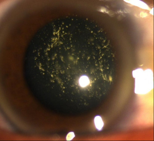
Múltiples opacidades brillantes suspendidas en el vítreo, consistentes con hialosis asteroidea.

Imagen: “Asteroid hyalosis: multiple yellow mobile vitreous particles” por University of Mohamed V souissi, hôpital des Spécialités, Ophtalology A Department. Licencia: CC BY 2.0, editado por Lecturio.

Tratamiento

  • Rara vez se necesita tratamiento.
  • La vitrectomía puede estar indicada en EN Erythema nodosum is an immune-mediated panniculitis (inflammation of the subcutaneous fat) caused by a type IV (delayed-type) hypersensitivity reaction. It commonly manifests in young women as tender, erythematous nodules on the shins. Erythema Nodosum casos de pérdida grave de agudeza visual.

Vasculatura Fetal Persistente

Definición

La vasculatura fetal persistente, antes conocida como vítreo primario hiperplásico persistente, es una afección en EN Erythema nodosum is an immune-mediated panniculitis (inflammation of the subcutaneous fat) caused by a type IV (delayed-type) hypersensitivity reaction. It commonly manifests in young women as tender, erythematous nodules on the shins. Erythema Nodosum la que los LOS Neisseria vasos sanguíneos embrionarios no involucionan.

Etiología

  • Fallo en EN Erythema nodosum is an immune-mediated panniculitis (inflammation of the subcutaneous fat) caused by a type IV (delayed-type) hypersensitivity reaction. It commonly manifests in young women as tender, erythematous nodules on the shins. Erythema Nodosum la involución del sistema vascular vítreo y hialoideo primario embrionario
  • La mayoría de los LOS Neisseria casos no son genéticos

Presentación clínica

Esta afección suele ser unilateral y puede presentarse con:

  • Leucocoria: reflejo pupilar blanco (también asociado al AL Amyloidosis retinoblastoma Retinoblastoma Retinoblastoma is a rare tumor but the most common primary intraocular malignancy of childhood. It is believed that the condition arises from a neuronal progenitor cell. Retinoblastoma can be heritable or non-heritable. Retinoblastoma)
  • Disminución de la agudeza visual
  • Microftalmia (ojo anormalmente pequeño por falta de desarrollo) del ojo afectado
  • Estrabismo

Diagnóstico

La vasculatura fetal persistente suele diagnosticarse justo después del nacimiento.

  • Fundoscopia y examen con lámpara de hendidura:
    • Proceso ciliar rotado y alargado
    • Tallo fibroso sobre el nervio óptico
  • El ultrasonido puede mostrar un tallo fibrovascular entre el cristalino y el nervio óptico.
  • La TC puede mostrar:
    • Cámara anterior poco profunda
    • Aumento de la densidad de la cámara vítrea
    • Realce del tejido intravítreo
    • Cristalino irregular
Vasculatura fetal persistente

Vasculatura fetal persistente:
En el examen con lámpara de hendidura, se identifica una tracción de los procesos ciliares hacia el centro de la cápsula posterior del cristalino en el ojo izquierdo y una masa retrolental.

Imagen: “Persistent fetal vasculature (PFV)” por Department of Ophthalmology, Hippokration General Hospital, 54642 Thessaloniki, Greece. Licencia: CC BY 3.0, editada por Lecturio.

Tratamiento

  • El tratamiento es sintomático
  • Puede ser necesario corregir la visión.
  • Se puede recomendar la cirugía para prevenir o tratar las complicaciones.

Complicaciones

  • Glaucoma Glaucoma Glaucoma is an optic neuropathy characterized by typical visual field defects and optic nerve atrophy seen as optic disc cupping on examination. The acute form of glaucoma is a medical emergency. Glaucoma is often, but not always, caused by increased intraocular pressure (IOP). Glaucoma
  • Cataratas
  • Hemorragia intraocular
  • Desprendimiento de retina Retina The ten-layered nervous tissue membrane of the eye. It is continuous with the optic nerve and receives images of external objects and transmits visual impulses to the brain. Its outer surface is in contact with the choroid and the inner surface with the vitreous body. The outermost layer is pigmented, whereas the inner nine layers are transparent. Eye: Anatomy

Referencias

  1. Ahmed F., Tripathy K. (2023). Posterior vitreous detachment. StatPearls. Retrieved August 5, 2025, from https://www.ncbi.nlm.nih.gov/books/NBK563273/
  2. Park, J. H., Yang, H., Kwon, H., & Jeon, S. (2021). Risk factors for onset or progression of posterior vitreous detachment at the vitreomacular interface after cataract surgery. Ophthalmology Retina, 5(3), 270–278. https://doi.org/10.1016/j.oret.2020.07.017
  3. Jena S, Tripathy K. (2023). Vitreous hemorrhage. StatPearls. Retrieved August 5, 2025, from https://www.ncbi.nlm.nih.gov/books/NBK559131/
  4. Johnson, B. B. (2025). Vitreous hemorrhage. EyeWiki. Retrieved August 5, 2025, from https://eyewiki.org/Vitreous_Hemorrhage
  5. Kuriakose A. (2025). Synchysis Scintillans. American Academy of Ophthalmology. Retrieved August 5, 2025, from https://eyewiki.org/Synchysis_Scintillans
  6. Man V. (2025). Asteroid hyalosis. American Academy of Ophthalmology. Retrieved August 5, 2025, from https://eyewiki.org/Asteroid_Hyalosis_(AH)
  7. Phillpotts B. A. (2023). Vitreous hemorrhage. Medscape. Retrieved August 5, 2025, from https://emedicine.medscape.com/article/1230216
  8. Ramasubramanian, A., Duff-Lynes, S. M. (2023). Persistent hyperplastic primary vitreous. EyeWiki. Retrieved August 5, 2025, from https://eyewiki.org/Persistent_Hyperplastic_Primary_Vitreous
  9. Goncalves, M.B. (2020). Asteroid hyalosis and synchysis scintillans. In: Rodrigues, E., Meyer, C., and Tomazoni, E. (Eds.), Trauma and Miscellaneous Disorders in Retina. Retina Atlas. Springer, Singapore, pp. 67–71. https://www.researchgate.net/publication/336072427_Asteroid_Hyalosis_and_Synchysis_Scintillans
  10. Ramkissoon, P. (2018). Asteroid hyalosis versus synchisis scintillans. Vision Magazine Online. http://visionmagazineonline.co.za/2018/04/01/asteroid-hyalosis-versus-synchysis-scintillans/
  11. Columbia University Department of Ophthalmology (n.d.), Asteroid hyalosis. Retrieved August 5, 2025, from https://www.columbiaeye.org/education/digital-reference-of-ophthalmology/vitreous-retina/tumor-others/asteroid-hyalosis.
  12. Sihota R, Tandon R. (2014). Parson’s Diseases of the Eye, 22nd ed., pp. 341–348.
  13. Vitreous degeneration. (n.d.). Retrieved August 5, 2025, from http://www.med.umich.edu/1libr/Ophthalmology/comprehensive/VitreousDegeneration.pdf
  14. Mai, A., et al. (2018). Persistent fetal vasculature. University of Iowa Health Care. Retrieved August 5, 2025, from  https://webeye.ophth.uiowa.edu/eyeforum/cases/270-persistent-fetal-vasculature.htm

¡Crea tu cuenta gratis o inicia una sesión para seguir leyendo!

Regístrate ahora y obtén acceso gratuito a Lecturio con páginas de concepto, videos médicos y cuestionarios para tu educación médica.

User Reviews

Que tengas una sesión de estudio alegre y navideña 🎁 Ahorra 50% en todos los planes >>

Details