Domina Conceptos Médicos

Estudia para la escuela de medicina y tus examenes con Lecturio.

Enfermedad Trofoblástica Gestacional

Las enfermedades trofoblásticas gestacionales son un espectro de trastornos placentarios que resultan de un crecimiento trofoblástico placentario anormal. Estos trastornos van desde embarazos molares benignos (molas completas y parciales) hasta afecciones neoplásicas como molas invasoras y coriocarcinoma. El diagnóstico se confirma por la gonadotropina coriónica humana (hCG, por sus siglas en EN Erythema nodosum is an immune-mediated panniculitis (inflammation of the subcutaneous fat) caused by a type IV (delayed-type) hypersensitivity reaction. It commonly manifests in young women as tender, erythematous nodules on the shins. Erythema Nodosum inglés) beta elevada en EN Erythema nodosum is an immune-mediated panniculitis (inflammation of the subcutaneous fat) caused by a type IV (delayed-type) hypersensitivity reaction. It commonly manifests in young women as tender, erythematous nodules on the shins. Erythema Nodosum suero y los LOS Neisseria hallazgos del ultrasonido, que dependen del trastorno. El tratamiento es principalmente mediante dilatación y legrado y/o metotrexato.

Last updated: Dec 15, 2025

Editorial responsibility: Stanley Oiseth, Lindsay Jones, Evelin Maza

Descripción General

Epidemiología

  • Ocurre en EN Erythema nodosum is an immune-mediated panniculitis (inflammation of the subcutaneous fat) caused by a type IV (delayed-type) hypersensitivity reaction. It commonly manifests in young women as tender, erythematous nodules on the shins. Erythema Nodosum aproximadamente 1 embarazo de cada 1 200 embarazos en EN Erythema nodosum is an immune-mediated panniculitis (inflammation of the subcutaneous fat) caused by a type IV (delayed-type) hypersensitivity reaction. It commonly manifests in young women as tender, erythematous nodules on the shins. Erythema Nodosum Estados Unidos. Mayor incidencia en EN Erythema nodosum is an immune-mediated panniculitis (inflammation of the subcutaneous fat) caused by a type IV (delayed-type) hypersensitivity reaction. It commonly manifests in young women as tender, erythematous nodules on the shins. Erythema Nodosum el sudeste asiático y Japón
  • Factores de riesgo:
    • Antecedente de enfermedad trofoblástica gestacional ( GTD GTD Gestational trophoblastic diseases are a spectrum of placental disorders resulting from abnormal placental trophoblastic growth. These disorders range from benign molar pregnancies (complete and partial moles) to neoplastic conditions such as invasive moles and choriocarcinoma. Gestational Trophoblastic Disease, por sus siglas en EN Erythema nodosum is an immune-mediated panniculitis (inflammation of the subcutaneous fat) caused by a type IV (delayed-type) hypersensitivity reaction. It commonly manifests in young women as tender, erythematous nodules on the shins. Erythema Nodosum inglés)
    • Antecedente de aborto espontáneo previo y/o infertilidad
    • Deficiencia de proteínas, ácido fólico y caroteno

Clasificación

  • Molas hidatiformes: consideradas premalignas debido a su potencial de malignidad
    • Mola completa
    • Mola parcial
  • Molas invasoras
  • Coriocarcinoma: incidencia en EN Erythema nodosum is an immune-mediated panniculitis (inflammation of the subcutaneous fat) caused by a type IV (delayed-type) hypersensitivity reaction. It commonly manifests in young women as tender, erythematous nodules on the shins. Erythema Nodosum EE. UU. de 1/40 000

Mola Hidatiforme

Las molas hidatiformes se caracterizan por inflamación quística de las vellosidades coriónicas y proliferación del epitelio coriónico. Hay 2 tipos: mola completa y mola parcial.

Mola completa Mola parcial
Cariotipo 46,XX o 46,XY Triploide (69,XXX, 69, XXY XXY Klinefelter syndrome is a chromosomal aneuploidy characterized by the presence of 1 or more extra X chromosomes in a male karyotype, most commonly leading to karyotype 47,XXY. Klinefelter syndrome is associated with decreased levels of testosterone and is the most common cause of congenital hypogonadism. Klinefelter Syndrome o 69,XYY)
Formado por Óvulo enucleado y un solo espermatozoide 2 espermatozoides y 1 óvulo
Partes fetales Ausente Presente
Nivel de gonadotropina coriónica humana (hCG, por sus siglas en EN Erythema nodosum is an immune-mediated panniculitis (inflammation of the subcutaneous fat) caused by a type IV (delayed-type) hypersensitivity reaction. It commonly manifests in young women as tender, erythematous nodules on the shins. Erythema Nodosum inglés) ↑↑↑
Hallazgos del ultrasonido
  • Útero en EN Erythema nodosum is an immune-mediated panniculitis (inflammation of the subcutaneous fat) caused by a type IV (delayed-type) hypersensitivity reaction. It commonly manifests in young women as tender, erythematous nodules on the shins. Erythema Nodosum panal de abeja
  • “Racimos de uvas”
  • Útero en “tormenta de nieve”
Revela partes fetales
Riesgo de malignidad Mayor riesgo de coriocarcinoma Raro

Presentación clínica

  • Hemorragia vaginal
  • Agrandamiento uterino (más de lo esperado para una edad gestacional determinada)
  • Presión o dolor Dolor Inflammation pélvico
  • Puede estar asociada con
    • Preeclampsia Preeclampsia A complication of pregnancy, characterized by a complex of symptoms including maternal hypertension and proteinuria with or without pathological edema. Symptoms may range between mild and severe. Pre-eclampsia usually occurs after the 20th week of gestation, but may develop before this time in the presence of trophoblastic disease. Hypertensive Pregnancy Disorders temprana (antes de las 20 semanas de gestación)
    • Quistes tecaluteínicos
    • Hiperemesis gravídica
    • Hipertiroidismo

Diagnóstico

  • Niveles de hCG: ↑ beta-hCG sérica
  • Ultrasonido:
    • Revela una distribución irregular de áreas hipoecoicas (quísticas) e hiperecoicas (sólidas) dentro del útero
    • Característicamente conocido como patrón de “tormenta de nieve
Transvaginal ultrasonography showing a molar pregnancy

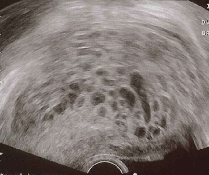
Ultrasonido transvaginal de una mola hidatiforme: En el ultrasonido se observa un característico “patrón en tormenta de nieve”.

Imagen: “Transvaginal ultrasonography showing a molar pregnancy” por Mikael Häggström. Licencia: CC0

Tratamiento

  • Dilatación y legrado (curetaje)
  • Se monitorizan los LOS Neisseria niveles de hCG (se espera una tendencia a la baja después del tratamiento)
  • Histerectomía (último recurso)
TC Axial de Mola

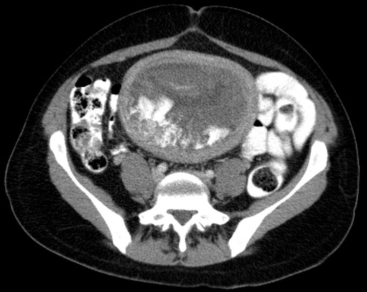
Hidatide en imagen de tomografía computarizada (TC) axial

Imagen: “Blasenmole Computertomographie axial” por Hellerhoff. Licencia: CC BY-SA 3.0

Related videos

Coriocarcinoma

El coriocarcinoma es una neoplasia maligna muy agresiva de células trofoblásticas que puede desarrollarse durante o después del embarazo en EN Erythema nodosum is an immune-mediated panniculitis (inflammation of the subcutaneous fat) caused by a type IV (delayed-type) hypersensitivity reaction. It commonly manifests in young women as tender, erythematous nodules on the shins. Erythema Nodosum la madre o el bebé.

Etiología

Puede ser precedido por:

  • Mola hidatiforme (50%)
  • Aborto de un embarazo ectópico (20%)
  • Embarazo a término normal (30%)

Presentación clínica

  • Hemorragia uterina anormal o amenorrea
  • Si existe propagación hematógena a los LOS Neisseria pulmones: dificultad para respirar y/o hemoptisis

Diagnóstico

  • Niveles de hCG: ↑ beta-hCG sérica
  • Ultrasonido pélvico: hipervascularidad
  • Radiografía de tórax: metástasis pulmonares con forma de “bala de cañón”

Tratamiento

  • Depende del estadio y la clasificación de riesgo
  • Riesgo bajo: monoterapia con metotrexato o combinación con actinomicina D
  • Alto riesgo: régimen de múltiples agentes
    • Etopósido
    • Metotrexato
    • Actinomicina D
    • Leucovorina cálcica
    • Ciclofosfamida
    • Vincristina
Interfaz entre coriocarcinoma con necrosis central y placenta normal

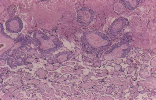
Coriocarcinoma con necrosis central

Imagen: “Interface between choriocarcinoma with central necrosis and normal placenta” por Department of Obstetrics and Gynecology, St. Louis University, Missouri, USA. Licencia: CC BY 2.0

Diagnóstico Diferencial

Las siguientes afecciones son diagnósticos diferenciales de la enfermedad trofoblástica gestacional:

  • Embarazo ectópico: o embarazo extrauterino, refiere a la implantación del blastocisto fuera de la cavidad uterina. El sitio más común es la trompa de Falopio. Las pacientes afectadas sufren de dolor Dolor Inflammation abdominal agudo. El diagnóstico se realiza mediante ultrasonido y análisis de laboratorio, que confirma el embarazo con implantación fuera del útero.

Referencias

  1. Berkowitz, R. S., Horowitz, N. S., & Elias, K. M. (2024). Gestational trophoblastic neoplasia: Epidemiology, clinical features, diagnosis, staging, and risk stratification. In B. Goff & D. S. Dixon (Section Eds.), A. Chakrabarti & S. R. Vora (Deputy Eds.), UpToDate. Retrieved March 16, 2025, from https://www.uptodate.com/contents/gestational-trophoblastic-neoplasia-epidemiology-clinical-features-diagnosis-staging-and-risk-stratification
  2. Horowitz, N. S., Eskander, R. N., Adelman, M. R., & Burke, W. (2021). Epidemiology, diagnosis, and treatment of gestational trophoblastic disease: A Society of Gynecologic Oncology evidenced-based review and recommendation. Gynecologic 1 Oncology, 163, 605–613.
  3. Bruce, S., & Sorosky, J. (2024). Gestational Trophoblastic Disease. In StatPearls. StatPearls Publishing.
  4. Bruce S, Sorosky J. Gestational Trophoblastic Disease. [Updated 2024 Feb 25]. In: StatPearls [Internet]. Treasure Island (FL): StatPearls Publishing; 2025 Jan-. Available from: https://www.ncbi.nlm.nih.gov/books/NBK470267/
  5. Hernandez, E. (2024, August 21). Gestational Trophoblastic Neoplasia. eMedicine. https://emedicine.medscape.com/article/279116-overview

¡Crea tu cuenta gratis o inicia una sesión para seguir leyendo!

Regístrate ahora y obtén acceso gratuito a Lecturio con páginas de concepto, videos médicos y cuestionarios para tu educación médica.

User Reviews

Que tengas una sesión de estudio alegre y navideña 🎁 Ahorra 50% en todos los planes >>

Details