Playlist
Show Playlist
Hide Playlist
Splenic Abnormalities

-
Slides Gallbladder and Spleen.pdf
-
Download Lecture Overview
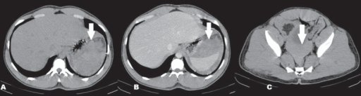
00:01 So let's move on to splenic abnormalities. 00:03 It's actually very uncommon to find focal lesions within the spleen. 00:07 So it's rare to find a splenic abnormality. 00:09 The most common splenic lesion is a cyst and it's usually an incidentaloma or an incidental finding on a CT. 00:18 So splenic cyst are well defined. 00:21 They're smoothly marginated low density lesions that are found within the spleen as you can see right here. 00:27 There's no enhancement after contrast administration. 00:30 So what do you see on this image? There's an abnormality adjacent to the spleen. 00:37 This is also a very common finding and usually found as an incidental finding. 00:42 So what is that structure right there? It has the same density as the spleen and it's located right next to it. 00:56 So this is actually a splenule. This is an accessory spleen. 01:01 It's a very common incidental finding and it's again, a well circumscribed mass that has the same exact density as the spleen. 01:08 It's found in close proximity to the spleen and often a patient may have multiple splenules that are seen. 01:14 And again, these are asymptomatic and there's nothing that needs to be done about this. 01:18 They're just a normal finding that we see on a CT. 01:21 So we've reviewed multiple abnormalities of the gall bladder and the biliary system. 01:26 It's important to recognize some of these abnormalities because ultrasound is often performed to evaluate for these. 01:31 And hopefully this gives you a good background for the next talk.
About the Lecture
The lecture Splenic Abnormalities by Hetal Verma, MD is from the course Abdominal Radiology.
Included Quiz Questions
What is TRUE regarding a splenule?
- It is an accessory spleen.
- It is a benign splenic cyst.
- It is a mass that needs to be removed.
- It is a rare finding.
- It is of higher density than the spleen.
Which of the following is NOT true regarding splenic cysts?
- They are precancerous lesions.
- They are well-defined structures.
- There is no enhancement on contrast administration.
- They are low-density lesions seen within the spleen.
- They have smooth margins.
Author of lecture Splenic Abnormalities

Hetal Verma, MD
Customer reviews
5,0 of 5 stars
| 5 Stars |
|
5 |
| 4 Stars |
|
0 |
| 3 Stars |
|
0 |
| 2 Stars |
|
0 |
| 1 Star |
|
0 |
