Playlist
Show Playlist
Hide Playlist
Spleen

-
Slides 11 Human Organ Systems Meyer.pdf
-
Reference List Histology.pdf
-
Download Lecture Overview
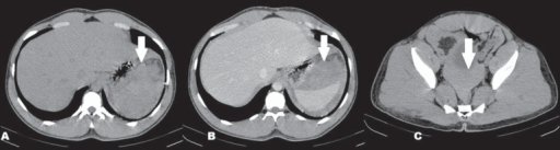
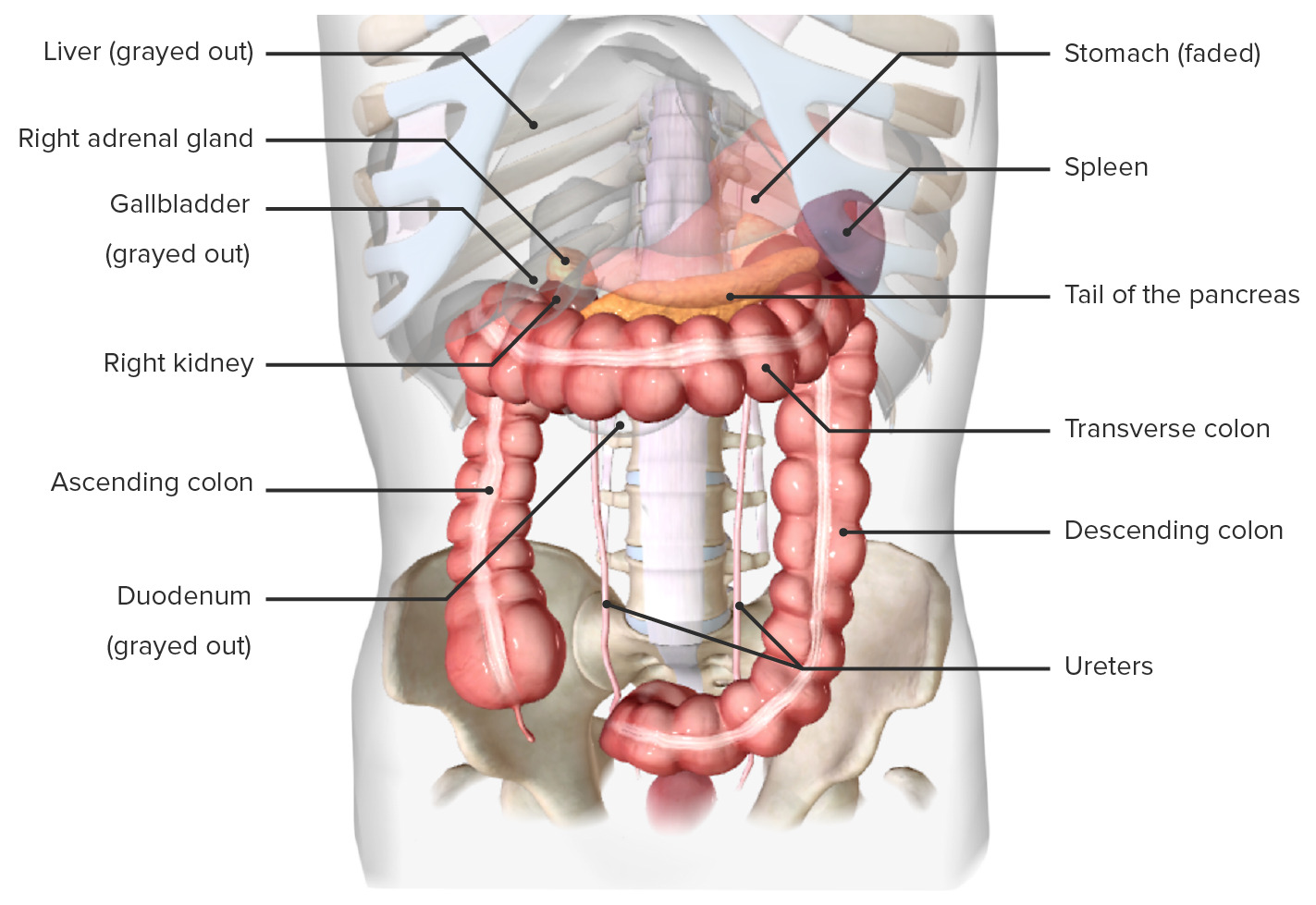
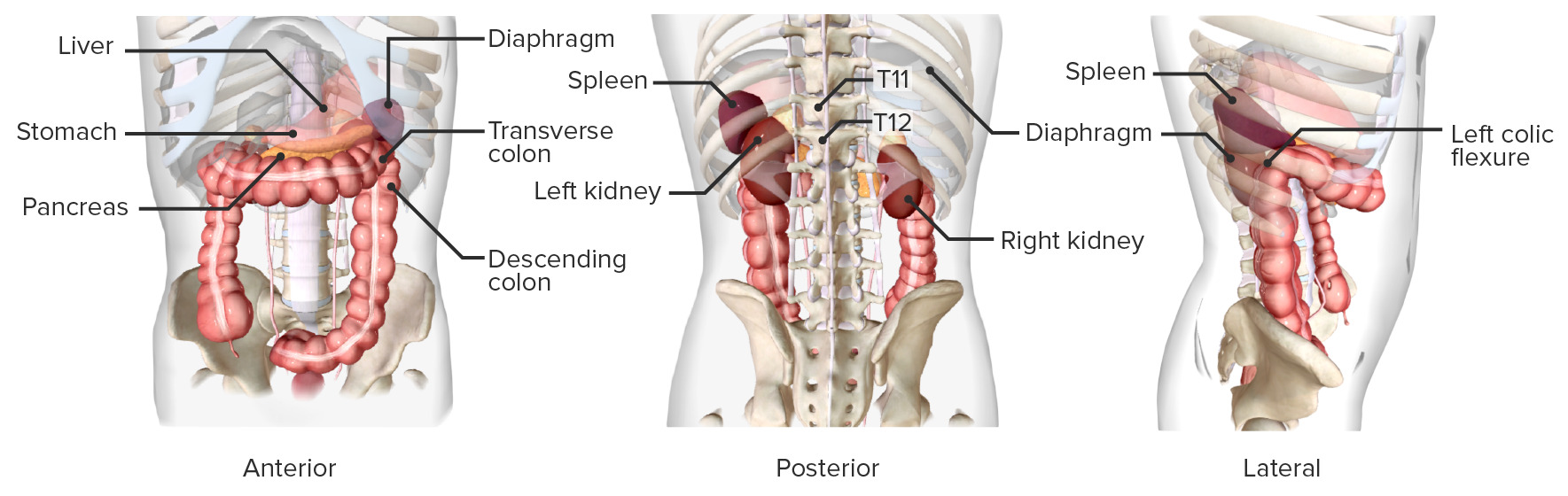
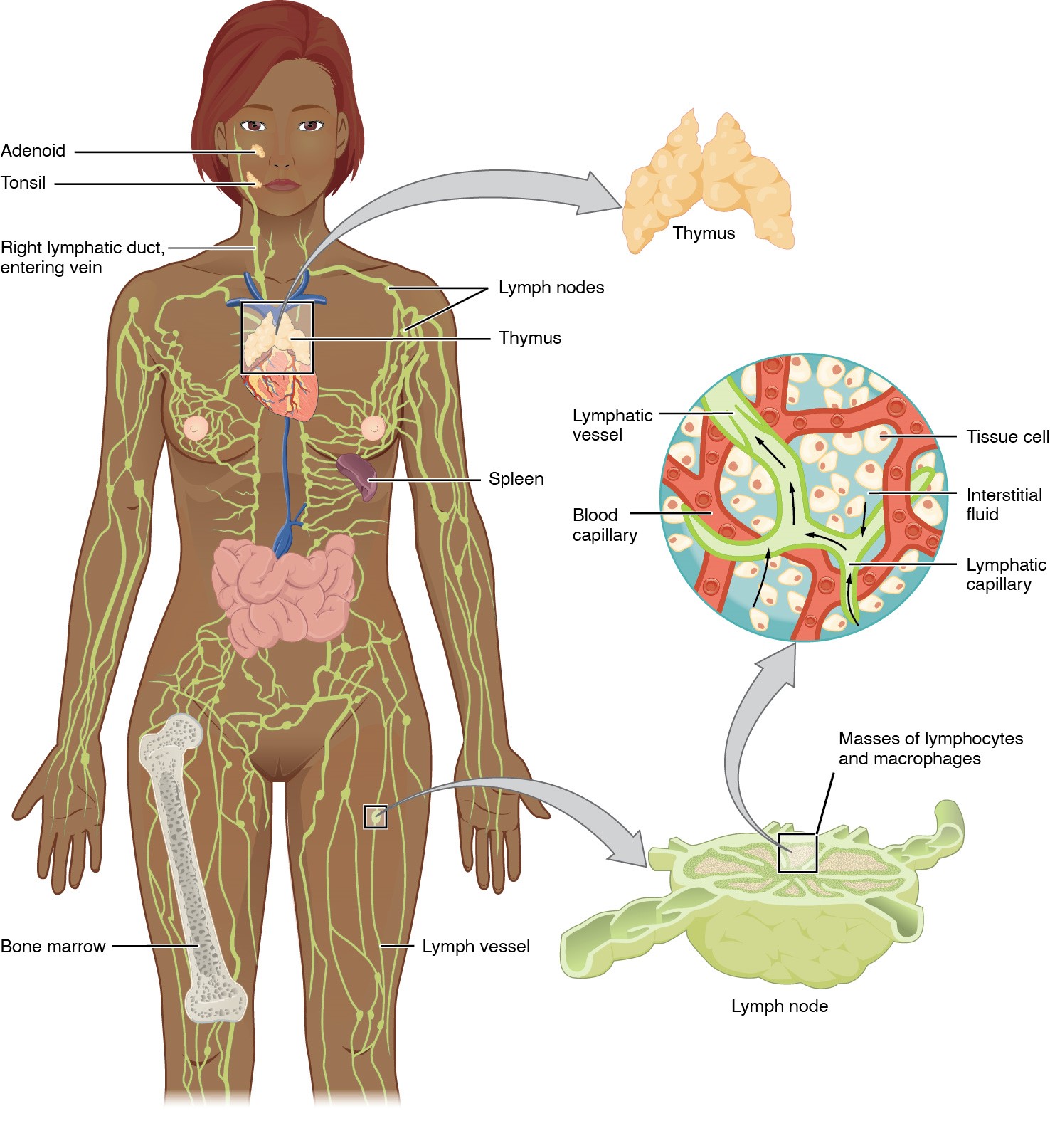
00:01 Finally, let’s look at the spleen. A lymph node filters lymph and the spleen filters blood. They’re very similar organs really. It’s just that they just clean different components of the body, blood or lymph. When you look at the spleen which is about the size of a fist, clenched fist, it has got essentially two major components to it, when you look at the components in a histological section. 00:29 You have white pulp which here stained as tiny circular blue-stained components. And you have the red pulp. This name is from what you see when you cut spleen freshly. The white pulp is where lymphocytes are. The red pulp is where all the vascular components are. 00:49 The red pulp is the component that cleans the blood. You need to know a little bit about the blood supply to the spleen. On the left-hand diagram, trabecular arteries come into the spleen, and they form central arteries. These central arteries, shown on the right-hand side, move into the body or the substance of the spleen, and they get surrounded by lymphoid tissue. They’re called central arteries because they’re actually in the centre of the splenic lobule, although you don’t really appreciate the lobular structure of the spleen when you look at histological sections. And those central arteries then pass out to little radial arteries, which then open into sinuses. And those sinuses eventually are surrounded by macrophages that can phagocytose old red blood cells. Those sinuses also open to the free space where the circulation then goes into an open circulation. And the blood meanders through the reticular network just like lymph meanders through the lymph node. 02:01 And then the blood can be checked for contents of antigens, foreign cells etc, and cleaned. That blood can then flow back into the venule system through sinusoids, and then back into the circulation. On the right-hand side, you can see the central artery, and it’s surrounded by a high load of lymphoid tissue. That lymphoid tissue I’ve abbreviated here as being PALS. It stands for peri, around, arterial, the central artery, periarterial lymphatic sheath. And it’s essentially T cells. Down below that, you can see a nodule, a lymph nodule. And what happens when blood passes out of these central arteries through the radial arteries into the blood spaces of the spleen, it flows along the length of these arteries. And along the length of all these lymphoid tissue, that lymphoid tissue has got reticular cells around it that attract all the lymphocytes into it. And then, if an antigen is detected, then an immune response is created, as you see here. The germinal centre starts, plasma cells are formed, memory B cells are formed, antibodies are formed, and also the T-cells that work together with the B cells, but the T cells can also ingest cells etc, that happens to have percolated entered via the blood stream. The arteries in the spleen, the trabecular arteries are shown here with a bit of smooth muscle around them, and they’re supported by connective tissue of the trabeculae. Sometimes, these trabeculae have smooth muscle in them that helps to contract the spleen in certain animals, and therefore, return blood cells quickly into the circulation. The vein, on the other hand, is just an endothelial lining around some of these trabecular collagenous sort of units, separating components of the spleen, trabeculae coming in from the capsule. And you also see the pulp vein, very thin-walled vein coming in, and eventually, joining out and being part of one of these large trabecular veins. The blood passing out of these blood vessels filter through the reticular network, as I explained. It filters through splenic cords lined by reticular cells and macrophages. 04:47 And again, these reticular cells can hone in all the sorts of accessory cells I’ve mentioned before. Macrophages can break down red blood cells that happen to have passed through that network, they are aged. They can’t whine their way through the very fine meshwork of the reticular network, and therefore, they haven’t got the elasticity to do that. 05:10 Therefore, the macrophages detect that and then destroy them. On the left-hand side, you can see some little bright red components that represent trabeculae, connective tissue. You can see the red pulp or the blood cells passing through that area, the splenic cords. And then you see the whitish spaces. They are the sinusoids that eventually, those cells will pass into and return to the rest of the vascular system. Higher power, you can see on the left-hand side the splenic sinusoids. And if you look very, very closely, you can see little gaps in the cell wall. Large spaces in the endothelial wall that allow these cells to return back into the vascular system, and then leave again in the same area, but mostly to return into the vascular system, and as I said before, rejoin the circulation. And those endothelial linings are wrapped up by incomplete reticular fibres, very fine little fibres you see just underneath the endothelial cells. And the diagram on the left-hand side shows you one of these capillaries, one of these sinusoids. The endothelial cells are elongated along the length of the sinusoid. And there are gaps between them as I pointed out, an incomplete basement membranes, basal lamina. And you can see the cells moving across the wall of these very leaky sinusoids. 06:51 On the right-hand side, you can see the reticular network stained, and there're little gaps in that network, again, creating space for the cells to move in and out of the lining of the vessel.
About the Lecture
The lecture Spleen by Geoffrey Meyer, PhD is from the course Lymphoid Histology.
Included Quiz Questions
Which of the following statements regarding the spleen is INCORRECT?
- The lymphocytes in the periarterial lymphatic sheath are mostly B lymphocytes.
- The spleen mainly filters the blood.
- The white pulp of the spleen mainly consists of lymphatic tissue.
- Red pulp is characterized by the presence of splenic sinusoids and splenic cords.
- The periarterial lymphatic sheath surrounds the central artery in the spleen.
Which of the following is the main function of splenic cords?
- Blood filtration of damaged erythrocytes
- Antigen presentation
- T-cell maturation
- Forms a mesh for supporting macrophages
- Blood supply to the organ
Which of the following statements regarding the periarteriolar lymphoid sheaths within the spleen is MOST ACCURATE?
- They consist of portions of the white pulp, populated largely by T cells, that surround central arteries
- They consist of portions of the red pulp, populated largely by T cells, that surround central arteries.
- They consist of portions of the red pulp, populated largely by B cells, that surround central arteries.
- They consist of portions of the white pulp, populated largely by B cells, that surround central arteries.
- They consist of portions of the white pulp, populated largely by T cells, that surround peripheral arteries.
What type of muscle is seen around the trabecular arteries in the spleen?
- Smooth muscle
- Cardiac muscle
- Smooth and skeletal muscle
- Skeletal muscle
- There is no muscle around the trabecular arteries in the spleen.
Which of the following is not part of the red pulp?
- All of the options provided may be found in the red pulp of the spleen.
- Macrophages
- Splenic cords
- Splenic cells
- Red blood cells
Author of lecture Spleen

Geoffrey Meyer, PhD
Customer reviews
3,0 of 5 stars
| 5 Stars |
|
1 |
| 4 Stars |
|
0 |
| 3 Stars |
|
0 |
| 2 Stars |
|
0 |
| 1 Star |
|
1 |
the video is in depth explanation explaining structures and function
you have to write everything to the material.only pictures present
