Playlist
Show Playlist
Hide Playlist
Introduction to Inflammatory Myopathies with Case

-
Slides Inflammatory Myopathies.pdf
-
Download Lecture Overview
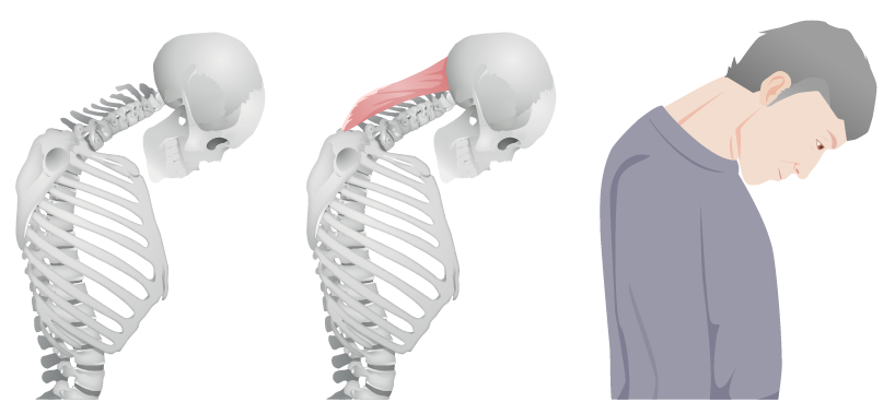
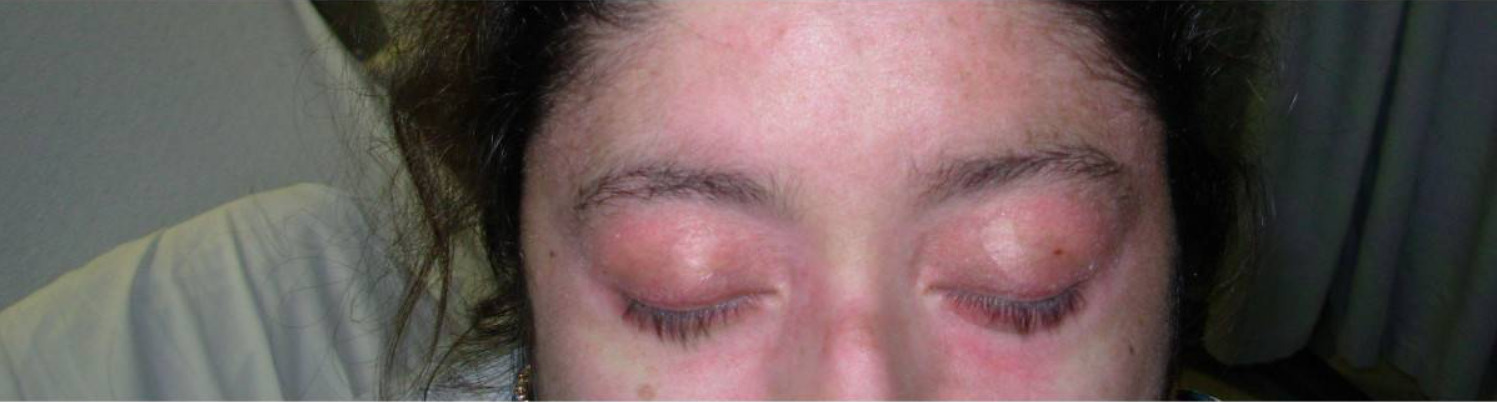
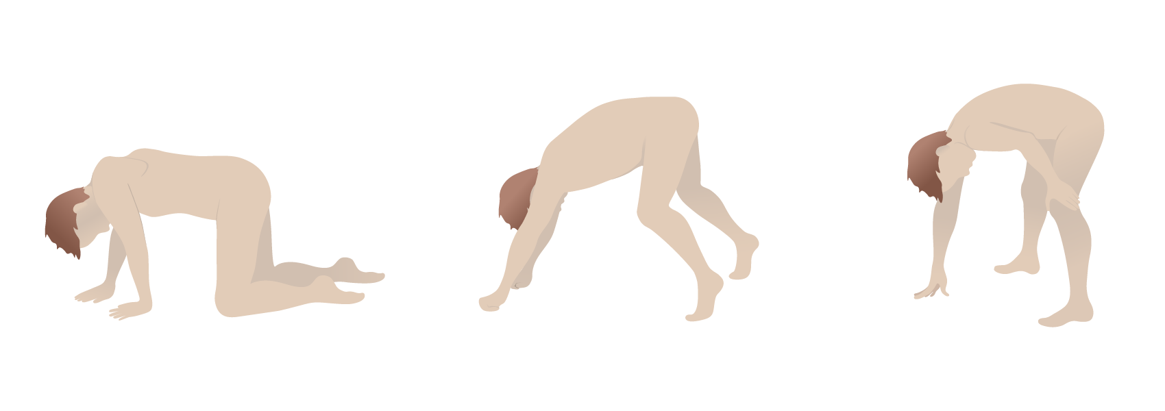
00:00 In this lecture, we're going to talk about the inflammatory myopathies, one of the most important and common types of myopathies. And let’s start with a case; 54-year-old female who presents with weakness, a common chief complaint for a muscle disorder. This person has been a long-time patient and has a history of thyroid disease treated with levothyroxine, as well as hyperlipidemia managed with atorvastatin. For the past 2 months, the patient has noticed progressive weakness. This is most prominent in the legs where the patient now has difficulty arising out of a chair in the morning after breakfast, and has had to use arms to help walking upstairs at times. More recently, the patient has noticed difficulty with arm strength as well. There are no sensory symptoms. The patient denies swallowing difficulty and does not have signs of ptosis, diplopia, dysarthria or dysphagia. On exam, we see proximal weakness in the legs, and normal deep tendon reflexes. So, where do we diagnose this? Where do we localize and diagnose this case? Let’s think about those 3 key features of a peripheral nervous system disorder. What's the distribution? What's the sensory exam? And what are the reflex findings? Here the distribution is clearly proximal; difficulty walking upstairs, getting up of a chair, proximal weakness on exam. The distribution supports a proximal disorder like a muscle disorder or perhaps neuromuscular junction condition. There are no sensory findings, so we would less favor a peripheral nerve disorder. Reflex examination is normal, so again we don't hear findings that are concerning or consistent with a peripheral nerve disorder. And our important wild card, our pertinent negative. There's no ptosis, diplopia, dysarthria or dysphagia. We don't see bulbar findings like we would see with a neuromuscular junction disorder. We're thinking about a myopathy. There also are some important clinical features present in this case. The patient has thyroid disease, is on a statin, has a progressive course and is in her middle age. And those will be important for differentiating some of the final diagnoses. This patient underwent work-up and the work-up included a creatine kinase, looking for muscle inflammation which was substantially elevated. The CK was 3200, normal being 50 to 160. An EMG and nerve conduction study was performed and the result was myopathic muscle units indicating a muscle disorder, also showing spontaneous activity with fibrillation potentials and positive sharp waves. The key thing about spontaneous activity is that is telling us there may be inflammation within the muscle. Thyroid study showed normal thyroid stimulating hormone and free thyroxine levels. There's no signs of thyroid dysfunction. 02:45 The patient is doing well on her levothyroxine. And the general exam did not show a rash. No facial heliotrope rash. No Gottron’s papules. So again, what's the diagnosis? Is this dermatomyositis, hypothyroid myopathy, polymyositis or inclusion body myositis? Well, dermatomyositis typically presents with a rash and we don't see those typical dermatologic findings that point us in that direction. We don't like that diagnosis. The thyroid studies were normal. And we expect those to be abnormal in a patient with hypothyroid myopathy, and so this is unlikely to explain this patient’s weakness. Inclusion body myositis actually follows a different pattern. There's proximal lower extremity weakness, but patients with IBM present with a distal upper extremity weakness and that's not the pattern that we see in this case. So, this is a good presentation and a typical presentation for a patient with polymyositis. 03:46 Proximal weakness, no sensory complaints, normal deep tendon reflexes, no rash to suggest dermatomyositis and all of the findings to suggest that this is an inflammatory myopathy. 03:58 Elevated CK, myopathic findings on the EMG, and that spontaneous activity that is suggesting muscle inflammation. Importantly, this patient would need to undergo evaluation to exclude other potential causes of inflammatory myopathy that overlap with polymyositis, including statin-associated myopathy.
About the Lecture
The lecture Introduction to Inflammatory Myopathies with Case by Roy Strowd, MD is from the course Acquired Neuromuscular Diseases.
Included Quiz Questions
Which of the following lab findings is most associated with polymyositis?
- Elevated CK
- Elevated ESR
- Low T3
- Normal ESR
- Normal CK
In an electromyogram (EMG) test & nerve conduction study (NCS), the finding of spontaneous activity suggests the presence of...?
- ...muscle inflammation.
- ...a nerve abnormality.
- ...hypothyroidism.
- ...a toxic myopathy.
- ...normal muscle and nerve function.
Author of lecture Introduction to Inflammatory Myopathies with Case

Roy Strowd, MD
Customer reviews
5,0 of 5 stars
| 5 Stars |
|
5 |
| 4 Stars |
|
0 |
| 3 Stars |
|
0 |
| 2 Stars |
|
0 |
| 1 Star |
|
0 |
