Playlist
Show Playlist
Hide Playlist
Introduction – Joints of Lower Limb


-
Slides 09 LowerLimbAnatomy Pickering.pdf
-
Download Lecture Overview
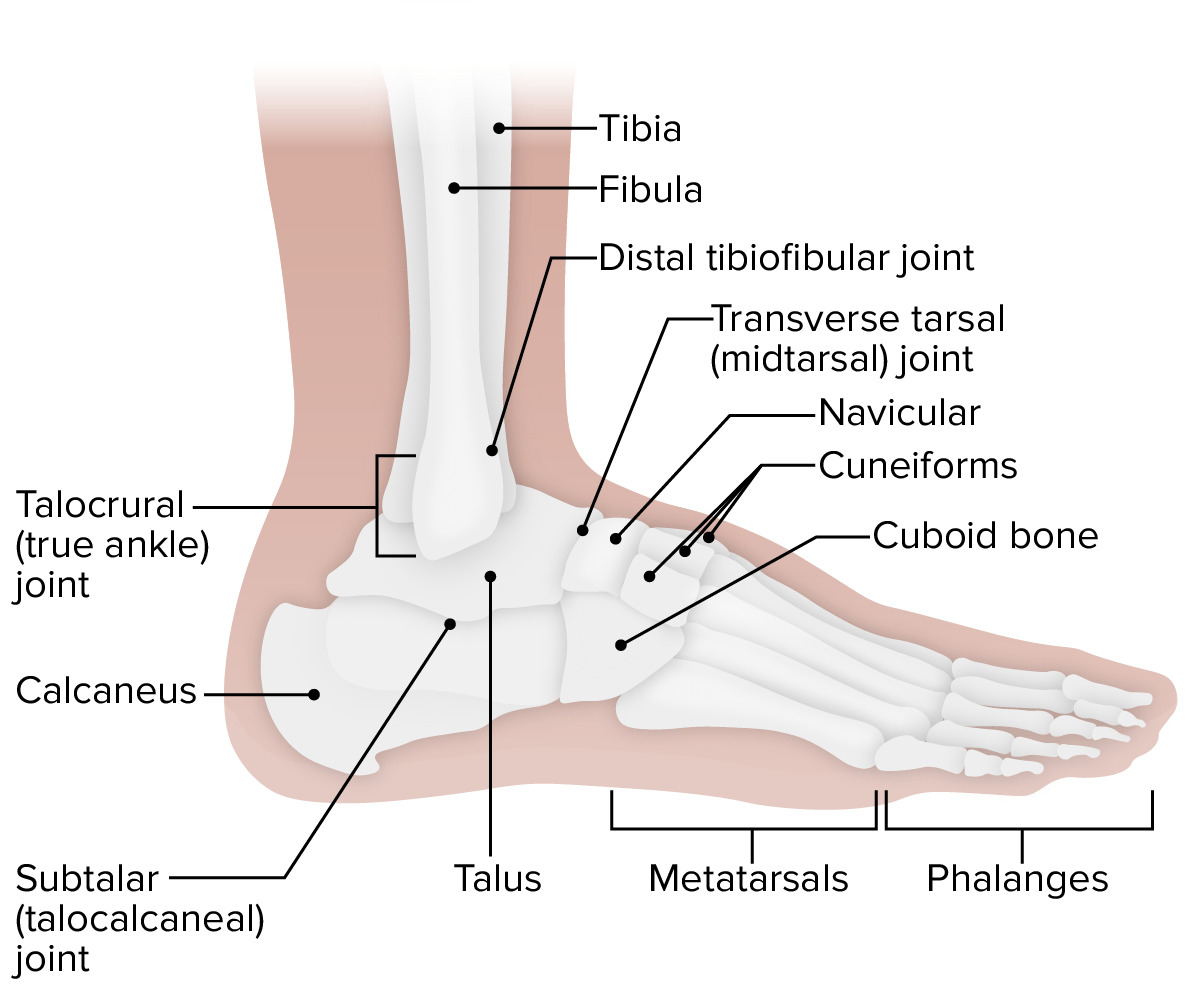
00:01 In this lecture, we’re going to look at the joints of the lower limb. So we’re going to look at numerous joints that are located within the inferior appendicular skeleton. 00:12 We’re going to look at the hip joints, the knee joints, the talofibular joints, the ankle joints, the various joints of the foot, and then finish by looking briefly at the arches of the foot. So to start off with, we’re going to look at the hip joints. Here on the screen, we can see a number of images which are really going to highlight various features of the hip joint. So here, can see the anterior aspect where we’ve got the head of the femur going to articulate with the acetabulum. Here, you can see the actual deep acetabular fossa with the head sitting within it. Here, we can see various parts of the joint capsule and some ligaments that are going to strengthen this joint. So the hip joint connects the lower limb, the femur, to the pelvis forming the pelvic girdle. The articulation is a ball and socket joint that is very stable with the large head of the femur sitting in the deep acetabulum formed by the three bones of the pelvis. As we mentioned in the first lecture, these are the pubis, the ilium, and the ischium. The increased stability of the hip joint compared to the shoulder joint in the upper limb is important to support the body weight, but whereas, the glenohumeral joint was unstable but offered a wide range of movement. The stability of the hip joint means that some movements are compromised and there’s not as much an extensive array of movement possible at the hip joint. 01:50 So here, we can see the hip joint has been opened up, and we can see here on this right view of the hip joints, we have the head of the femur. The head of the femur is covered by this articular cartilage except at the fovea which has the ligament of the head of the femur attaching. The depth of the acetabulum is increased by way of this acetabular labrum which is running around the surface, the perimeter of the acetabulum. And that increases the depth. So the rim of the acetabulum formed by the semilunar articular portion of articular cartilage, here we can see this lunate surface, this semilunar portion here, this lunate surface. 02:38 And this is articular cartilage which allows the head of the femur to rotate within the acetabulum. As I mentioned, the depth of the acetabulum is increased by this acetabular labrum. And we also have more inferiorly we have the transverse acetabular ligament, which we can see here. So if we look at the joint capsule of the hip joints, then it passes from the rim of the acetabulum, which we can see here, of the pelvis to the intertrochanteric line ateriorly and proximal to the intertrochanteric crest posteriorly. So here we have the anterior view and it’s passing to the intertrochanteric line between the greater and the lesser trochantus. 03:29 And posteriorly here, we can see the joint capsule is passing just proximal to the intertrochanteric crest, which we can see here. So we can see exposed is the neck of the femur. If we were to look at the hip joint in more detail, then we would see we have these thickenings of the fibrous capsule and a spiral across the joint and form specific ligaments. We have the iliofemoral ligament. This is running anteriorly and superiorly. It attaches to the anterior inferior iliac spine and the acetabular rim passing to the intertrochanteric line. So we can see the iliofemoral ligament passing down here. We can see we have it passing in this direction. This ligament prevents hyperextension. We also have the pubofemoral ligaments, and this is positioned anteriorly and inferiorly. We can see it here. 04:29 It’s running from the obturator crest of the pubic bone, and this merges with the fibrous capsule of the hip joint. It prevents excessive abduction. Here, we can see the pubofemoral ligament. 04:42 We can see it here. The final ligament is the ischiofemoral, and this is located posteriorly. 04:48 We can see it here in the posterior view. It’s running from the ischial part of the acetabular rim to the neck of the femur at the base of the great trochanter. So here we can see it running to the neck of the femur and towards the base of the great trochanter. 05:03 We can see it here. This ligament limits the extension and medial rotation.
About the Lecture
The lecture Introduction – Joints of Lower Limb by James Pickering, PhD is from the course Lower Limb Anatomy [Archive].
Included Quiz Questions
Which type of articulation is used by the hip joint?
- Ball-and-socket joint
- Saddle joint
- Condyloid joint
- Hinge joint
- Pivot joint
Which surface of the hip joint is not covered by articular cartilage?
- Ligament of the head of the femur
- Acetabular labrum
- Acetabular ligament
- Rim of the acetabulum
- Lunate surface of the acetabulum
What is the function of the iliofemoral ligament?
- Prevents hyperextension
- Prevents flexion
- Lubricates joints
- Gives rise to white blood cells
- Prevents abduction
Author of lecture Introduction – Joints of Lower Limb

James Pickering, PhD
Customer reviews
5,0 of 5 stars
| 5 Stars |
|
5 |
| 4 Stars |
|
0 |
| 3 Stars |
|
0 |
| 2 Stars |
|
0 |
| 1 Star |
|
0 |
