Playlist
Show Playlist
Hide Playlist
Blood/Lymphatic Circulation and High Endothelial Venules – Lymphocyte Recirculation and Homing

-
06 Slides Lymphocyte Recirculation and Homing.pdf
-
Reference List Immune System.pdf
-
Download Lecture Overview
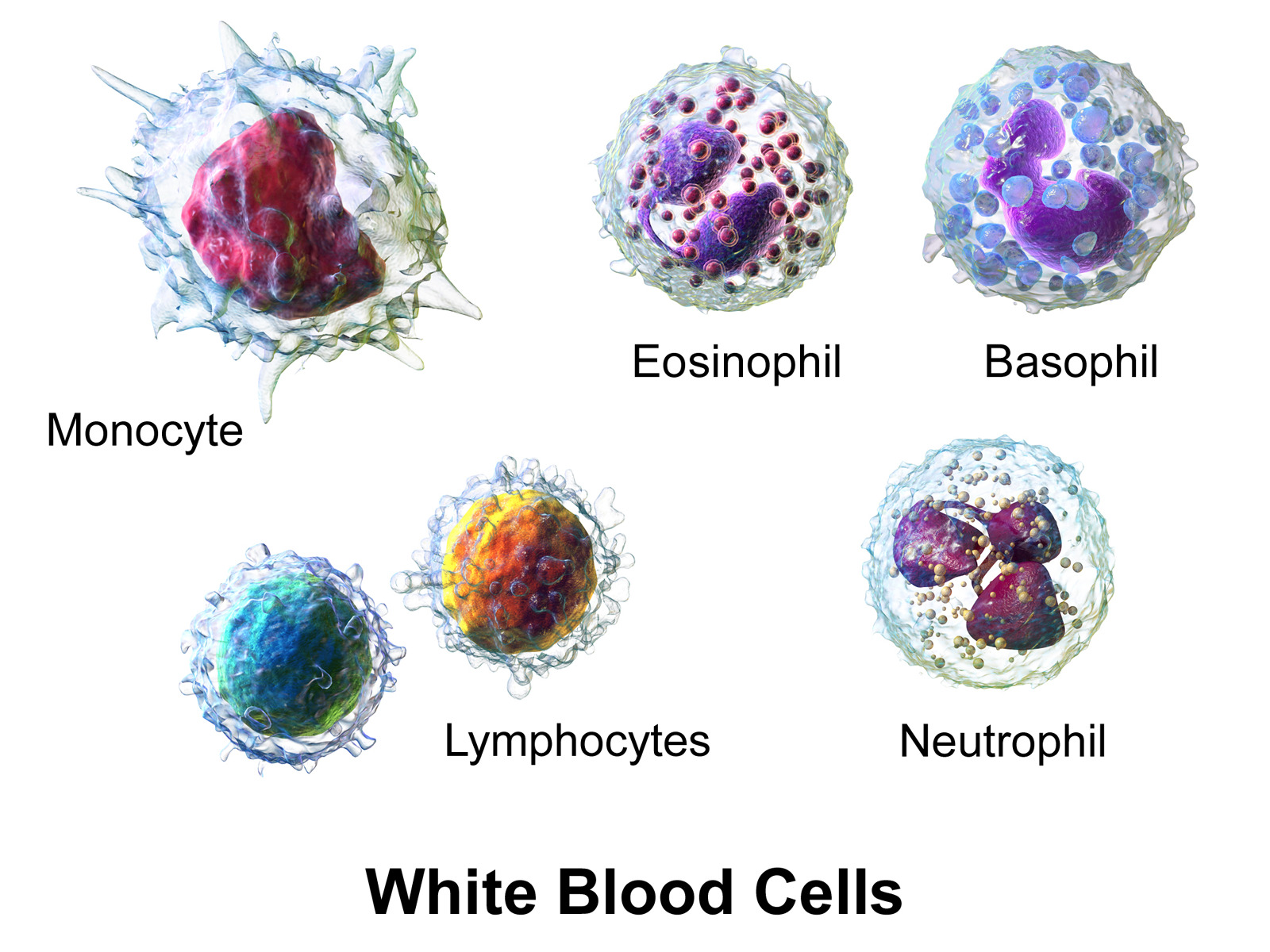
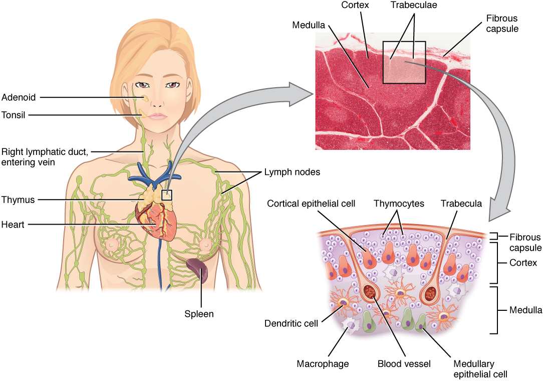
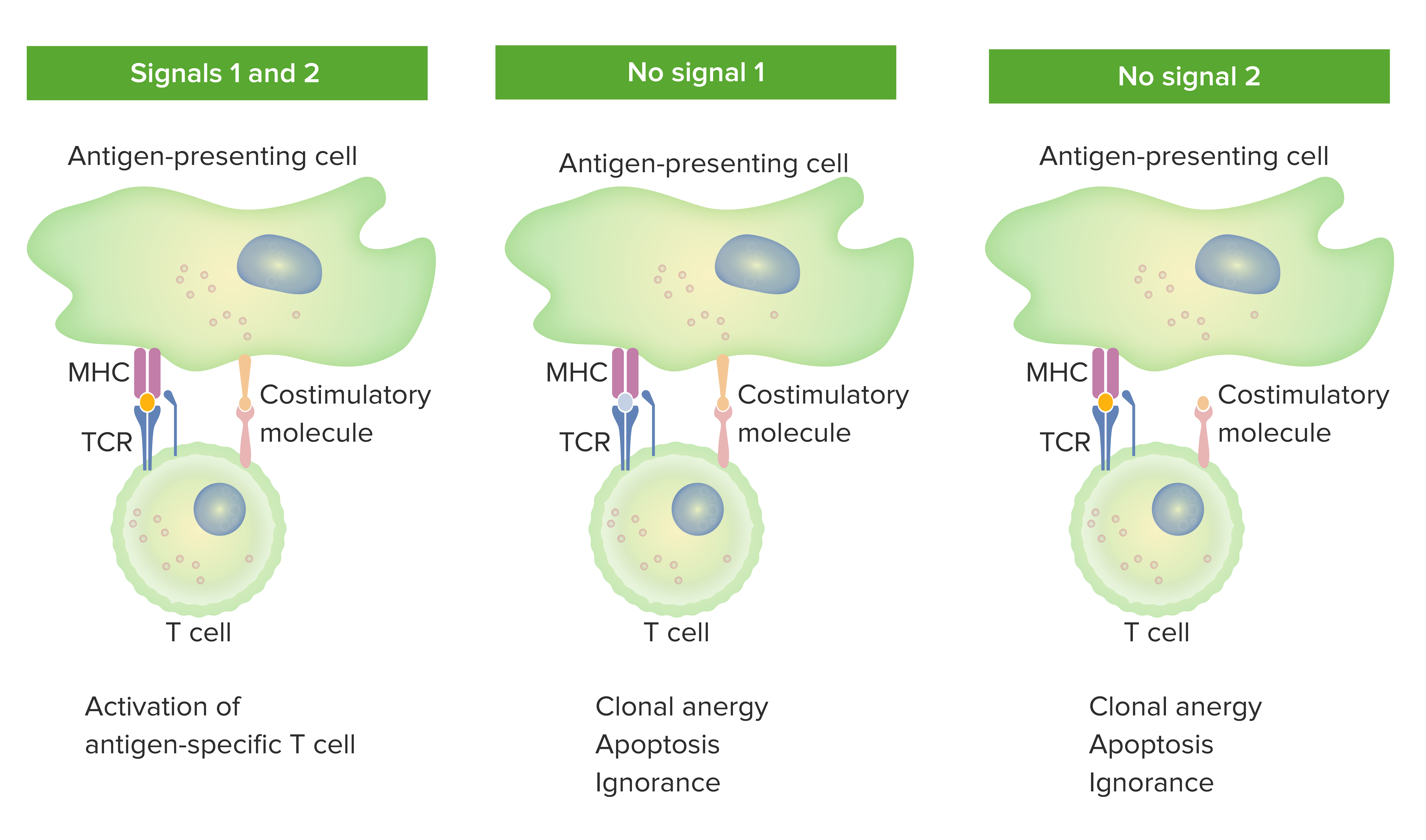
00:01 Let’s look at the blood and lymphatic circulation. 00:05 So here we can see the blood circulation. 00:08 And lymphocytes and other blood cells will be traveling around in the blood circulation. 00:14 And they may go to the liver, they may go to the skin, they may go to the kidney or they might go to the gut. 00:21 Following their arrival at these locations, they can look for antigen. 00:29 And they can ultimately leave these locations and go to the local lymph nodes. 00:38 Whether they’re coming from the liver, the skin or the kidney, they can end up in nearby lymph nodes. 00:48 Lymphocytes that are activated in the Peyer’s patches in the gut can also travel to the local lymph node and in this case, it is the mesenteric lymph nodes. 01:01 Ultimately, the lymphocytes can leave those lymph nodes and travel via the lymphatic vessels to other lymph nodes, because the lymph nodes are connected to each other via the lymphatics. 01:15 So they’ll leave via the efferent lymphatics for those particular nodes and then later on enter via the afferent lymphatics to downstream nodes. 01:28 The lymphocytes can also directly enter into the lymph nodes. 01:33 They don’t have to enter via the lymphatic vessels; they can actually go straight there from the blood circulation. 01:38 And they do this by structures within the lymph nodes called high endothelial venules, HEV. 01:45 Ultimately, after seeing whether or not their antigen is present in the lymph node, the lymphocytes can leave via the efferent lymphatics, again travel through the lymphatic vessels and ultimately they can rejoin the blood circulation via the thoracic duct. 02:06 So let’s have a look at these two systems. 02:08 Really they’re very much like a road system and a rail system; two different systems but interconnected, just like you can drive your car to the station, park at the station and hop on a train. 02:20 So lymphocytes can travel around both of these systems and use the interconnections such as the high endothelial venules, such as the thoracic duct to get between one system and another. 02:34 Looking now in a little bit more detail at the events that occur in the lymph node and particularly, the events occurring as lymphocytes leave the blood vessels and enter the lymph node. 02:47 So at the top, we have a lymphocyte, it’s a naïve T-cell, it’s not seen antigen before. 02:54 And it’s going to enter a lymph node. 02:56 But this lymph node in this particular example, it doesn’t have the antigen that this T-cell is specific for. 03:04 So the lymphocyte will enter the lymph node via the high endothelial venules. 03:10 It will have a look, see whether its antigen is there or not. 03:13 And if the antigen isn’t there, it will actually not hang around in that lymph node. 03:17 It will move quite rapidly out of the lymph node via the efferent lymphatic vessels. 03:24 Then it can go to the next lymph node in the chain. 03:29 And maybe that lymph node has got antigen present that the T-cell is specific for, because maybe there’s an infection in the tissues. 03:38 And pathogens picked up by dendritic cells can be carried from the location of the infection via the afferent lymphatics into the lymph node. 03:53 And when the lymphocyte arrives, lo and behold, its antigen is there. 03:57 So it will stay in that lymph node and become activated. 04:03 It may also be that the lymphocyte directly entered that node and will immediately be activated because its antigen is present. 04:12 So at the top we have a lymph node without the antigen, in the middle we have a lymph node where the antigen is present that that particular T-cell is specific for. 04:20 And that T-cell may have entered via the lymphatics because it’s come from another node, or it may enter directly through the HEV from the blood circulation. 04:32 Following interaction with dendritic cells, and stimulation of the lymphocyte, this naïve lymphocyte then becomes activated. 04:42 And the activated lymphocyte can leave the lymph node via the efferent lymphatic vessels and ultimately rejoin the blood circulation via the thoracic duct. 04:53 Remember the thoracic duct it’s a connec-- one of the connections between the lymphatic circulation and the blood circulation. 05:01 Now this T-cell is activated, and it’s responding to a particular infection. 05:06 But it’s no good at being in the lymphatic vessels or in the blood circulation, it needs to actually get to where the infection is, so it can carry out its job of eliminating the infection. 05:16 So it will recirculate back to the location where the antigen originated from and therefore be able to attack the pathogen and hopefully get rid of the infection. 05:28 In terms of the high endothelial venules, this is a very specific process whereby molecules associated with the high endothelial venules interact with molecules on the surface of the lymphocyte to facilitate binding of the lymphocyte to the high endothelial venule. And then exit from the venule into the body of the lymph node. This is mediated by cell surface molecules present on the lymphocyte. So for example, the adhesion molecule, L-selectin on the surface of the naïve T-cell interacts with a molecule called PNAd (Peripheral Node Adresin). Peripheral Node Adresin - PNAd, on the surface of the high endothelial venules. So this is a cell-cell interaction, one molecule on the surface of the T-cell, L-selectin interacting with a molecule on the surface of the endothelial cell, PNAd. Also there will be soluble cytokines that can bind to the surface of the high endothelial venule. And in particular, one type of cytokine, the chemokine, is key in this process. As the name suggests, these are chemotactic cytokines. And two of them are particularly important. Those chemokines are called CCL19 and CCL21. And they are recognized by a particular chemokine receptor on the surface of the naïve T-cell. And that receptor is called CCR7. 07:06 Having then entered the lymph node, those naïve T-cells can become activated by interaction with dendritic cells. 07:14 And then, as we just saw they can leave the lymph node via the efferent lymphatic vessels.
About the Lecture
The lecture Blood/Lymphatic Circulation and High Endothelial Venules – Lymphocyte Recirculation and Homing by Peter Delves, PhD is from the course Adaptive Immune System. It contains the following chapters:
- The Blood and Lymphatic Circulation
- High Endothelial Venules
Included Quiz Questions
Which of the following is a specific marker on the surface of naive T cells?
- CCR7
- CD 80
- CD 28
- CD 40
- CD 4
Where does the lymph in Peyer's patches drain?
- Mesenteric lymph nodes
- Subcutaneous lymph nodes
- Spleen
- Renal lymph nodes
- Cervical lymph nodes
What is the main role of high endothelial venules?
- Allow lymphocytes to enter lymph nodes directly from blood vessels
- Allow lymphocytes to travel out of the efferent lymphatic vessels
- Allow lymphocytes to directly enter the thoracic duct
- Allow drainage of lymphocytes from lymph nodes into blood
- Recognize antigens in lymph nodes
Which of the following statements regarding cell markers are CORRECT?
- Peripheral node addressin (PNAd) marks high endothelial venules (HEV)
- Peripheral node addressin (PNAd) marks naive T cells
- L selectin marks high endothelial venules (HEV)
- C-C Chemokine receptor type 7 marks high endothelial venules (HEV)
Author of lecture Blood/Lymphatic Circulation and High Endothelial Venules – Lymphocyte Recirculation and Homing

Peter Delves, PhD
Customer reviews
5,0 of 5 stars
| 5 Stars |
|
5 |
| 4 Stars |
|
0 |
| 3 Stars |
|
0 |
| 2 Stars |
|
0 |
| 1 Star |
|
0 |
