Domina Conceptos Médicos

Estudia para la escuela de medicina y tus examenes con Lecturio.

Tipos Celulares: Eucariotas y Procariotas

Los LOS Neisseria organismos pueden clasificarse principalmente en EN Erythema nodosum is an immune-mediated panniculitis (inflammation of the subcutaneous fat) caused by a type IV (delayed-type) hypersensitivity reaction. It commonly manifests in young women as tender, erythematous nodules on the shins. Erythema Nodosum 2 grupos, procariotas y eucariotas, que presentan diferencias fundamentales a nivel celular. Los LOS Neisseria procariotas son organismos unicelulares que incluyen 2 de los LOS Neisseria 3 dominios de la vida: las bacterias y las arqueas. Los LOS Neisseria eucariotas pueden ser organismos unicelulares o pluricelulares e incluyen plantas, animales, hongos y protozoos. Las células procariotas constan de un único compartimento lleno de citoplasma encerrado por una membrana y una pared celular, mientras que las células eucariotas contienen un núcleo bien organizado contenido por una membrana, junto con otros orgánulos unidos a ella.

Last updated: Dec 15, 2025

Editorial responsibility: Stanley Oiseth, Lindsay Jones, Evelin Maza

Organismos Eucariotas

Los LOS Neisseria organismos eucariotas incluyen:

  • Protozoos
  • Algas
  • Hongos
  • Plantas
  • Animales

Organización celular

  • Unicelular
  • Multicelular

Características celulares

  • Tamaño:
    • Grande
    • 0,5–100 μm de diámetro
  • Organización:
    • Compartimentado en EN Erythema nodosum is an immune-mediated panniculitis (inflammation of the subcutaneous fat) caused by a type IV (delayed-type) hypersensitivity reaction. It commonly manifests in young women as tender, erythematous nodules on the shins. Erythema Nodosum orgánulos unidos a la membrana
    • Forma determinada por el citoesqueleto
    • Sistema de endomembranas (retículo endoplásmico, Golgi, lisosomas) que contiene grandes ribosomas para la síntesis de proteínas
  • El ADN:
    • Lineal
    • Estructurado en EN Erythema nodosum is an immune-mediated panniculitis (inflammation of the subcutaneous fat) caused by a type IV (delayed-type) hypersensitivity reaction. It commonly manifests in young women as tender, erythematous nodules on the shins. Erythema Nodosum múltiples cromosomas en EN Erythema nodosum is an immune-mediated panniculitis (inflammation of the subcutaneous fat) caused by a type IV (delayed-type) hypersensitivity reaction. It commonly manifests in young women as tender, erythematous nodules on the shins. Erythema Nodosum forma de bastón
    • Organizado y empaquetado con histonas
    • Los LOS Neisseria cromosomas pueden ser haploides (no apareados) o diploides (apareados).
    • Almacenado en EN Erythema nodosum is an immune-mediated panniculitis (inflammation of the subcutaneous fat) caused by a type IV (delayed-type) hypersensitivity reaction. It commonly manifests in young women as tender, erythematous nodules on the shins. Erythema Nodosum el núcleo contenido por una membrana
    • La transcripción se produce dentro del núcleo; la traducción, en EN Erythema nodosum is an immune-mediated panniculitis (inflammation of the subcutaneous fat) caused by a type IV (delayed-type) hypersensitivity reaction. It commonly manifests in young women as tender, erythematous nodules on the shins. Erythema Nodosum el citosol.
  • Membrana celular: bicapa lipídica
  • Pared celular:
    • Solo presente en EN Erythema nodosum is an immune-mediated panniculitis (inflammation of the subcutaneous fat) caused by a type IV (delayed-type) hypersensitivity reaction. It commonly manifests in young women as tender, erythematous nodules on the shins. Erythema Nodosum algunas células
    • Compuesto por celulosa (plantas y algas) o quitina (moluscos, insectos, crustáceos, hongos)
  • Motilidad:
    • Flagelos
    • Proyecciones flexibles hechas de microtúbulos
  • La división celular:
    • Meiosis Meiosis The creation of eukaryotic gametes involves a DNA replication phase followed by 2 cellular division stages: meiosis I and meiosis II. Meiosis I separates homologous chromosomes into separate cells (1n, 2c), while meiosis II separates sister chromatids into gametes (1n, 1c). Meiosis
    • Mitosis Mitosis A type of cell nucleus division by means of which the two daughter nuclei normally receive identical complements of the number of chromosomes of the somatic cells of the species. Cell Cycle
  • Transporte de materia a través de la membrana celular:
    • Difusión simple y facilitada
    • Fagocitosis
Célula eucariota

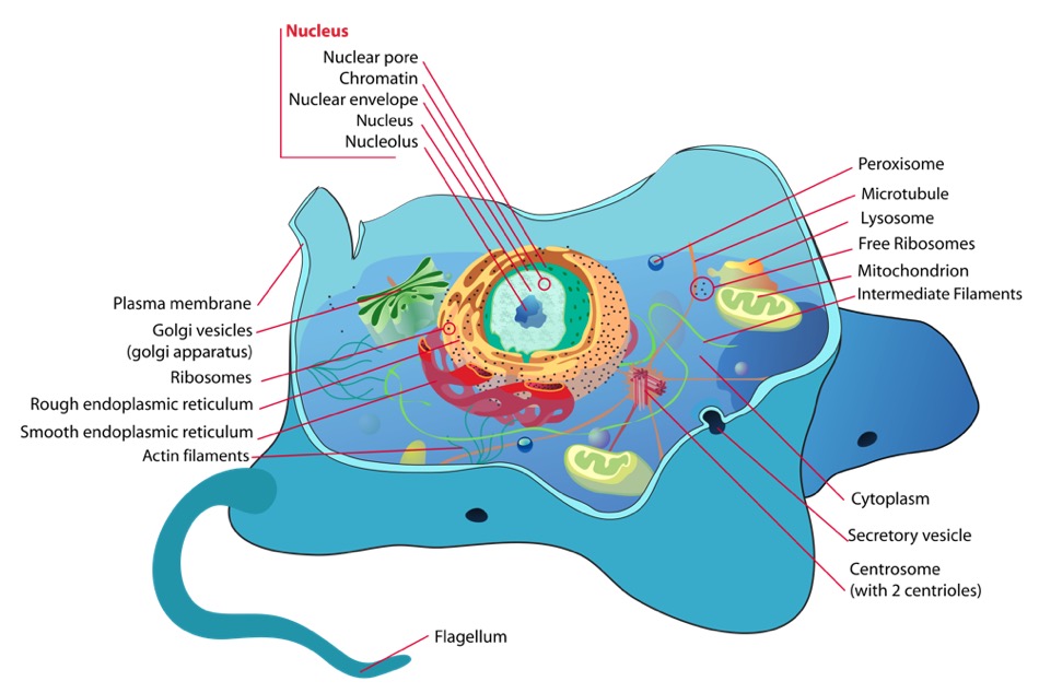
Una célula eucariota y sus componentes

Imagen: “Animal cell structure” por Mariana Ruiz. Licencia: Dominio Público

Organismos Procariotas

Los LOS Neisseria organismos procariotas incluyen las bacterias y las arqueas.

La organización celular es unicelular.

Características Celulares

  • Tamaño:
    • Pequeño
    • 0.1–5 μm de diámetro
  • Organización:
    • No hay una verdadera compartimentación
    • Las reacciones metabólicas se producen libremente en EN Erythema nodosum is an immune-mediated panniculitis (inflammation of the subcutaneous fat) caused by a type IV (delayed-type) hypersensitivity reaction. It commonly manifests in young women as tender, erythematous nodules on the shins. Erythema Nodosum el citosol.
    • Los LOS Neisseria ribosomas se encuentran libres en EN Erythema nodosum is an immune-mediated panniculitis (inflammation of the subcutaneous fat) caused by a type IV (delayed-type) hypersensitivity reaction. It commonly manifests in young women as tender, erythematous nodules on the shins. Erythema Nodosum el citosol
    • Falta de citoesqueleto
    • Puede tener cuerpos de inclusión: agregados densos polimerizados de proteínas o nutrientes
  • El ADN:
    • Circular
    • Organizado y empaquetado con proteínas asociadas a nucleoides
    • Los LOS Neisseria cromosomas son exclusivamente haploides (no apareados).
    • Cromosoma único
    • Puede tener segmentos adicionales de ADN extracromosómico llamados plásmidos
    • Se encuentra en EN Erythema nodosum is an immune-mediated panniculitis (inflammation of the subcutaneous fat) caused by a type IV (delayed-type) hypersensitivity reaction. It commonly manifests in young women as tender, erythematous nodules on the shins. Erythema Nodosum el nucleoide (zona no contenida por una membrana)
    • Tanto la transcripción como la traducción se producen en EN Erythema nodosum is an immune-mediated panniculitis (inflammation of the subcutaneous fat) caused by a type IV (delayed-type) hypersensitivity reaction. It commonly manifests in young women as tender, erythematous nodules on the shins. Erythema Nodosum el citosol.
  • Membrana celular: bicapa lipídica
  • Pared celular:
    • Se encuentra en EN Erythema nodosum is an immune-mediated panniculitis (inflammation of the subcutaneous fat) caused by a type IV (delayed-type) hypersensitivity reaction. It commonly manifests in young women as tender, erythematous nodules on the shins. Erythema Nodosum casi todas las células
    • A menudo compuesto de peptidoglicano
  • Motilidad: flagelos espirales rígidos
  • División celular: fisión binaria
  • Transporte de materia a través de la membrana celular: difusión simple y facilitada
Estructura de las bacterias

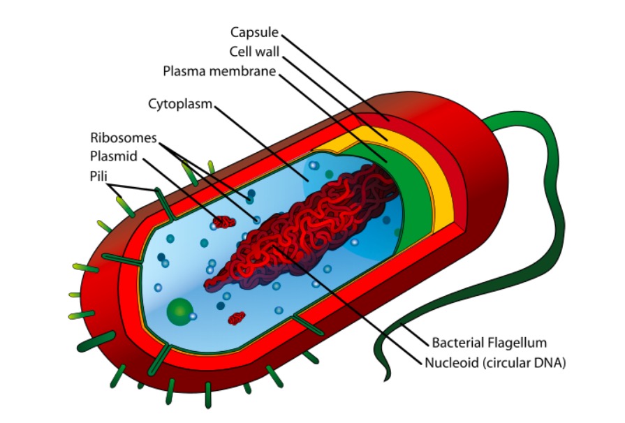
Estructura de una célula procariota

Imagen: “Average prokaryote cell” por Mariana Ruiz Villarreal. Licencia: Dominio Público
Eucariotas vs procariotas

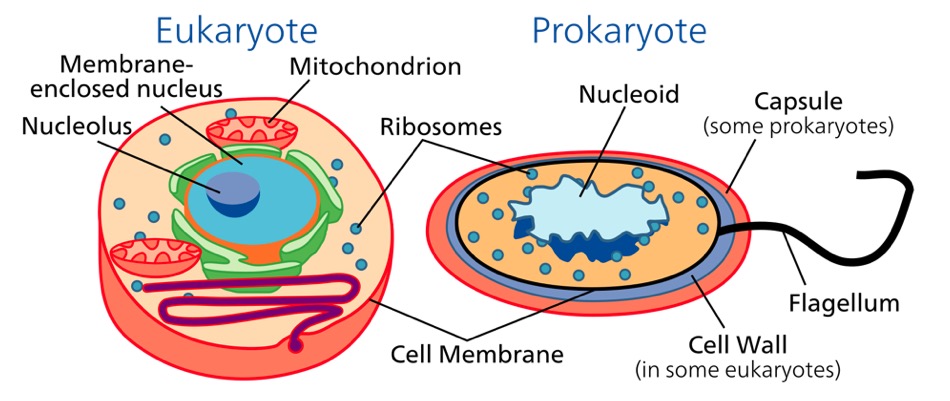
Comparación de células eucariotas y procariotas

Imagen: “The cells of eukaryotes (left) and prokaryotes (right)” por Science Primer. Licencia: Dominio Público

Relevancia Clínica

  • Bacterias: microorganismos unicelulares procariotas metabólicamente activos que se dividen por fisión binaria. Algunos de estos organismos desempeñan un papel importante en EN Erythema nodosum is an immune-mediated panniculitis (inflammation of the subcutaneous fat) caused by a type IV (delayed-type) hypersensitivity reaction. It commonly manifests in young women as tender, erythematous nodules on the shins. Erythema Nodosum la patogénesis de las enfermedades. El tratamiento de la enfermedad bacteriana suele incluir antibióticos; sin embargo, la elección del antibiótico puede variar en EN Erythema nodosum is an immune-mediated panniculitis (inflammation of the subcutaneous fat) caused by a type IV (delayed-type) hypersensitivity reaction. It commonly manifests in young women as tender, erythematous nodules on the shins. Erythema Nodosum función de la estructura y el metabolismo de la bacteria Bacteria Bacteria are prokaryotic single-celled microorganisms that are metabolically active and divide by binary fission. Some of these organisms play a significant role in the pathogenesis of diseases. Bacteriology.
  • Hongos: estos organismos pertenecen al AL Amyloidosis dominio de los LOS Neisseria eucariotas y, al AL Amyloidosis igual que las plantas, tienen paredes celulares y vacuolas, presentan flujo citoplasmático y son inmóviles. Sin embargo, casi todos los LOS Neisseria hongos tienen paredes celulares compuestas de quitina, no de celulosa. Los LOS Neisseria hongos no realizan la fotosíntesis, sino que obtienen sus sustratos para el metabolismo como saprófitos (i.e., obtienen su alimento de la materia muerta). Una infección causada por un hongo se denomina micosis.

Referencias

  1. Alberts, B., Johnson, A., Lewis, J., Morgan, D., Raff, M., Roberts, K., & Walter, P. (2024). Molecular Biology of the Cell (7th ed.). W. W. Norton & Company.
  2. Lodish, H., Berk, A., Zipursky, S. L., Matsudaira, P., Baltimore, D., & Darnell, J. (2000). Molecular Cell Biology (4th ed.). W. H. Freeman. Section 1.3, The Architecture of Cells. https://www.ncbi.nlm.nih.gov/books/NBK21665/
  3. Sato, M., Kakisaka, K., & Suzuki, T. (2025). Comparative studies on eukaryotic and prokaryotic cell organelles: Recent advances and clinical implications. Journal of Cellular Architecture, 45(1), 17-32. https://doi.org/10.1007/s10123-024-0089-2
  4. Tocheva, E. I., Ortega, D. R., & Jensen, G. J. (2023). Electron cryotomography of bacterial cell structure and function. Annual Review of Microbiology, 77, 343-364. https://doi.org/10.1146/annurev-micro-032723-041300
  5. Vellai, T., & Vida, G. (1999). The origin of eukaryotes: the difference between prokaryotic and eukaryotic cells. Proceedings of the Royal Society B: Biological Sciences, 266(1428), 1571-1577. https://doi.org/10.1098/rspb.1999.0817
  6. Yang, L., & Zhang, R. (2024). Structural and functional comparison of prokaryotic and eukaryotic cellular components: Implications for synthetic biology. Nature Reviews Microbiology, 22(3), 178-195. https://doi.org/10.1038/s41579-023-0912-6

¡Crea tu cuenta gratis o inicia una sesión para seguir leyendo!

Regístrate ahora y obtén acceso gratuito a Lecturio con páginas de concepto, videos médicos y cuestionarios para tu educación médica.

User Reviews

Que tengas una sesión de estudio alegre y navideña 🎁 Ahorra 50% en todos los planes >>

Details