Domina Conceptos Médicos

Estudia para la escuela de medicina y tus examenes con Lecturio.

Reflujo Vesicoureteral

El reflujo vesicoureteral (RVU) es el flujo retrógrado de orina desde la vejiga hacia el tracto urinario superior. El RVU primario suele ser consecuencia del cierre incompleto de la unión ureterovesical, mientras que el RVU secundario se debe a una obstrucción anatómica o fisiológica. El reflujo vesicoureteral no provoca síntomas específicos, pero se sospecha tras detectar una hidronefrosis en EN Erythema nodosum is an immune-mediated panniculitis (inflammation of the subcutaneous fat) caused by a type IV (delayed-type) hypersensitivity reaction. It commonly manifests in young women as tender, erythematous nodules on the shins. Erythema Nodosum el ultrasonido prenatal o en EN Erythema nodosum is an immune-mediated panniculitis (inflammation of the subcutaneous fat) caused by a type IV (delayed-type) hypersensitivity reaction. It commonly manifests in young women as tender, erythematous nodules on the shins. Erythema Nodosum un niño pequeño que presenta una infección urinaria. Debe realizarse una cistouretrografía miccional para diagnosticar la condición y evaluar su gravedad. La mayoría de los LOS Neisseria pacientes tendrán una resolución espontánea del RVU. Algunos pacientes pueden requerir tratamiento quirúrgico, especialmente los LOS Neisseria individuos con reflujo de alto grado.

Last updated: Dec 15, 2025

Editorial responsibility: Stanley Oiseth, Lindsay Jones, Evelin Maza

Descripción General

Definición

El reflujo vesicoureteral (RVU) es el flujo retrógrado de orina desde la vejiga hacia el tracto urinario superior.

Epidemiología

  • Es el hallazgo urinario más común en EN Erythema nodosum is an immune-mediated panniculitis (inflammation of the subcutaneous fat) caused by a type IV (delayed-type) hypersensitivity reaction. It commonly manifests in young women as tender, erythematous nodules on the shins. Erythema Nodosum los LOS Neisseria niños:
    • Ocurre en EN Erythema nodosum is an immune-mediated panniculitis (inflammation of the subcutaneous fat) caused by a type IV (delayed-type) hypersensitivity reaction. It commonly manifests in young women as tender, erythematous nodules on the shins. Erythema Nodosum 1%–2% de los LOS Neisseria niños < 2 años de edad
    • La prevalencia es del 15% en EN Erythema nodosum is an immune-mediated panniculitis (inflammation of the subcutaneous fat) caused by a type IV (delayed-type) hypersensitivity reaction. It commonly manifests in young women as tender, erythematous nodules on the shins. Erythema Nodosum niños con hidronefrosis prenatal
  • Es 3 veces más frecuente en EN Erythema nodosum is an immune-mediated panniculitis (inflammation of the subcutaneous fat) caused by a type IV (delayed-type) hypersensitivity reaction. It commonly manifests in young women as tender, erythematous nodules on the shins. Erythema Nodosum niños blancos que en EN Erythema nodosum is an immune-mediated panniculitis (inflammation of the subcutaneous fat) caused by a type IV (delayed-type) hypersensitivity reaction. It commonly manifests in young women as tender, erythematous nodules on the shins. Erythema Nodosum niños negros
  • Es 2 veces más frecuente en EN Erythema nodosum is an immune-mediated panniculitis (inflammation of the subcutaneous fat) caused by a type IV (delayed-type) hypersensitivity reaction. It commonly manifests in young women as tender, erythematous nodules on the shins. Erythema Nodosum las niñas que en EN Erythema nodosum is an immune-mediated panniculitis (inflammation of the subcutaneous fat) caused by a type IV (delayed-type) hypersensitivity reaction. It commonly manifests in young women as tender, erythematous nodules on the shins. Erythema Nodosum los LOS Neisseria niños

Etiología

RVU primario:

  • Predisposición genética
  • Defecto congénito de la porción terminal del uréter (e.g., uréter intravesical corto)

RVU secundario:

  • Causas anatómicas:
    • Válvulas uretrales posteriores
    • Duplicación ureteral congénita
    • Obstrucción de la salida de la vejiga
  • Obstrucción funcional de la vejiga:
    • Vejiga neurógena (e.g., espina bífida)
    • Inestabilidad del detrusor

Fisiopatología

Fisiología normal

  • Los LOS Neisseria uréteres distales atraviesan la pared de la vejiga en EN Erythema nodosum is an immune-mediated panniculitis (inflammation of the subcutaneous fat) caused by a type IV (delayed-type) hypersensitivity reaction. It commonly manifests in young women as tender, erythematous nodules on the shins. Erythema Nodosum un ángulo oblicuo.
  • Durante la micción, la vejiga y los LOS Neisseria músculos circundantes se contraen y comprimen el uréter intravesical → prevención del reflujo de orina hacia los LOS Neisseria uréteres
Funcionamiento normal de la vejiga y los uréteres

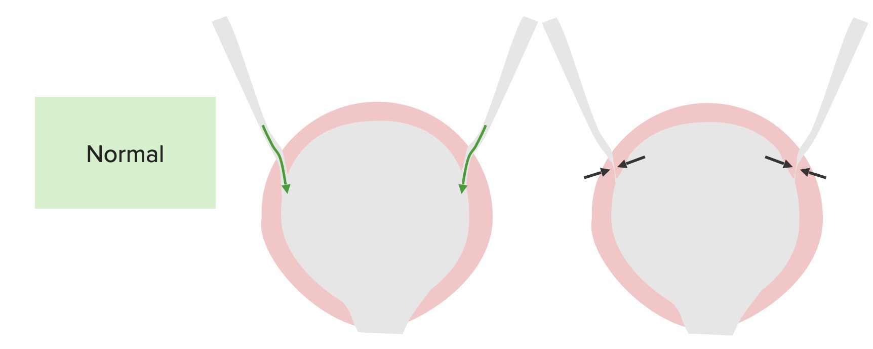
Función normal de la vejiga y los uréteres:
Los uréteres entran en un ángulo, lo cual permite que la vejiga comprima el orificio ureteral, que se cierra durante la micción para evitar el reflujo.

Imagen por Lecturio.

RVU primario

  • Un uréter intravesical corto → el uréter entra en EN Erythema nodosum is an immune-mediated panniculitis (inflammation of the subcutaneous fat) caused by a type IV (delayed-type) hypersensitivity reaction. It commonly manifests in young women as tender, erythematous nodules on the shins. Erythema Nodosum un ángulo diferente
  • Mal funcionamiento del mecanismo valvular durante la micción → reflujo de la orina hacia el uréter y el sistema colector
Fisiopatología del reflujo vesicoureteral primario

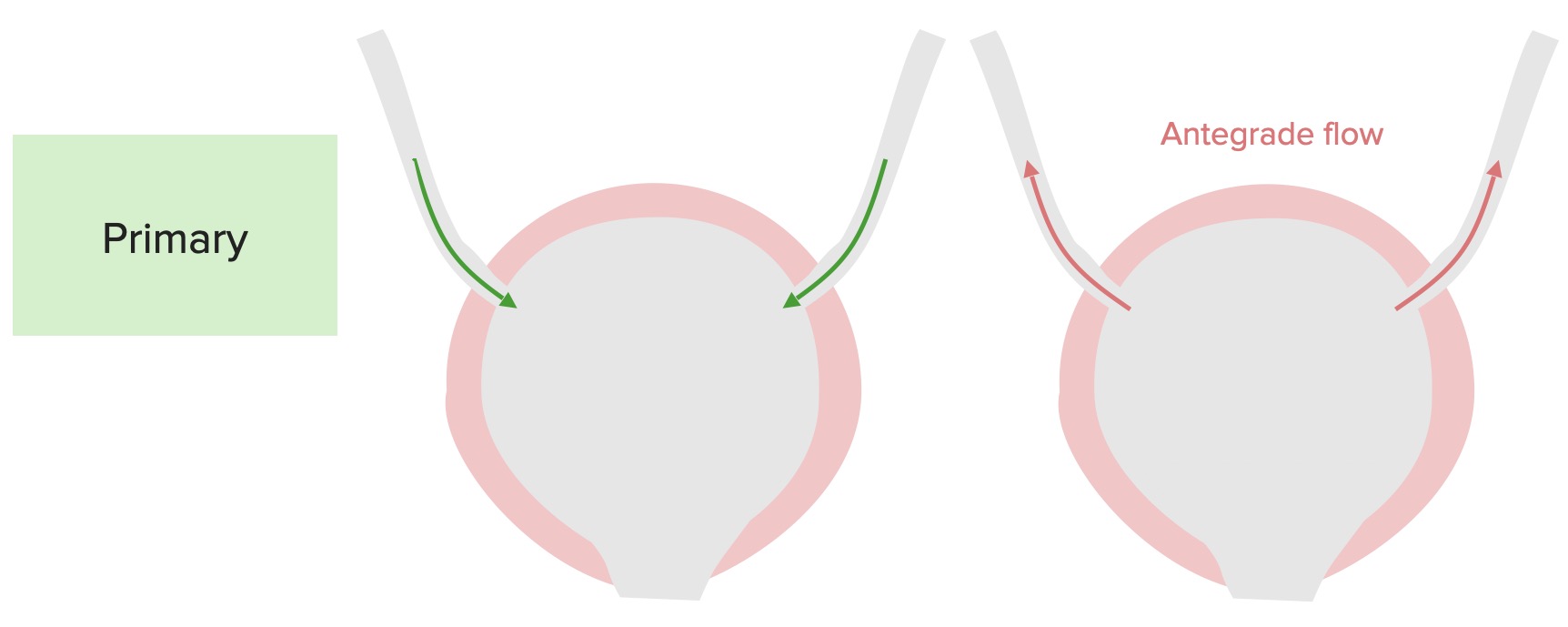
Fisiopatología del reflujo vesicoureteral primario:
Un defecto en el uréter terminal y en la capacidad de cerrar la unión ureterovesical da lugar al flujo anterógrado hacia arriba de los uréteres durante la micción.

Imagen por Lecturio.

Consecuencias potenciales

  • Reflujo intrarrenal persistente → cicatrización renal → nefropatía por reflujo
  • RVU → ↑ riesgo de infecciones del tracto urinario (ITU), incluida la pielonefritis.

Presentación Clínica y Diagnóstico

Presentación clínica

No existen signos o síntomas específicos para el RVU, y se puede sospechar de esta enfermedad en EN Erythema nodosum is an immune-mediated panniculitis (inflammation of the subcutaneous fat) caused by a type IV (delayed-type) hypersensitivity reaction. It commonly manifests in young women as tender, erythematous nodules on the shins. Erythema Nodosum las siguientes circunstancias:

  • Período prenatal: hidronefrosis observada en EN Erythema nodosum is an immune-mediated panniculitis (inflammation of the subcutaneous fat) caused by a type IV (delayed-type) hypersensitivity reaction. It commonly manifests in young women as tender, erythematous nodules on the shins. Erythema Nodosum el ultrasonido prenatal
  • Período postnatal:
    • ITU (a menudo recurrente)
    • Disfunción intestinal o vesical

Evaluación de exámenes de laboratorio

No hay pruebas de laboratorio que puedan diagnosticar el RVU. Se pueden realizar las siguientes evaluaciones para valorar las complicaciones:

  • BUN y creatinina → disfunción renal
  • Análisis de orina con cultivo → ITU

Imagenología

Ultrasonido renal y vesical:

  • Indicaciones:
    • Después de la 1era infección urinaria febril en EN Erythema nodosum is an immune-mediated panniculitis (inflammation of the subcutaneous fat) caused by a type IV (delayed-type) hypersensitivity reaction. It commonly manifests in young women as tender, erythematous nodules on the shins. Erythema Nodosum todos los LOS Neisseria niños de 2–24 meses
    • En EN Erythema nodosum is an immune-mediated panniculitis (inflammation of the subcutaneous fat) caused by a type IV (delayed-type) hypersensitivity reaction. It commonly manifests in young women as tender, erythematous nodules on the shins. Erythema Nodosum todos los LOS Neisseria niños con casos recurrentes de ITU
  • Posibles hallazgos:
    • Uréteres dilatados
    • Hidronefrosis

Cistouretrograma miccional:

  • Prueba de elección para el diagnóstico y el grado de RVU
  • Indicaciones:
    • Hidronefrosis bilateral o cicatrización renal encontrada en EN Erythema nodosum is an immune-mediated panniculitis (inflammation of the subcutaneous fat) caused by a type IV (delayed-type) hypersensitivity reaction. It commonly manifests in young women as tender, erythematous nodules on the shins. Erythema Nodosum el ultrasonido
    • 1era ITU con ultrasonido anormal
    • ITU recurrentes
  • Técnica:
    • El contraste se coloca en EN Erythema nodosum is an immune-mediated panniculitis (inflammation of the subcutaneous fat) caused by a type IV (delayed-type) hypersensitivity reaction. It commonly manifests in young women as tender, erythematous nodules on the shins. Erythema Nodosum la vejiga a través de una sonda.
    • Se toman imágenes fluoroscópicas durante la micción.
    • Reflujo de contraste hacia el uréter durante la micción → RVU
Hallazgos de imagen en el reflujo vesicoureteral

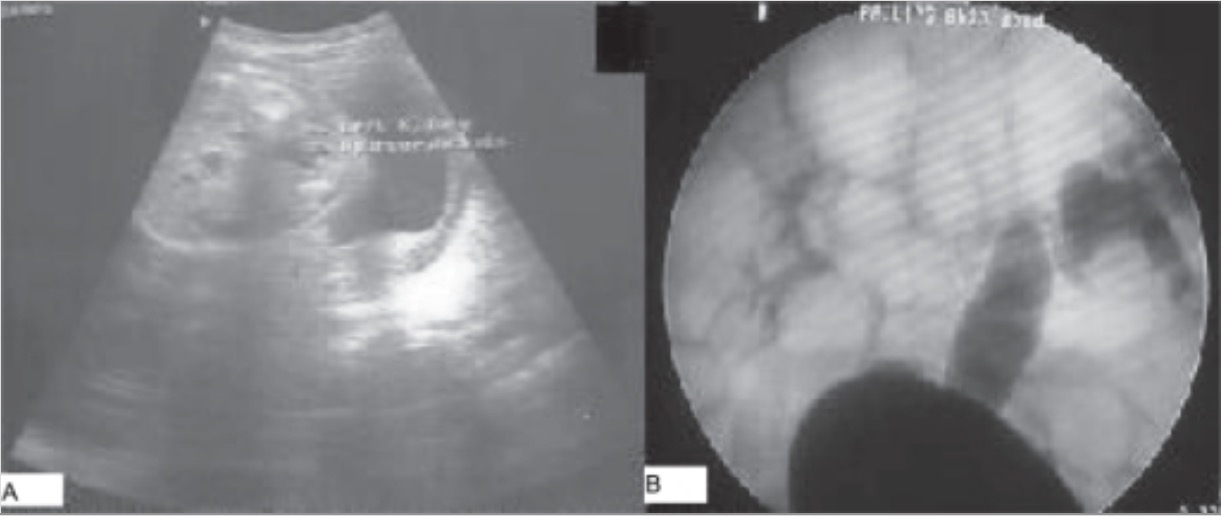
Hallazgos imagenológicos en el reflujo vesicoureteral:
A: Ultrasonido postnatal que muestra una hidronefrosis unilateral
B: Cistouretrograma miccional que muestra reflujo de contraste en los uréteres y sistemas colectores derecho e izquierdo (derecho: grado III, izquierdo: grado V)

Imagen: “Imaging findings in vesicoureteral reflux” por Department of Urology, Kles Kidney Foundation, Nehru Nagar, Belgaum, India. Licencia: CC BY 2.0

Clasificación de la gravedad

La clasificación de la gravedad ayuda a cuantificar el grado del RVU basándose en EN Erythema nodosum is an immune-mediated panniculitis (inflammation of the subcutaneous fat) caused by a type IV (delayed-type) hypersensitivity reaction. It commonly manifests in young women as tender, erythematous nodules on the shins. Erythema Nodosum los LOS Neisseria hallazgos imagenológicos.

  • Grado I: reflujo hacia el uréter
  • Grado II: reflujo en EN Erythema nodosum is an immune-mediated panniculitis (inflammation of the subcutaneous fat) caused by a type IV (delayed-type) hypersensitivity reaction. It commonly manifests in young women as tender, erythematous nodules on the shins. Erythema Nodosum el uréter, la pelvis Pelvis The pelvis consists of the bony pelvic girdle, the muscular and ligamentous pelvic floor, and the pelvic cavity, which contains viscera, vessels, and multiple nerves and muscles. The pelvic girdle, composed of 2 “hip” bones and the sacrum, is a ring-like bony structure of the axial skeleton that links the vertebral column with the lower extremities. Pelvis: Anatomy renal y los LOS Neisseria cálices sin dilatación
  • Grado III: el reflujo llena y dilata ligeramente el uréter con un ligero embotamiento de los LOS Neisseria cálices.
  • Grado IV: el reflujo resulta en EN Erythema nodosum is an immune-mediated panniculitis (inflammation of the subcutaneous fat) caused by a type IV (delayed-type) hypersensitivity reaction. It commonly manifests in young women as tender, erythematous nodules on the shins. Erythema Nodosum uréteres y cálices moderadamente dilatados. También se observa cierta tortuosidad del uréter.
  • Grado V: el reflujo masivo que provoca una dilatación importante del sistema colector urinario y un reflujo intrarrenal (“megauréter”)
Clasificación del reflujo vesicoureteral

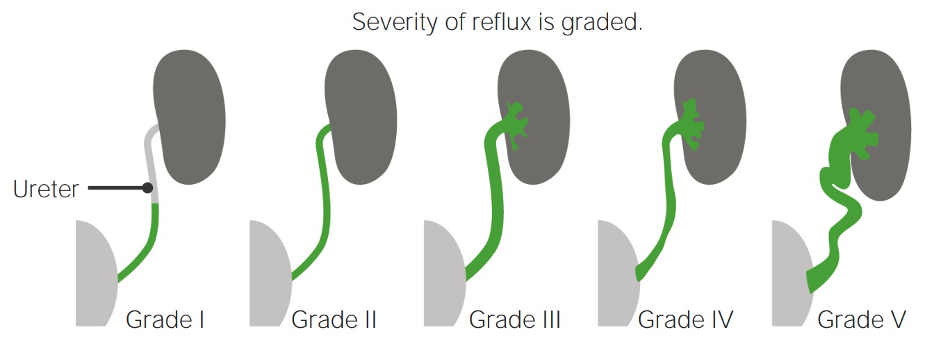
Clasificación de la gravedad del reflujo vesicoureteral

Imagen por Lecturio.

Tratamiento y Pronóstico

Tratamiento

Tratamiento conservador:

  • Vigilancia:
  • Tratamiento rápido de las ITU
  • Profilaxis antibiótica:
    • Controversial
    • El objetivo es prevenir las ITU recurrentes.
    • Opciones:
      • Trimetoprim-sulfametoxazol
      • Nitrofurantoína

Tratamiento invasivo

  • El objetivo es mejorar el ángulo del uréter → ↓ reflujo
  • Indicaciones:
    • Grados IV o V
    • ITU recurrentes a pesar de la profilaxis antibiótica
    • Disminución de la función renal
  • Procedimientos:
    • Inyección transuretral subureteral
    • Reimplantación ureteral quirúrgica abierta

Pronóstico

  • La mayoría de los LOS Neisseria pacientes muestran una resolución espontánea del RVU.
  • Factores que aumentan la probabilidad de resolución:
    • Menor gravedad del reflujo
    • Edad de diagnóstico < 2 años
    • Enfermedad unilateral

Diagnóstico Diferencial

  • Obstrucción de la unión ureteropélvica: la causa más común de hidronefrosis pediátrica. Una lesión anatómica o una alteración funcional provoca una obstrucción en EN Erythema nodosum is an immune-mediated panniculitis (inflammation of the subcutaneous fat) caused by a type IV (delayed-type) hypersensitivity reaction. It commonly manifests in young women as tender, erythematous nodules on the shins. Erythema Nodosum la entrada del uréter en EN Erythema nodosum is an immune-mediated panniculitis (inflammation of the subcutaneous fat) caused by a type IV (delayed-type) hypersensitivity reaction. It commonly manifests in young women as tender, erythematous nodules on the shins. Erythema Nodosum el riñón. La obstrucción de la unión ureteropélvica suele presentarse como una masa abdominal palpable, dolor Dolor Inflammation de costado, hematuria Hematuria Presence of blood in the urine. Renal Cell Carcinoma, ITU o retraso en EN Erythema nodosum is an immune-mediated panniculitis (inflammation of the subcutaneous fat) caused by a type IV (delayed-type) hypersensitivity reaction. It commonly manifests in young women as tender, erythematous nodules on the shins. Erythema Nodosum el crecimiento. El diagnóstico se realiza con imagenología. Debe realizarse una cistouretrografía miccional para descartar el RVU. El tratamiento puede incluir la vigilancia o la cirugía.
  • ITU: un amplio espectro de enfermedades, que van desde la cistitis simple autolimitada hasta la pielonefritis grave que puede provocar la sepsis Sepsis Systemic inflammatory response syndrome with a proven or suspected infectious etiology. When sepsis is associated with organ dysfunction distant from the site of infection, it is called severe sepsis. When sepsis is accompanied by hypotension despite adequate fluid infusion, it is called septic shock. Sepsis and Septic Shock y la muerte. Dependiendo de la localización de la infección, los LOS Neisseria pacientes pueden presentar disuria, urgencia urinaria, aumento de la frecuencia urinaria, dolor Dolor Inflammation suprapúbico y fiebre. El análisis de orina y el urocultivo, junto con la presentación clínica, ayudan a diagnosticar las ITU. Se debe solicitar imagenología para descartar el RVU en EN Erythema nodosum is an immune-mediated panniculitis (inflammation of the subcutaneous fat) caused by a type IV (delayed-type) hypersensitivity reaction. It commonly manifests in young women as tender, erythematous nodules on the shins. Erythema Nodosum los LOS Neisseria niños. Las opciones de tratamiento incluyen antibióticos orales o IV.
  • Válvulas uretrales posteriores: pliegues membranosos que obstruyen el lumen de la uretra posterior. Los LOS Neisseria bebés suelen presentar ITU, disminución del chorro urinario, incontinencia urinaria y RVU. El diagnóstico de las válvulas uretrales posteriores puede realizarse mediante un ultrasonido o una cistouretrografía miccional. El tratamiento es quirúrgico.
  • Megacaliosis congénita: un hallazgo incidental que resulta del desarrollo inadecuado de las pirámides medulares renales. El agrandamiento de los LOS Neisseria cálices puede simular una hidronefrosis. El aspecto radiográfico de los LOS Neisseria cálices junto con el calibre normal de la pelvis Pelvis The pelvis consists of the bony pelvic girdle, the muscular and ligamentous pelvic floor, and the pelvic cavity, which contains viscera, vessels, and multiple nerves and muscles. The pelvic girdle, composed of 2 “hip” bones and the sacrum, is a ring-like bony structure of the axial skeleton that links the vertebral column with the lower extremities. Pelvis: Anatomy renal y el uréter ayudan al AL Amyloidosis diagnóstico. No se requiere ningún tratamiento.

Referencias

  1. Baskin, L.S. (2025). Congenital ureteropelvic junction obstruction. UpToDate. Retrieved June 13, 2025, from https://www.uptodate.com/contents/congenital-ureteropelvic-junction-obstruction
  2. Holmes, N. (2025). Clinical presentation and diagnosis of posterior urethral valves. UpToDate. Retrieved June 13, 2025, from https://www.uptodate.com/contents/clinical-presentation-and-diagnosis-of-posterior-urethral-valves
  3. Mattoo, T.K., Greenfield, S.P. (2025). Clinical presentation, diagnosis, and course of primary vesicoureteral reflux. UpToDate. Retrieved June 13, 2025, from https://www.uptodate.com/contents/clinical-presentation-diagnosis-and-course-of-primary-vesicoureteral-reflux
  4. Mattoo, T.K., Greenfield, S.P. (2023). Management of vesicoureteral reflux. In Kim, M.S. (Ed.), UpToDate. Retrieved June 13, 2025, from https://www.uptodate.com/contents/management-of-vesicoureteral-reflux
  5. Van Why, S.K., Avner, E.D. (2016). Vesicoureteral Reflux. Nelson Textbook of Pediatrics, 2562–67.
  6. Estrada, Jr., C.R., Cendron, M. (2021). Vesicoureteral reflux. In Kim, E.D. (Ed.), Medscape. Retrieved June 13, 2025, from https://emedicine.medscape.com/article/439403-overview
  7. Banker, H., Aeddula, N.R. (2024). Vesicoureteral reflux. [online] StatPearls. Retrieved June 13, 2025, from https://www.ncbi.nlm.nih.gov/books/NBK563262/

¡Crea tu cuenta gratis o inicia una sesión para seguir leyendo!

Regístrate ahora y obtén acceso gratuito a Lecturio con páginas de concepto, videos médicos y cuestionarios para tu educación médica.

User Reviews

Que tengas una sesión de estudio alegre y navideña 🎁 Ahorra 50% en todos los planes >>

Details