El recto y el canal anal son las partes más terminales del tracto gastrointestinal inferior/intestino grueso que forman una unidad funcional y controlan la defecación. La incontinencia fecal puede ocurrir si se altera esta función. La continencia fecal se mantiene gracias a varias estructuras anatómicas importantes, incluidos los LOS Neisseria pliegues rectales, válvulas anales, músculo puborrectal en EN Erythema nodosum is an immune-mediated panniculitis (inflammation of the subcutaneous fat) caused by a type IV (delayed-type) hypersensitivity reaction. It commonly manifests in young women as tender, erythematous nodules on the shins. Erythema Nodosum forma de cabestrillo y los LOS Neisseria esfínteres anales internos y externos. Las ondas peristálticas dentro de la capa muscular rectal, la relajación involuntaria del esfínter anal interno (controlado por el sistema nervioso autónomo (SNA)) y la relajación voluntaria del esfínter anal externo (controlado por la corteza cerebral) son esenciales para que ocurra la defecación. El rico plexo de venas que rodean el canal anal puede convertirse en EN Erythema nodosum is an immune-mediated panniculitis (inflammation of the subcutaneous fat) caused by a type IV (delayed-type) hypersensitivity reaction. It commonly manifests in young women as tender, erythematous nodules on the shins. Erythema Nodosum hemorroides si se dilata.
Last updated: Dec 15, 2025
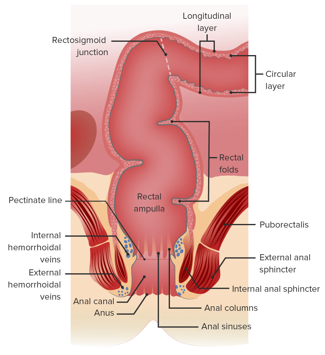
Anatomía macroscópica del recto y canal anal
Imagen por Lecturio.El recto es el órgano visceral más posterior de la cavidad pélvica.
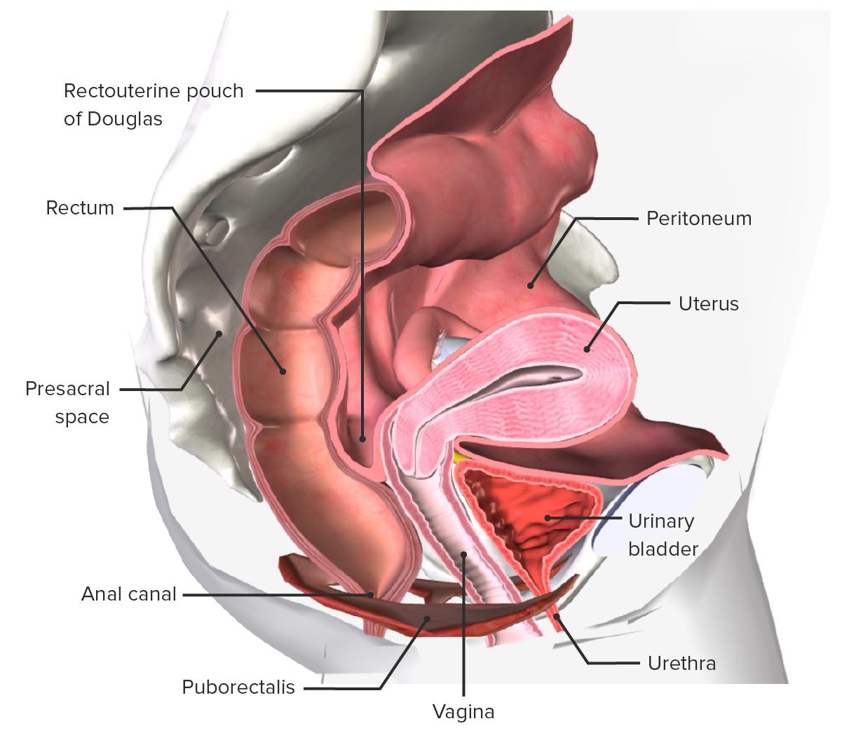
Anatomía de la pelvis interna femenina
Imagen por BioDigital, editado por Lecturio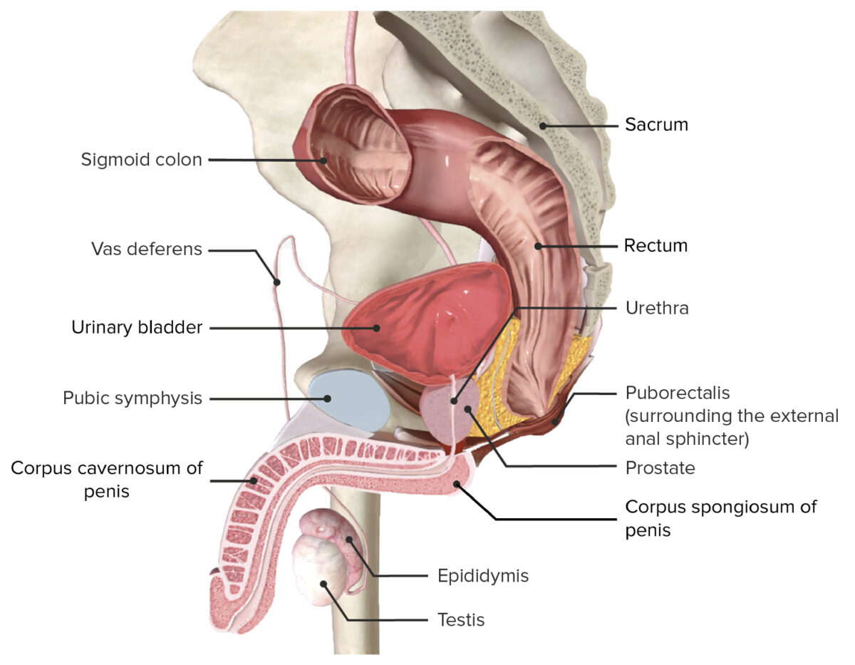
Anatomía de la pelvis interna masculina
Imagen por BioDigital, editado por LecturioDe manera similar a otros segmentos del tracto gastrointestinal, las capas de la pared anorrectal (desde la luz interna hacia afuera) son mucosa → submucosa → capa muscular → serosa. No hay vellosidades o pliegues circulares en EN Erythema nodosum is an immune-mediated panniculitis (inflammation of the subcutaneous fat) caused by a type IV (delayed-type) hypersensitivity reaction. It commonly manifests in young women as tender, erythematous nodules on the shins. Erythema Nodosum la pared anorrectal como los LOS Neisseria del intestino delgado.
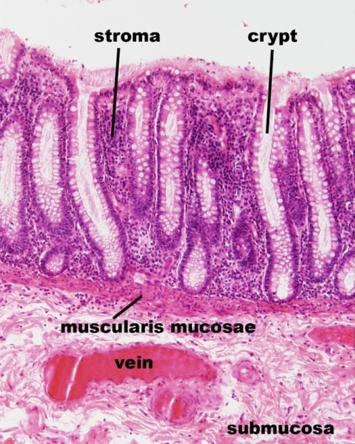
Imagen histológica de la mucosa rectal (corte longitudinal):
Las criptas rectales con epitelio cilíndrico simple son visibles y el estroma (lámina propia) se ve envolviendo las criptas.
Formada por 2 capas de músculo liso:
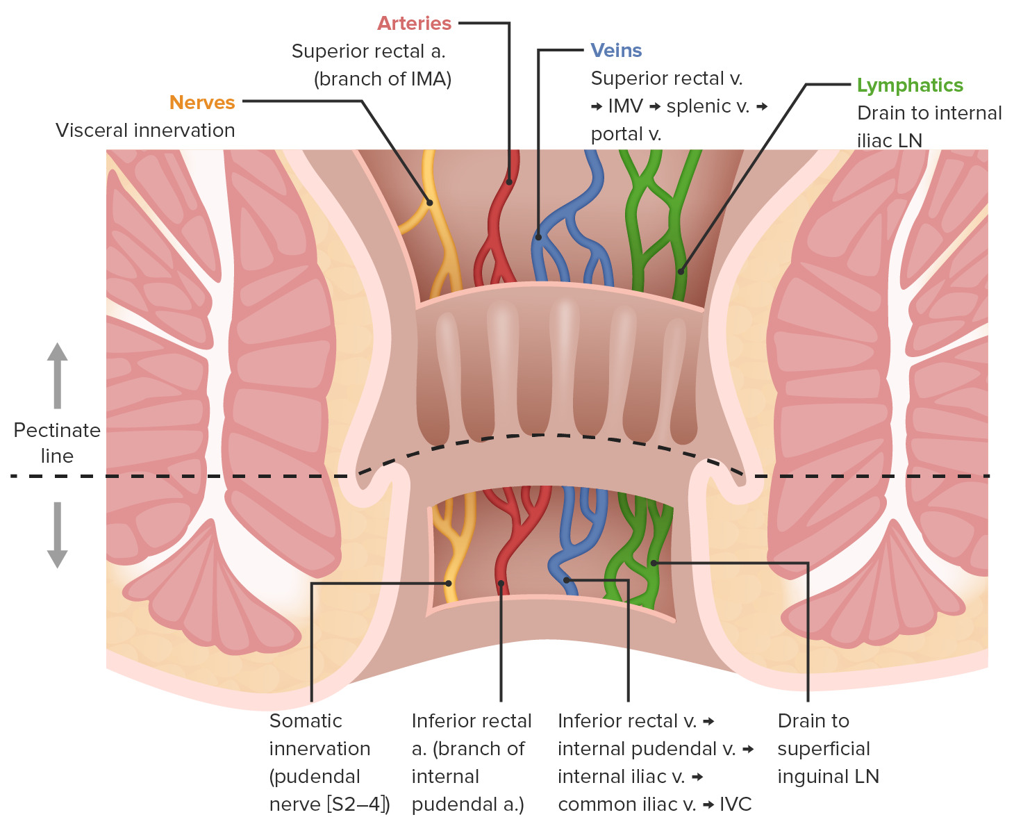
Diferencias en la irrigación e inervación anal por encima y por debajo de la línea pectínea:
IMA: arteria mesentérica inferior
IMV: vena mesentérica inferior
LN: ganglio linfático
IVC: vena cava inferior
Las funciones principales del recto y el canal anal implican la defecación controlada.
El recto contiene receptores de estiramiento que estimulan el reflejo de defecación cuando el recto comienza a llenarse de heces.
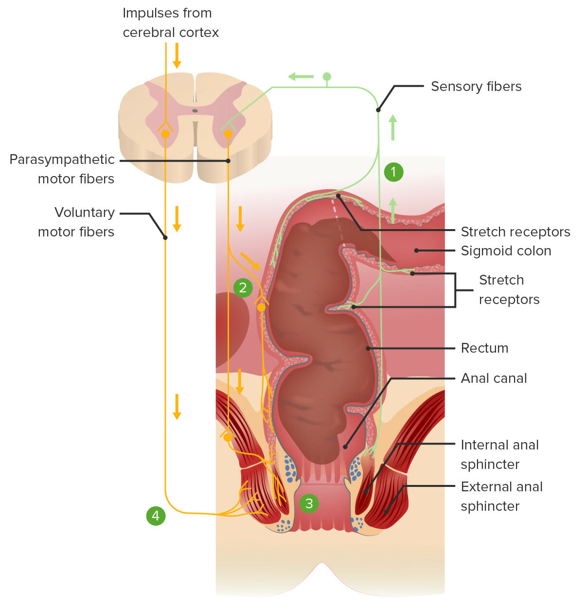
Reflejo de defecación:
1. Las heces estiran el recto y estimulan los receptores de estiramiento, transmitiendo la señal a la médula espinal.
2. Un reflejo espinal envía señales motoras parasimpáticas al plexo nervioso mientérico, lo que da como resultado la contracción de los músculos lisos dentro del recto, empujando las heces hacia abajo.
3. El mismo reflejo espinal también envía señales motoras parasimpáticas para relajar el esfínter anal interno.
4. Los impulsos voluntarios del cerebro evitan la defecación al mantener contraído el esfínter anal externo. La defecación ocurrirá si las señales voluntarias permiten que el esfínter anal externo se relaje.