El peritoneo es una membrana serosa que recubre la cavidad abdominopélvica. Este revestimiento está formado por tejido conectivo y se origina en EN Erythema nodosum is an immune-mediated panniculitis (inflammation of the subcutaneous fat) caused by a type IV (delayed-type) hypersensitivity reaction. It commonly manifests in young women as tender, erythematous nodules on the shins. Erythema Nodosum el mesodermo. Esta membrana recubre tanto las paredes abdominales (como peritoneo parietal Parietal One of a pair of irregularly shaped quadrilateral bones situated between the frontal bone and occipital bone, which together form the sides of the cranium. Skull: Anatomy) como todos los LOS Neisseria órganos viscerales (como peritoneo visceral). El peritoneo sostiene y suspende los LOS Neisseria órganos dentro de la cavidad abdominal y proporciona un importante conducto para la irrigación e inervación de estos órganos. Existen varios pliegues peritoneales, conocidos como mesenterios, epiplones y ligamentos. El epiplón mayor y menor dividen la cavidad peritoneal en EN Erythema nodosum is an immune-mediated panniculitis (inflammation of the subcutaneous fat) caused by a type IV (delayed-type) hypersensitivity reaction. It commonly manifests in young women as tender, erythematous nodules on the shins. Erythema Nodosum sacos mayores y menores, que son espacios anatómicos importantes dentro de la cavidad. Los LOS Neisseria órganos situados detrás del peritoneo parietal Parietal One of a pair of irregularly shaped quadrilateral bones situated between the frontal bone and occipital bone, which together form the sides of the cranium. Skull: Anatomy posterior se conocen como retroperitoneales, mientras que los LOS Neisseria órganos que sobresalen en EN Erythema nodosum is an immune-mediated panniculitis (inflammation of the subcutaneous fat) caused by a type IV (delayed-type) hypersensitivity reaction. It commonly manifests in young women as tender, erythematous nodules on the shins. Erythema Nodosum la cavidad y están totalmente cubiertos por el peritoneo visceral se conocen como intraperitoneales.
Last updated: Dec 15, 2025
El peritoneo es una membrana serosa que recubre la cavidad abdominopélvica y cubre sus órganos.
El peritoneo se desarrolla a partir del mesodermo del embrión trilaminar:
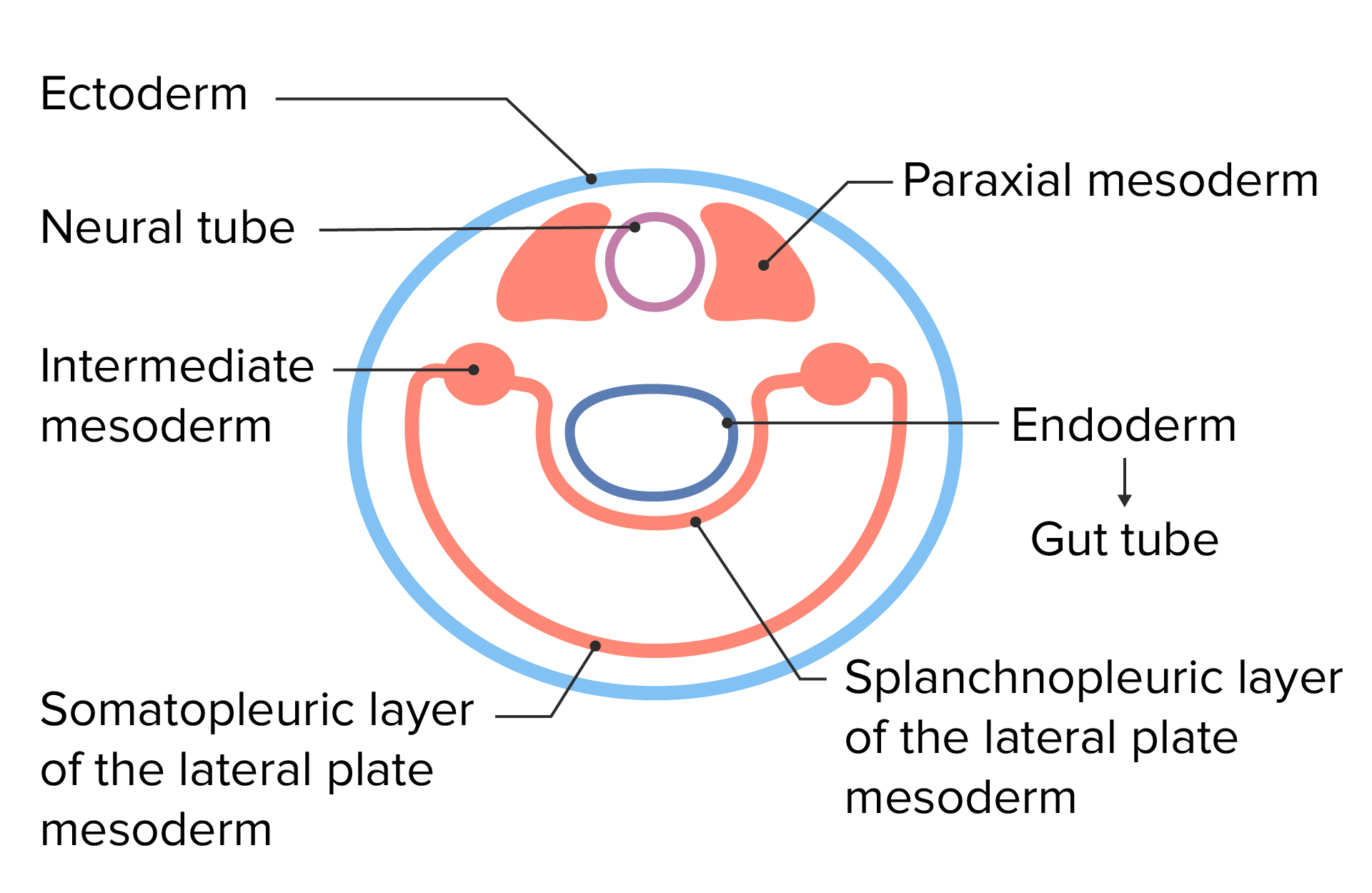
Capas celulares embrionarias tras el plegado lateral del disco trilaminar
Imagen por Lecturio.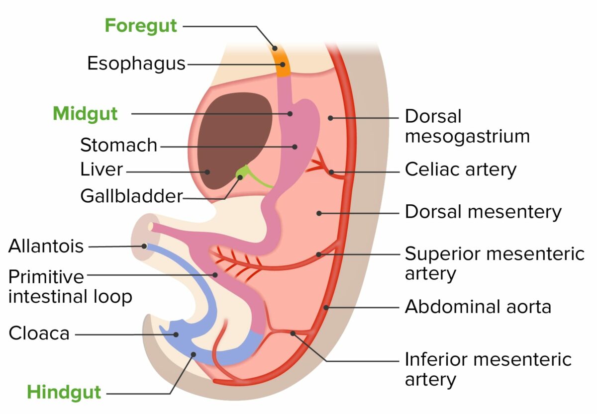
Desarrollo del mesenterio posterior con el tubo intestinal primitivo
Imagen por Lecturio.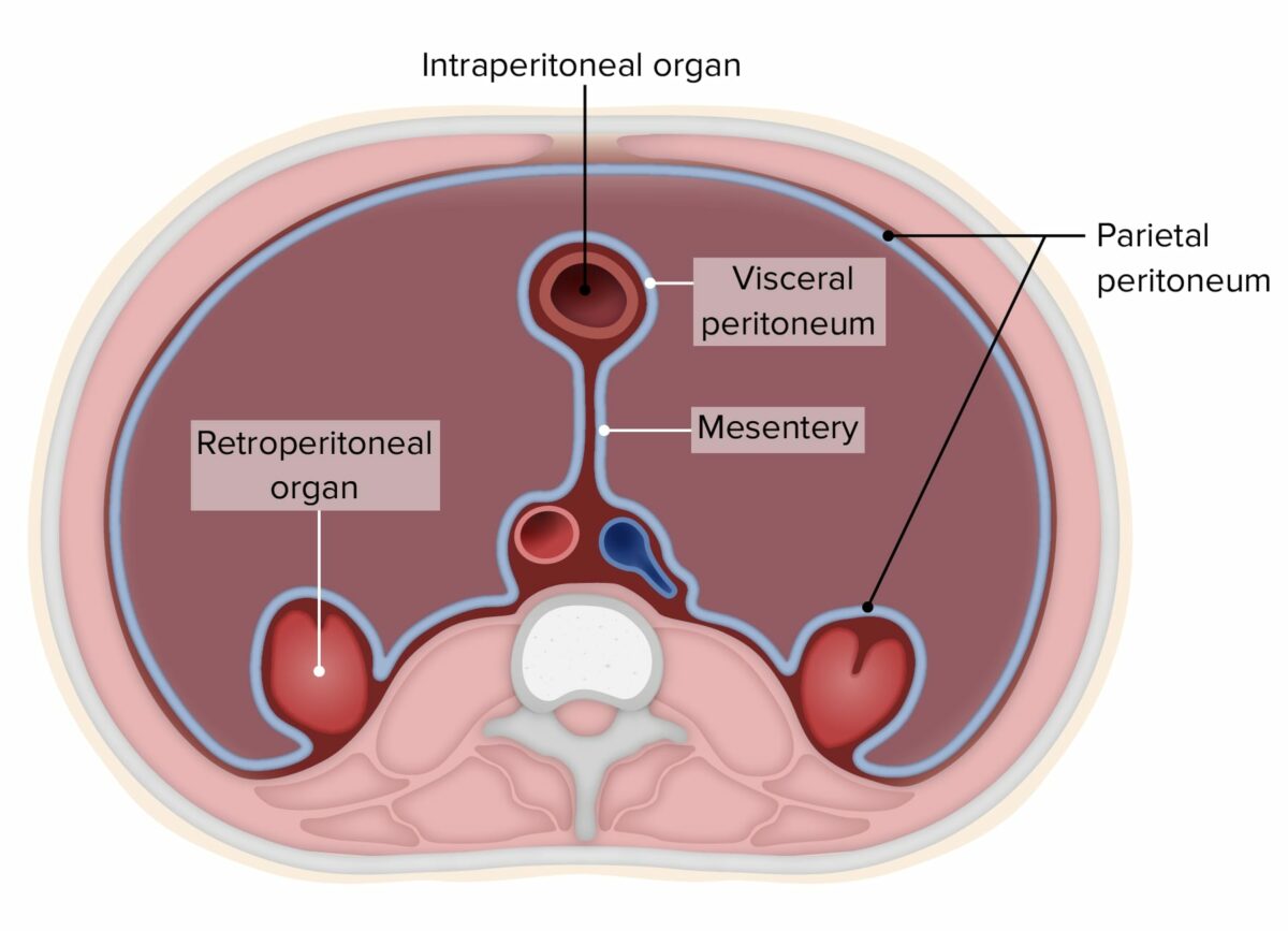
Diagrama del peritoneo (se han quitado los órganos abdominales para demostrar más claramente los conceptos)
Imagen por Lecturio.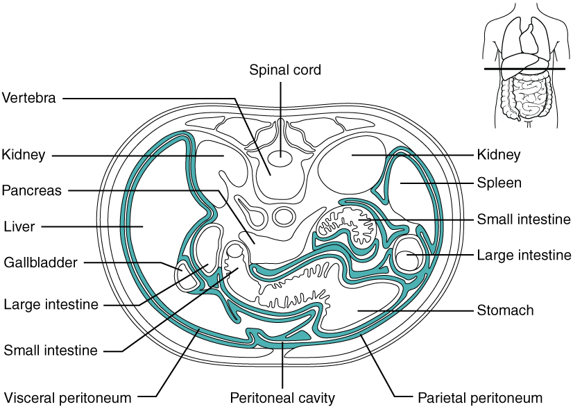
Diagrama de la cavidad peritoneal
Imagen: “Illustration from Anatomy & Physiology, Connexions Web site” por OpenStax College. Licencia: CC BY 3.0Los LOS Neisseria órganos pueden clasificarse según su cobertura peritoneal:
| Localización | Órganos |
|---|---|
| Intraperitoneal Intraperitoneal Peritoneum: Anatomy |
|
| Secundariamente retroperitoneales |
|
| Retroperitoneales primarios |
|
| Subperitoneales |
|
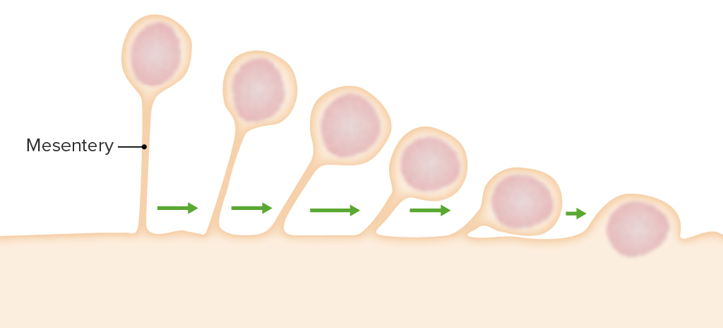
Desarrollo de órganos secundariamente retroperitoneales:
Estos órganos comienzan suspendidos por un mesenterio. Sin embargo, a medida que el órgano se desarrolla, el mesenterio retrocede hasta que el órgano queda a ras de la pared posterior y acaba siendo un órgano retroperitoneal.
Los LOS Neisseria epiplones son láminas de peritoneo en EN Erythema nodosum is an immune-mediated panniculitis (inflammation of the subcutaneous fat) caused by a type IV (delayed-type) hypersensitivity reaction. It commonly manifests in young women as tender, erythematous nodules on the shins. Erythema Nodosum capas. El epiplón mayor y el epiplón menor son los LOS Neisseria 2 epiplones.
Epiplón mayor:
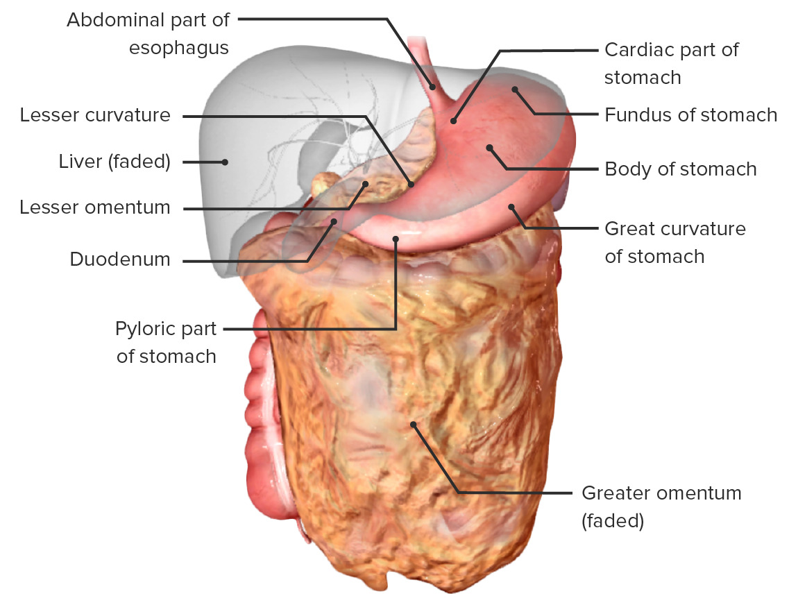
El estómago in situ y las relaciones con sus estructuras vecinas:
Observe el epiplón mayor y menor que continúa desde las curvaturas mayor y menor del estómago, respectivamente.
Epiplón menor:
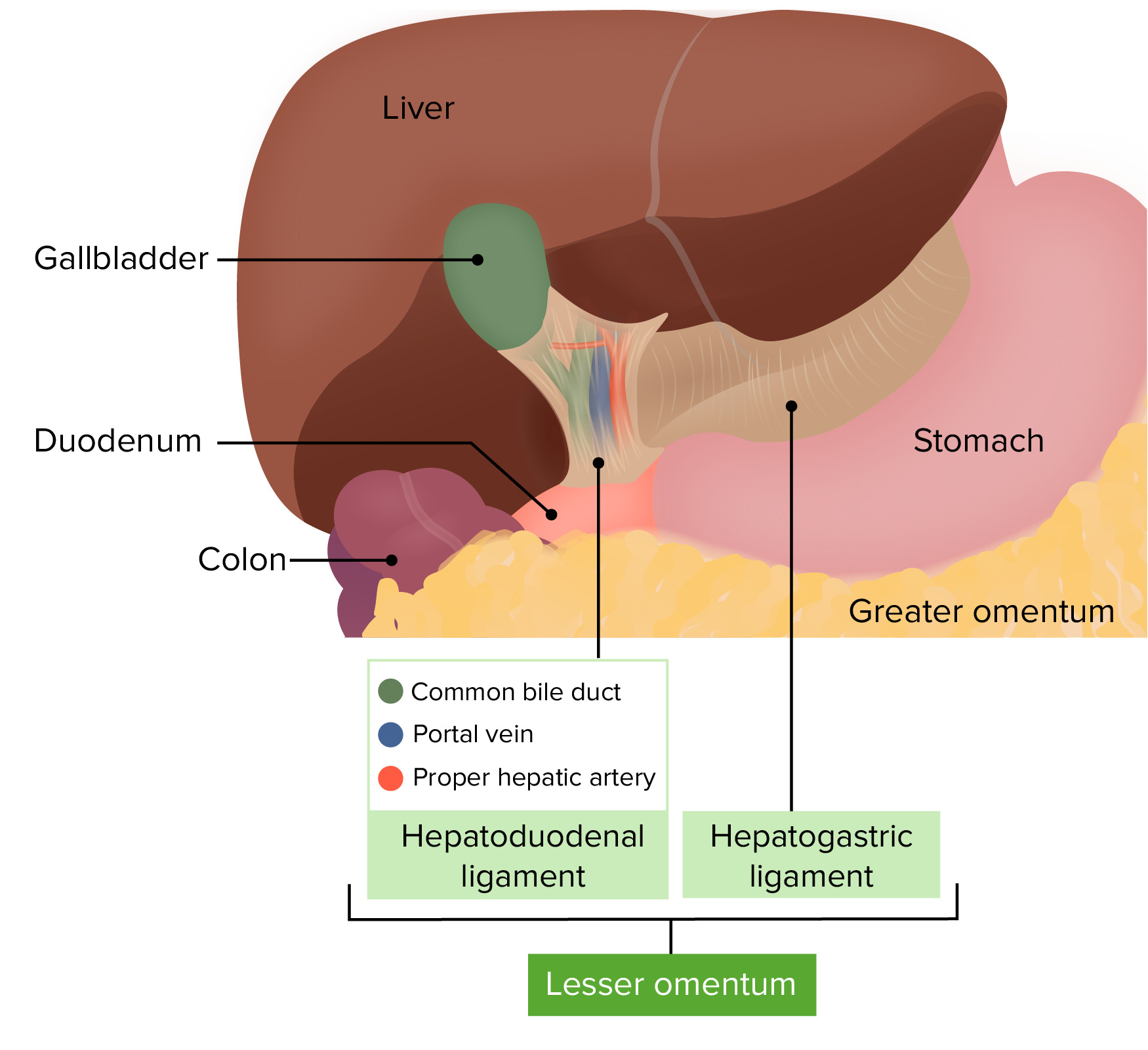
Epiplón menor y foramen epiploico in situ
Imagen por Lecturio.La cavidad peritoneal está dividida en EN Erythema nodosum is an immune-mediated panniculitis (inflammation of the subcutaneous fat) caused by a type IV (delayed-type) hypersensitivity reaction. It commonly manifests in young women as tender, erythematous nodules on the shins. Erythema Nodosum varios compartimentos diferentes por los LOS Neisseria epiplones y el mesocolon transversal:
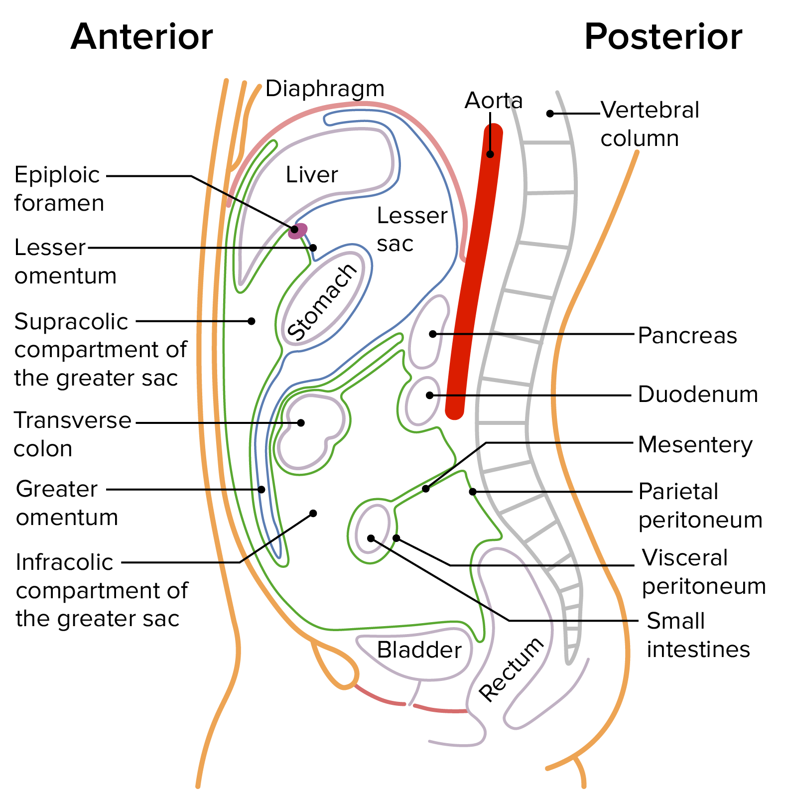
Una sección sagital a través del abdomen que muestra los sacos mayores y menores:
El saco mayor está delineado en verde, y el saco menor está delineado en azul.
El saco mayor está dividido en EN Erythema nodosum is an immune-mediated panniculitis (inflammation of the subcutaneous fat) caused by a type IV (delayed-type) hypersensitivity reaction. It commonly manifests in young women as tender, erythematous nodules on the shins. Erythema Nodosum 2 compartimentos por el mesocolon transversal.
El saco menor también se conoce como bursa epiploica.
| Peritoneo parietal Parietal One of a pair of irregularly shaped quadrilateral bones situated between the frontal bone and occipital bone, which together form the sides of the cranium. Skull: Anatomy | Peritoneo visceral | |
|---|---|---|
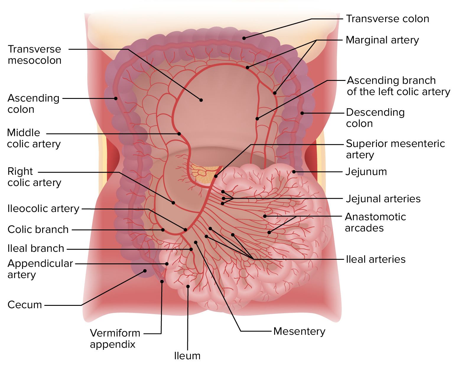
| Irrigación arterial | De la irrigación de la pared abdominal | Arterias mesentéricas superior e inferior |
| Drenaje venoso | Las venas drenan en EN Erythema nodosum is an immune-mediated panniculitis (inflammation of the subcutaneous fat) caused by a type IV (delayed-type) hypersensitivity reaction. It commonly manifests in young women as tender, erythematous nodules on the shins. Erythema Nodosum la vena cava inferior | Las venas drenan en EN Erythema nodosum is an immune-mediated panniculitis (inflammation of the subcutaneous fat) caused by a type IV (delayed-type) hypersensitivity reaction. It commonly manifests in young women as tender, erythematous nodules on the shins. Erythema Nodosum la vena porta |
| Inervación | Inervación somática de los LOS Neisseria nervios espinales T10-L1 (el dolor Dolor Inflammation puede ser localizado) | Inervación autonómica (el dolor Dolor Inflammation es difícil de localizar) |