El bazo es el órgano linfoide más grande del cuerpo, localizado en EN Erythema nodosum is an immune-mediated panniculitis (inflammation of the subcutaneous fat) caused by a type IV (delayed-type) hypersensitivity reaction. It commonly manifests in young women as tender, erythematous nodules on the shins. Erythema Nodosum el cuadrante superior izquierdo del abdomen, superior al AL Amyloidosis riñón izquierdo y posterior al AL Amyloidosis estómago, al AL Amyloidosis nivel de la 9na‒11va costilla, justo debajo del diafragma. El bazo está muy vascularizado y actúa como un importante filtro sanguíneo, limpiando la sangre de patógenos y eritrocitos dañados. El bazo también puede activar respuestas inmunológicas, producir anticuerpos y funcionar como reservorio para el almacenamiento de plaquetas. Hay 2 tipos principales de tejido esplénico: la pulpa roja, que contiene redes fibrovasculares densas para filtrar la sangre, y la pulpa blanca, que se compone principalmente de tejido linfoide que rodea a los LOS Neisseria vasos más grandes. El bazo tiene una cápsula relativamente débil; por lo tanto, puede romperse más fácilmente que otros órganos abdominales y provocar una hemorragia potencialmente mortal.
Last updated: Dec 15, 2025
El bazo es el órgano linfático más grande del cuerpo.
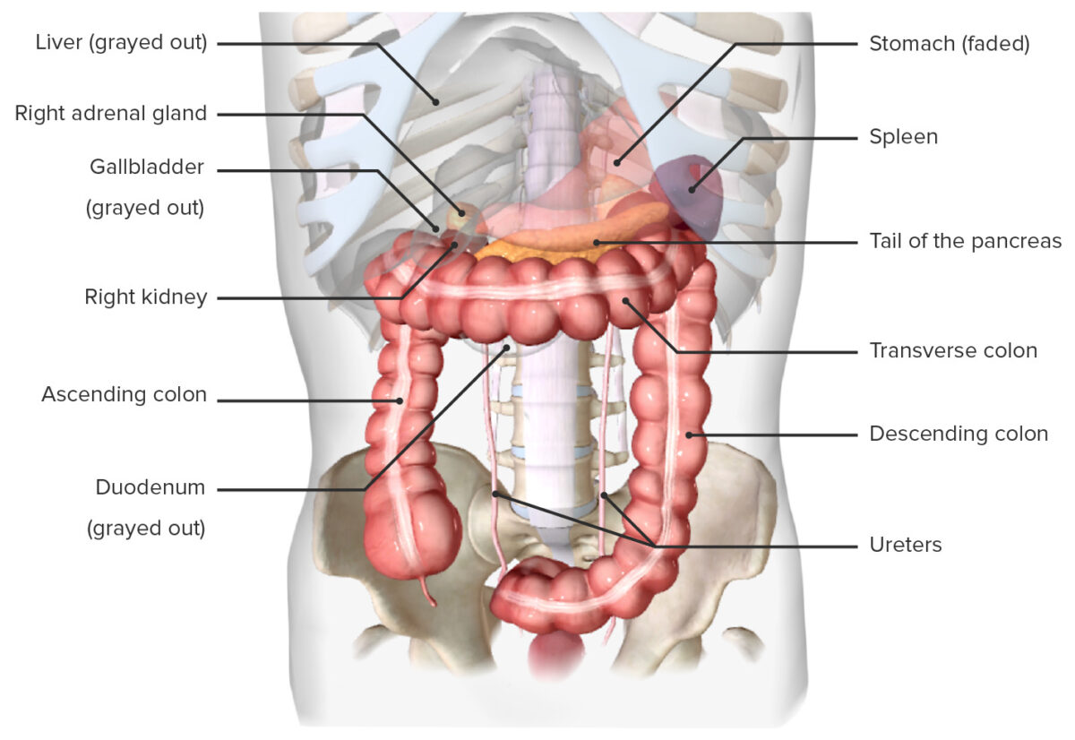
Localización del bazo, in situ (vista anterior)
Imagen por BioDigital, editado por Lecturio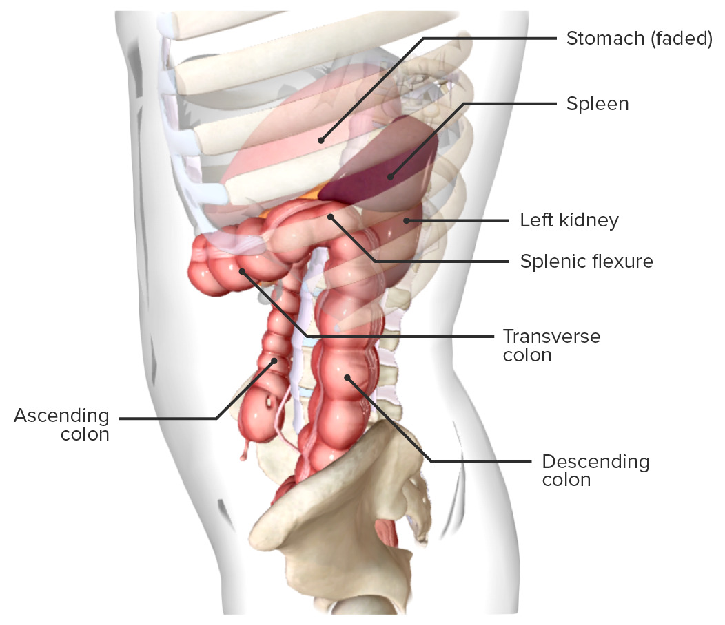
Localización del bazo, in situ (vista lateral)
Imagen por BioDigital, editado por Lecturio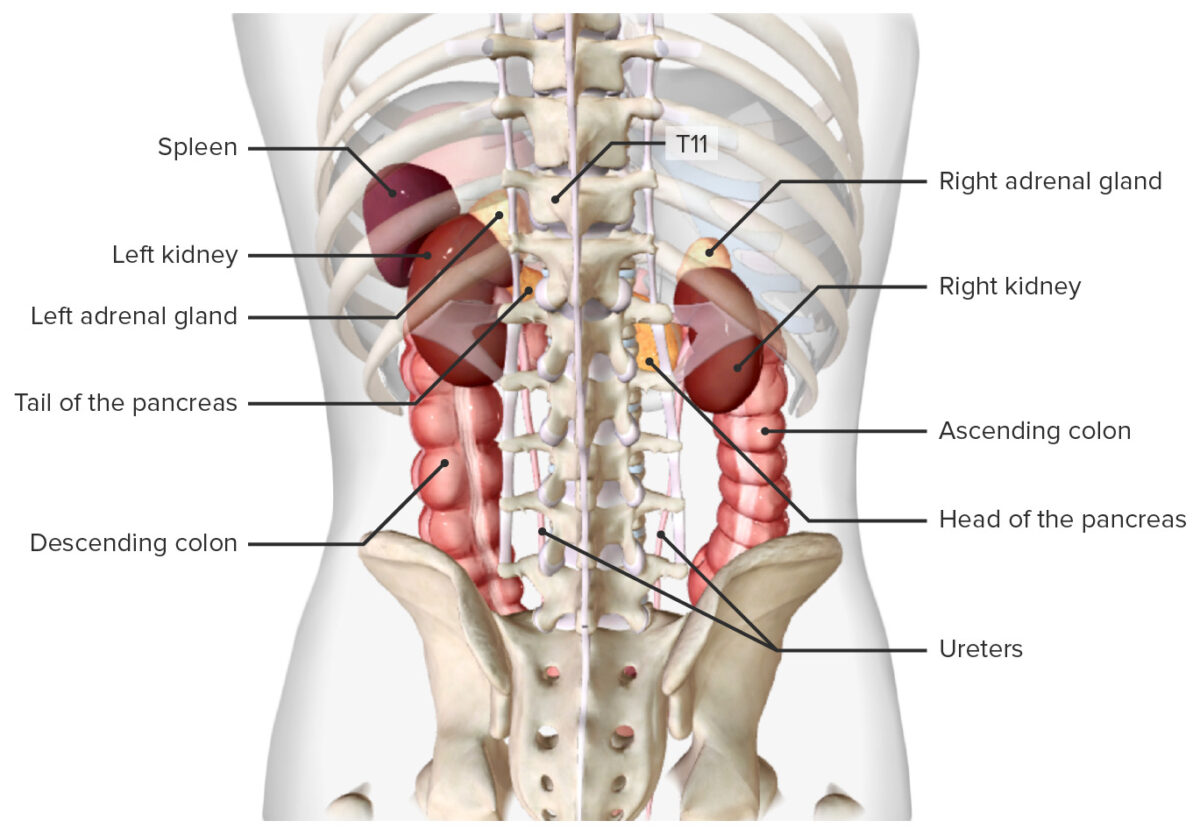
del bazo, in situ (vista posterior)
Imagen por BioDigita, editado por LecturioMediciones promedio en EN Erythema nodosum is an immune-mediated panniculitis (inflammation of the subcutaneous fat) caused by a type IV (delayed-type) hypersensitivity reaction. It commonly manifests in young women as tender, erythematous nodules on the shins. Erythema Nodosum adultos sanos normales:
El bazo está conectado a órganos adyacentes a través de varios ligamentos importantes.
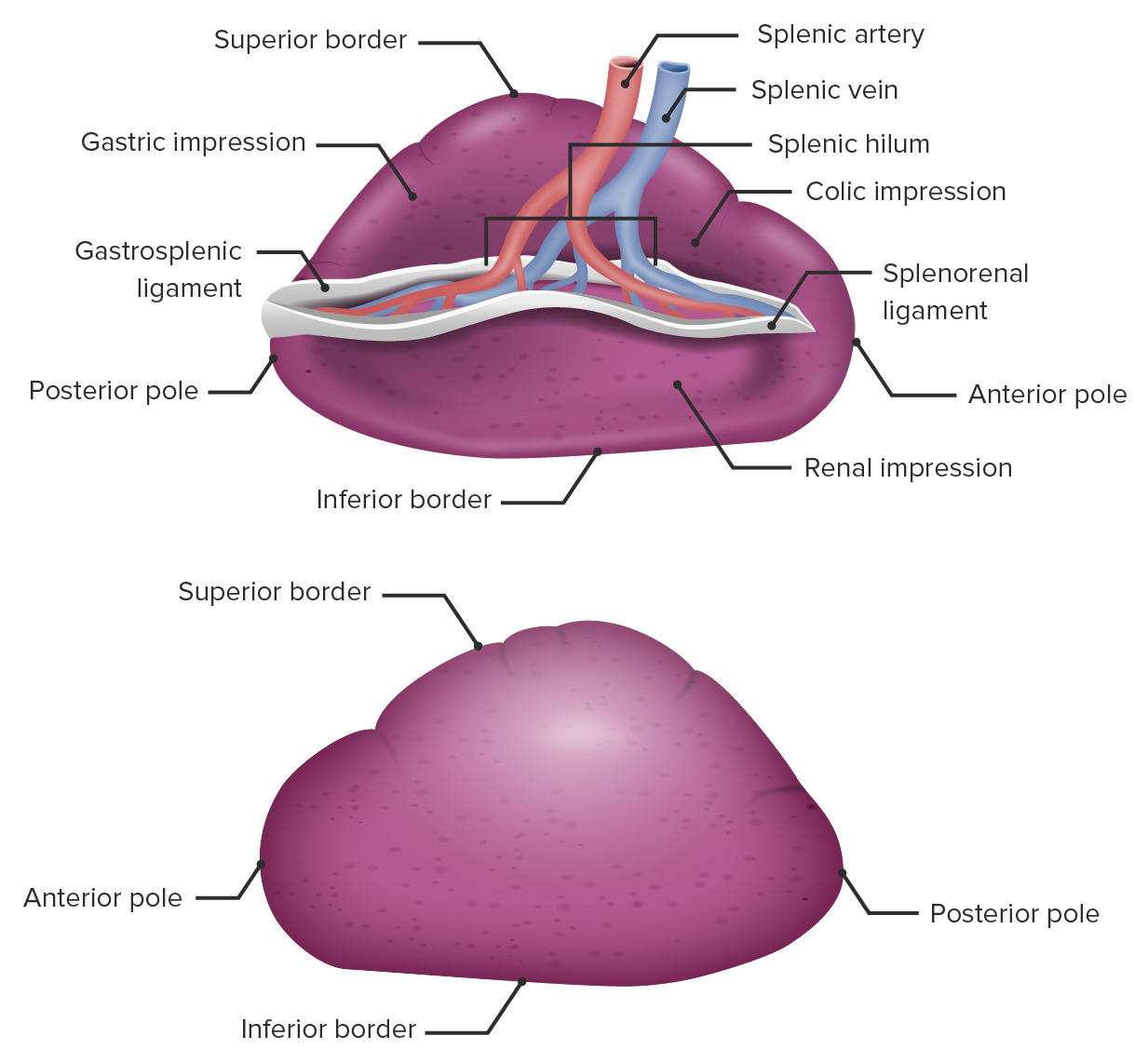
Anatomía macroscópica del bazo
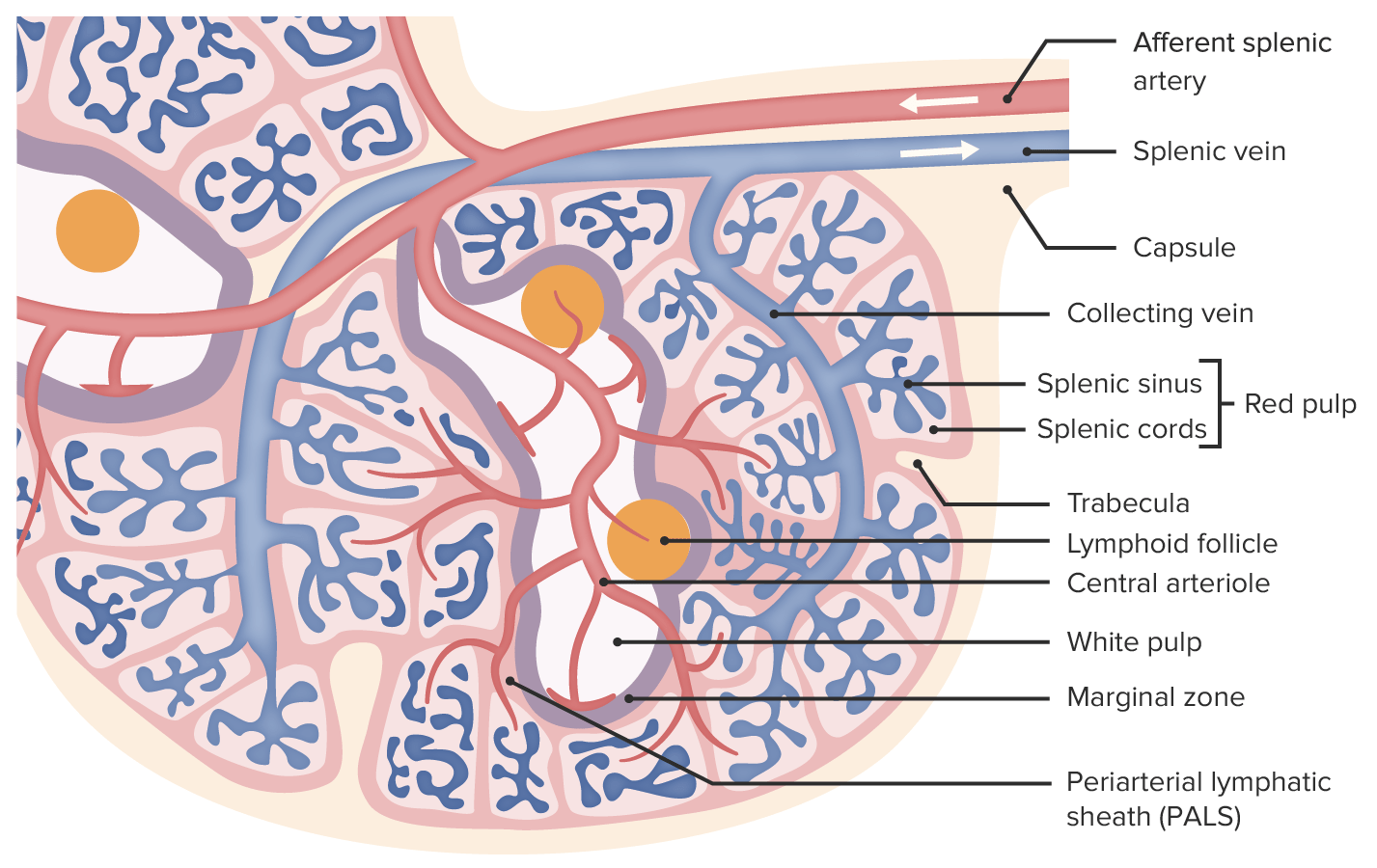
Imagen por Lecturio.El bazo consta de una cápsula y un tejido interno conocido como parénquima. El parénquima consta de 2 tipos de tejidos, pulpa blanca y pulpa roja.
La pulpa blanca constituye el 25% del bazo, rodea las arteriolas más grandes del bazo y contiene:
La pulpa roja constituye el 75% del bazo y está presente entre la pulpa blanca que rodea los LOS Neisseria vasos más grandes.
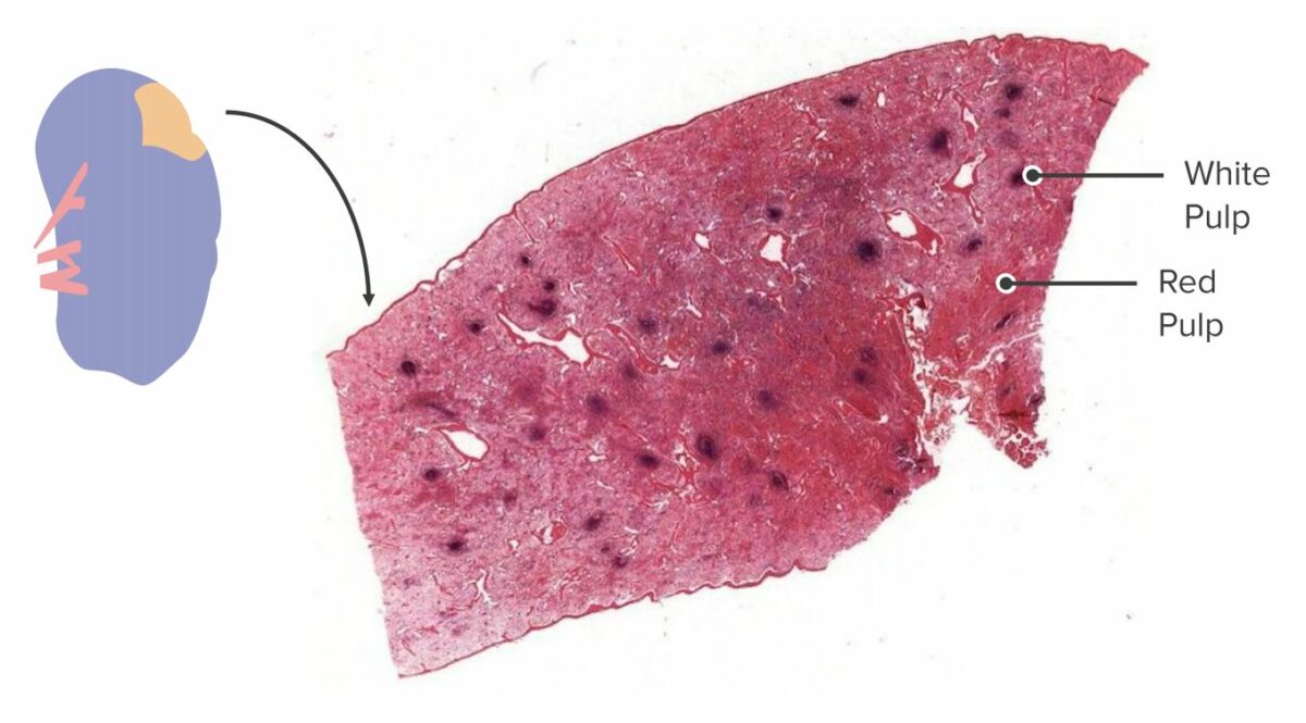
Sección transversal de un bazo:
En un espécimen macroscópico, la pulpa blanca y la pulpa roja se pueden identificar y diferenciar debido a su color y localización. La pulpa blanca se aglomera alrededor de los vasos sanguíneos, mientras que la pulpa roja constituye el resto del parénquima.

Corte histológico del bazo:
La vaina linfoide periarteriolar (PALS, por sus siglas en inglés) envuelve una arteriola central. Se ha formado un nódulo linfoide, rodeado de linfocitos T, lo que indica que se detectó un antígeno en la irrigación esplénica y se inició una respuesta inmunológica.
Anatomía microscópica del bazo
Imagen por Lecturio.Alrededor del 10% de la sangre permanece dentro de los LOS Neisseria vasos, mientras que aproximadamente el 90% se filtra hacia el tejido esplénico. La sangre fluye hacia, a través y fuera del bazo de la siguiente manera:
La inervación del bazo tiene lugar a través del sistema nervioso autónomo (SNA).
El bazo es el órgano linfoide secundario más grande del cuerpo. Las funciones del bazo son principalmente inmunológicas y hematológicas. La supervivencia sin bazo es posible ya que no es un órgano vital.