Playlist
Show Playlist
Hide Playlist
Ventricular Tachycardia: Diagnosis


-
Slides Tachyarrhythmia VentricularTachycardia CardiovascularPathology.pdf
-
Download Lecture Overview
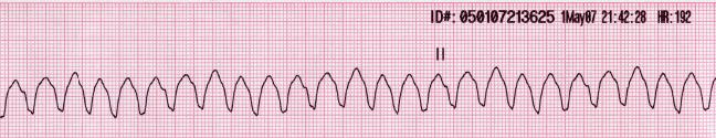
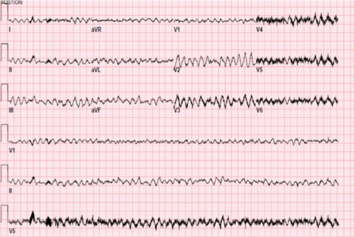
00:00 Alright so at this point, you think we were being redundant. Take a look at the category for arrhythmias, still dysarryhthmia and conduction system diseases but up until now we have only done SVTs. What is left? All important ventricles. Now here once again the way that I am setting up this for you is I'm going have you prepared for everything when you walk into your wards, but along the way though, we are going to then point out those incredibly important ventricular arrhythmias that you have to be aware of for any licensing exam. So let us begin and by the time we are down with the session, you will be extremely comfortable with just about any type of arrhythmia taking place due to a conduction system disease. Anything that will be left thereafter will be arrhythmias that are then secondary to vascular diseases. 01:00 Causes of ventricular tachycardia. Before we begin, since we were doing the ventricles, then you tell me as to what particular complex we are paying attention to. QRS complex only. 01:10 Here under ventricular, we will speed things up in which a QRS complex are closer to one another. The R wave are closer to one another and hence, you have an increase in heart rate. Remember greater than 100 beats per minute. Causes include ischemia, right ventricular outflow tract obstruction, structural heart diseases or medication induced. So, nothing really specific and you really making sure or you are trying to prevent from ventricular arrhythmias to take place to begin with because you are always worried about going into v.fib and hence death. What is the definition of ventricular tachy? What must you find? You must find greater than three consecutive premature ventricular contractions. In other words, your PVCs. And by that we mean that after the P wave, you should be able to see a QRS complex in correlation. But every once in a while, if we find that a ventricular contraction or QRS complex is taking place too early such as the PVC. Then this is the problem especially you can find greater than three consecutive PVCs. 02:20 The wide complex tachycardia. This is the one that you are paying attention to on and for your boards and or the wards. The wide complex tachy, QRS greater than 120 milliseconds. 02:34 What does that mean in terms of seconds? 0.12. The QRS complex is what we're referring to here. But you had memorized at least 0.12 seconds for QRS complex. Sustained if lasting greater than 30 seconds. AV dissociation, what does that mean? This would mean that the P waves are independent of the QRS. Fusion beats, appearance of typically differentiated. All you do here and this last little point is extremely important for you. RBBB and LBBB. How would you then be able to identify or understand that your patient is suffering from LBBB or RBBB? While you are paying attention apart from the EKG, which I will show you, you also want to pay attention to the history. Correct? And so therefore if it is RBBB, then you might then have what is known as a wide split of your S2. If it is an LBBB, what does that do? Well, you have an impulse that is passing through depolarization of the right bundle branch first. So, therefore, you have paradoxical split. Remember those discussions. Those are things which you want to have firmly understood before coming here into EKGs.
About the Lecture
The lecture Ventricular Tachycardia: Diagnosis by Carlo Raj, MD is from the course Arrhythmias: Basic Principles with Carlo Raj.
Included Quiz Questions
Which of the following is not a component of ventricular tachycardia?
- QRS complex < 120 milliseconds
- QRS complex > 120 milliseconds
- > 3 consecutive premature ventricular contractions
- Wide complex tachycardia
- Fusion beats
What is AV dissociation?
- The QRS complex is independent of the P wave.
- The QRS complex corresponds to each P wave.
- The QRS complex is independent of the T wave.
- The QRS complex corresponds to each T wave.
- The QRS complex is independent of theST segment.
Author of lecture Ventricular Tachycardia: Diagnosis

Carlo Raj, MD
Customer reviews
5,0 of 5 stars
| 5 Stars |
|
5 |
| 4 Stars |
|
0 |
| 3 Stars |
|
0 |
| 2 Stars |
|
0 |
| 1 Star |
|
0 |
