Playlist
Show Playlist
Hide Playlist
Protein-secreting, Mucus-secreting & Steroid-secreting Cells

-
Basic Histology 03.pdf
-
Reference List Histology.pdf
-
Download Lecture Overview
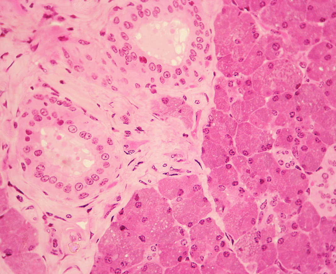
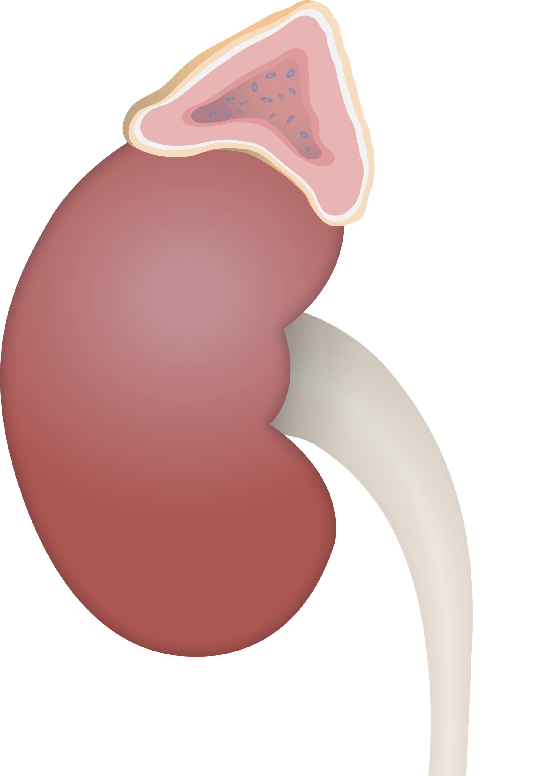
00:01 Let's look now at protein-secreting cells. 00:04 Here is a diagram showing you the basis of lots of salivary glands, lots of glands in the body that secrete protein enzymes that bring about the breakdown of food that we ingest On the left-hand side, colored green here is a representation of protein-secreting cells or acinar cells. 00:31 They're called acinar cells because the cells form a little ball like a grape and they all converge around the duct system you see labeled for the rest of the the diagram. 00:45 Intercalated cells form the beginnings of the duct system and then the striated duct cells form the rest, before these ducts join up to form large excretory ducts. 00:59 Well, this diagram illustrates on the left-hand side little tiny granules at the surface of the lumen of all these acinar cells, so the protein is made by the cell using huge amount of endoplasmic reticulum and those protein or digestive enzymes are then bound by membrane as secretory granules, and when they're stimulated to release these granules, the membrane fuses with the cell membrane and the product is released into that lumen. And as the product passes along with fluid, those striated duct cells can then modify the product and because of that, they often have lots and lots of mitochondria forming long columns at the base of the cell, and that's why they get their name striated ducts because you can see these striations in a light microscope preparations through these ducts. 02:03 Well, here's a real slide. 02:06 Forget the right-hand side where you get some pale-staining looking cells. 02:10 Focus on the structure in the middle. You can just make out a cluster of cells or an acinus. 02:18 You can make out a little tiny white space which is the lumen. The nuclei sit at the base of these cells and the bulk of the cytoplasm you see there is made up of these little tiny pink granules you can just make out in this particular slide. 02:37 These protein-secreting cells therefore are going to be full of rough or granular endoplasmic reticulum. 02:47 Mucus-secreting cells are similar. 02:51 Mucus-secreting cells are shown here mixed with protein or serous-secreting cells. 02:57 This happens to be typical of the submandibular gland, one of our salivary glands. 03:05 The mucus-secreting cells are those clear-stained ones you see. 03:10 I say clear-stained, they're not stained at all in fact, and that's because when these cells are processed for tissue preparation, the mucus leaks out during the alcohol dehydrations of the tissue, so there's nothing left in the cell unlike the protein components which are retained by fixative. 03:34 The fixative doesn't maintain the mucus. It doesn't fix the mucus and therefore, as I said, it gets flushed out during alcohol dehydration so because there's no stain, they appear as these white spaces within the cells. 03:53 A very common cell you're going to see in your histology study is the goblet cells shown here. 03:58 It lines many tubes. It's called a goblet cell because it looks like a wine goblet. 04:05 The nucleus is a very narrow structure at the base of the cell and as the cell makes mucus, it swells up at the surface or the apical end of the cell and just looks like a wine goblet, and then that mucus is then going to pass out through the surface of the cell into the lumen of where that epithelium sits. 04:33 Here on the left-hand side is a very low magnification of a part of the gut that happens to be the duodenum. 04:41 All those big folds are there to increase the surface area of this part of the gut and even on top of each of those folds, you have secondary folds called villi, shown in higher magnification on the right-hand side. 04:57 You don't need to know the details about the duodenum at this stage. 05:01 You'll learn about that when you look at the histology of the alimentary system, but it's just to show you that in the epithelium, most of those cells lining the epithelium you see on the right-hand side, the very tall cells or the very elongated nuclei, they're absorbing nutrients that are broken down inside the gut tube. They're called enterocytes, but if you carefully look through the epithelium, you can make out cells that look like wine goblets. They have a very pale-staining cytoplasm which is the mucus inside those cells. 05:41 Here's a better section that's stained specifically for the mucus. 05:46 A special preparation has been used in this particular section where the lipid has been preserved, the mucus has been preserved, and it takes up the stain so this part of the gut happens to be littered or dominated by goblet cells, and this is the part of the gut down towards the colon or the end of the gut where all that mucus is essentially used as a lubricant for the feces to pass through the gut tube. 06:20 Finally, let's look at steroid-secreting cells, and this is just a diagram that you don't need to memorize at this stage. 06:29 Leave that until you learn the histology of the adrenal gland, part of the endocrine system, but the adrenal gland sits on top of the kidney. It's divided into a number of regions. 06:44 A cortex which has those three zones you see there— zona glomerulosa, zona fasciculata, and zona reticularis and then there's the medulla. The medulla secretes adrenaline and noradrenaline or epinephrine and norepinephrine. The cortex, on the other hand, secretes all those hormones you see listed on the right-hand side, and again, please don't memorize them. 07:12 The key point is that that cortex, all those cells that make up those zones have within them a whole lot of lipid droplets, and that's why these cells you see in this part of the adrenal cortex has a lot of little clear, round, circular spots inside the cell. That lipid dominates these steroid-secreting cells and again, it's not retained during normal fixation and is washed out during alcohol dehydration of the tissue during tissue processing. 07:48 That's something I'll talk about in another lecture. 07:53 And all you're left with is this ghostly white granular appearance inside the cells. 08:00 Those lipid droplets are used as a precursor for making the steroid hormones, so they're a characteristic feature of not just the adrenal cortex but also the testes and the ovary, other glands in our body that make steroid hormones. 08:21 And then last of all and finally, this, I think is my favorite image. 08:27 What you see here is a little loop of epithelial cells looking into a clear space which is the lumen. 08:38 This happens to be a part of the gut, and down the bottom of that loop, you can just make out if you look carefully, 1, 2, about five mucus-secreting cells. 08:51 Look carefully into the cells and you'll just make out the foamy appearance of the mucus inside these cells that's not retained during the processing. 09:02 And then on the right-hand side up the top, you see this dark brown-staining cell. 09:09 That's an argentaffin cell. It's a steroid-secreting cell, and the steroid is in little granules you see there in the cell cytoplasm. But what's interesting is that look at the mucus cells. 09:28 The mucus is going to be secreted into the lumen that you see that white clear space whereas the granules of the argentaffin cell, the steroid hormone contained in these granules is on the abluminal side. It's on the other side of the nucleus. It's not near the lumen at all. 09:50 And the reason for that is that these steroids are not secreted into the lumen of the gut. 09:57 They're secreted into the basal part of the epithelium into the bloodstream, and those cells become what we call part of the Endocrine System, where cells make secretory products that don't get passed out through a duct or a lumen as you see here, but they're secreted internally into the bloodstream, where they circulate throughout the body and their target organ, their target cell is some distance away. 10:28 I think this is a marvelous slide to illustrate those key points about what we call exocrine secretion and what we call endocrine secretion.
About the Lecture
The lecture Protein-secreting, Mucus-secreting & Steroid-secreting Cells by Geoffrey Meyer, PhD is from the course The Mammalian Cell. It contains the following chapters:
- Protein-secreting Cells
- Mucus-secreting Cells
- Steroid-secreting Cells
Included Quiz Questions
Which of the following cells is a protein-secreting cell?
- Acinar cell
- Goblet cell
- Argentaffin cell
- Granulosa cell
- Leydig cell
Which of the following parts of the intestine has an abundance of goblet cells?
- Colon
- Rectum
- Jejunum
- Duodenum
- Ileum
Which of the following cells is an endocrine cell?
- Argentaffin cell
- Goblet cell
- Acinar cell
- Mature B cell
- Pancreatic acinar cell
Which of the following cells releases mineralocorticoids?
- Cells of the zona glomerulosa
- Cells of the zona reticularis
- Cells of the zona fasciculata
- Cells of the adrenal medulla
- Chromaffin cells
Which of the following is characteristic of endocrine glands?
- Secretion of products into the bloodstream
- Secretion of products into the duct
- Faster response from glands
- Working without central nervous system coordination
- The complex structure of the gland
Author of lecture Protein-secreting, Mucus-secreting & Steroid-secreting Cells

Geoffrey Meyer, PhD
Customer reviews
5,0 of 5 stars
| 5 Stars |
|
1 |
| 4 Stars |
|
0 |
| 3 Stars |
|
0 |
| 2 Stars |
|
0 |
| 1 Star |
|
0 |
Thank you, Dr. Meyer for this playlist, as an introduction in Histology! I was affraid about Histo, thinking that will be hard to learn, but your pedagogical skills and the excellent explanation helps me to study from a different perspective and, more important, with pleasure this discipline! Thank you!!! ????????
