Playlist
Show Playlist
Hide Playlist
Clinical Features of Primary Hyperparathyroidism & Osteitis Fibrosa Cystica

-
Slides Hypercalcemia.pdf
-
Reference List Pathology.pdf
-
Download Lecture Overview
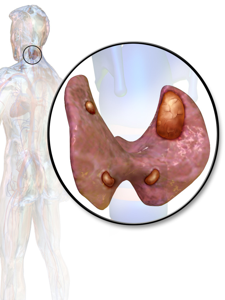
00:01 Primary hyperparathyroidism, asymptomatic, increased PTH-PTH, which will then drive the removal of calcium from your bone, may result in hypercalcuria. 00:14 So, you have, here, hypercalcemia and hypercalcuria. 00:18 There is a condition that I talked to you about earlier where it was your hypercalc-... 00:23 hypocalcuric hypercalcemia. 00:26 Thus familial, if you remember correctly. 00:29 There’s a decrease here in phosphorus because that excess PTH is working on the kidney and the PCT to then flush out the phosphate. 00:38 Speaking of the PCT, the PTH is then stimulating 1-alpha-hydroxylase resulting in activation of Vitamin D. There might be symptoms of primary hyperparathyroidism which includes your pathologic fractures and so forth that we talked about… brain, depression and that particular bone damage that’s taking place extensively is called osteitis fibrosa cystica a.k.a. Von Recklinghausen’s disease of the bone. 01:04 Next, what may then happen... remember we called as the brown tumor? Well, remember, this is not an actual tumor that you find in your bone; it’s a fact that you’re going to have replacement of this cavity being caused by excess PTH activity and osteoclasts in which you find haemosiderin within the cavity, you call this “a brown tumor”. 01:28 Yes, PTH works in the kidney to reabsorb the calcium, but as you know, majority of your calcium is stored in bone. 01:36 You start breaking down this bone, which PTH will do, all these calcium gets filtered through your glomerulus, may result in polyuria in males and result in calcium stones. 01:49 Symptoms of hypercalcemia, what are they again? In the brain, it will be depression and perhaps seizures; if it’s intestine and such, then you’re thinking about your peptic ulcer disease of the duodenal type maybe pancreatitis or gall stones; there might be constipation, pancreatitis, altered mental status and peptic ulcers. 02:07 Now what osteitis fibrosa cystica is is the following: take a look at the bone here. 02:11 Find it to be very, very lucent and the image that we’re seeing, looks like it’s a brown tumor but it’s not, those are just cavities. 02:22 You find X-ray characterized by subperiosteal bone resorption, extremely lucent, tapering at the distal clavicle, salt and pepper appearance on the skull and what that basically means is that because of the black areas that you would find on imaging, that means lucency, that means a bone is being broken down, that’s your pepper… where is the salt?... would be the areas of the bone that is remaining. 02:49 You call that “salt and pepper”, appearance. 02:52 And these are cysts and within the cysts, you might then develop your haemosiderin. 02:56 We now call this “brown tumor”. 03:02 The cavity is due to osteoclastic activity intermixed with fibrous tissue with very poorly mineralized bone and make sure you pay attention to the haemosiderin depositing in that area resulting in a brown-like appearance.
About the Lecture
The lecture Clinical Features of Primary Hyperparathyroidism & Osteitis Fibrosa Cystica by Carlo Raj, MD is from the course Parathyroid Gland Disorders.
Included Quiz Questions
What is the most important cause of hypercalciuria in hyperparathyroidism?
- Increased PTH
- Increased mineralized calcium
- Increased serum phosphate
- Decreased mineralized phosphorus
- Increased active vitamin D
What describes the pathogenesis of brown tumors?
- Fibrous replacement of resorbed bone
- Increased mineralization of bone
- An imbalance between calcium and phosphate in bone
- Improper deposition of bone
- Unregulated action of osteoblasts
What would NOT be seen in a characteristic x-ray of a patient with hyperparathyroidism?
- Increased cortical bone opacity
- "Salt and pepper" appearance
- Brown tumor
- Tapering of distal clavicle
- Subperiosteal bone resorption
Author of lecture Clinical Features of Primary Hyperparathyroidism & Osteitis Fibrosa Cystica

Carlo Raj, MD
Customer reviews
5,0 of 5 stars
| 5 Stars |
|
5 |
| 4 Stars |
|
0 |
| 3 Stars |
|
0 |
| 2 Stars |
|
0 |
| 1 Star |
|
0 |
