Playlist
Show Playlist
Hide Playlist
Posterior Segment of the Eye and Fundoscopic Exam – Anatomy Review
-
Slides Structures of the eye.pdf
-
Reference List Pathology.pdf
-
Download Lecture Overview
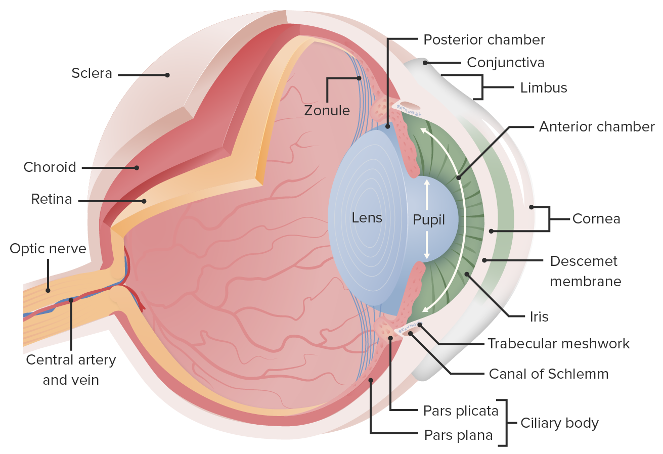
00:00 Now, we're going to go to the main part, the biggest part of the eye, which is more kind of a semi-solid gel. And this is the vitreous body. 00:08 The vitreous body is, again, kind of a semi-solid gel. 00:13 It is largely acellular, and it's avascular. 00:18 It's going to be where the majority of the light comes through, so it needs to be very transparent. 00:26 Okay, but there are diseases that can occur within the vitreous body. 00:29 You can have it detach from the retina at the back of the eye. 00:35 And that can cause some floaters. They can cause some shocks of light. 00:41 So, we have a posterior vitreous detachment. 00:43 Again, we'll talk about this in a whole separate session. 00:46 And you can get myodesopsias, these are just floaters. 00:49 So, there's the vitreous body, as I say, a semi-solid gel is not entirely completely homogeneous. 00:58 It has some non-homogeneous throughout it. 01:01 And those non-homogeneous, where it's a little bit of aggregation of that gel, is what are floaters and most of the time, we get used to looking right through them, we don't even notice them. 01:11 But if you're lying in bed, and you're kind of staring at the sun or whatever, you can see little floaters that move across your eye. 01:21 Those are the myodesopsias, that are so-called floaters. 01:25 Okay, non-homogeneous in vitreous body, and we'll talk more about those as well. 01:29 Alright, the sclera. 01:31 So, the sclera is this dense collagenous matrix that kind of sits all the way around the eye, that it's going to be responsible for allowing muscular insertion and maintaining the overall size and shape of the eye. 01:47 Next, inside is a highly vascularized structure called the choroid. 01:51 The choroid is continuous with the ciliary body and with the iris and we're going to call that entire structure in a moment, the uvea. 01:59 And we did right there, choroid and ciliary body and the iris. Uvea, okay. 02:06 And we can have diseases, inflammation of these, such as uveitis, or more specifically, chorioretinitis. 02:13 So, you don't necessarily have to have involvement of the iris, for example. 02:17 And then, finally, the innermost layer and the most delicate one that's responsible for all that vision, being able to turn light into electrical impulses that our brain can interpret as sight. 02:30 That's the retina. So, those three layers kind of go all the way around the eye. 02:35 What are they look like? Okay, so the sclera. So this is a trichrome stain. 02:39 The image on the right is a trichrome stain. 02:42 Things that are very, very blue have a lot of collagen in them, on a trichrome stain. 02:47 So, you can see the sclera, densely blue. 02:50 And so, it has a lot of strength, and integrity in terms of the structure, it provides the overall structure for the eye. 02:58 The next layer in is a highly, highly vascularized choroid. 03:02 And we'll spend more time talking about exactly what is going on in the choroid because a number of diseases such as macular degeneration, and things happen because of pathology in the choroid. 03:14 And the next layer in is the retina. 03:16 This is a vascular, it's got a lot of nerves, and neurons and cones and rods as you're well aware of and that's how we see. 03:25 But this is the structure that maintains that. 03:28 So, diseases that can occur as a result of injury to the sclera, choroid, retina. 03:33 So, you can have retinal detachment, the retina becomes detached from centerline membrane and away from its vasculature. 03:38 Ooh, not a good thing. 03:40 You can have diabetic retinopathy, which affects vasculature of the choroid. 03:44 You can have primary macular degeneration. 03:47 So, the cones and rods in the macula of the eye, which we'll talk about shortly, start failing. 03:55 And then, you can have other modes of failure in the retina, such as retinitis pigmentosa. 04:02 Again, we will come back to these. 04:03 So, we're just trying to give you an overall structure of what the eye - how it's put together. 04:08 And along the way, we've been giving you little red boxes to let you know, we're going to be coming back and talk about these in detail. 04:16 Okay, finally, all the information coming into the retina in terms of all those photons, hitting the rods and cones in the retina, have to go out somehow to get to the brain. 04:26 That's happening at the optic disc structure at the back of the eye. 04:31 And the optic disc coalesces all the nervous input from all the retinal rods and cones and then runs it out through an optic nerve. 04:38 At that same point is also where we have blood vessels coming in and out of the eye. 04:44 Okay, so if we have edema because there's increased pressure in the eye or in the brain, then we can get papilledema and that will cause compression of the nerves and make them nonfunctional. 05:00 We can also have inflammation of the optic nerve, so-called optic neuritis. 05:03 So we'll come back and talk about that. 05:05 And then, finally, important kind of area. This is geography of the retina. 05:12 Yeah, all the retina can see light and all the retina can help in terms of seeing the entire field that we look around and see. 05:21 But there are certain areas that give us the greatest visual acuity that allow us to read letters, to see colors the best, that's in an area in the eye, called the macula. 05:31 And the reason that we have the best visual acuity there is that we have a very dense collection of cones, the ones that see color. 05:39 The highest overall density is a little pit in the middle of the macula called the fovea. 05:44 And we can actually see these as we will in a moment, when we look into the eye, we can see these areas. 05:50 So, the macula is different than the optic disc. 05:54 Okay, the optic disc is actually a blind spot, you know, there is a blind spot, because there are no nerves there. That's just where rods and cones there. 06:03 It's where the big nerves come in and out, and the blood vessels come in and out. 06:06 So optic disc is a blind spot. 06:10 The macula is different, it's nearby, but that's our best visual acuity. 06:15 And the fovea is the absolute very best, it's a little pit with lots and lots of cones. 06:19 Okay, other things. 06:22 So there is a say coming in and out of the optic disc are the central retinal artery. 06:28 So these are going to be providing blood supply to the choroid which will then provide the necessary nutrition, oxygen, etc., to the retina. 06:38 There's a vein and there's an artery, we will see these when we look in with a fun - on an ophthalmologic fundoscopy. 06:46 You can have occlusion of the artery, you can have occlusion of the vein, clearly this is going to cause ischemia and infarction of the retina. Bad thing. 06:57 Okay, so let's see what all this really looks like as opposed to someone's drawing of it. 07:05 Here's the structures of the eye on fundoscopic exam. 07:08 So, we're looking at the fundus of the eye, the back of the eye, and the zone in the middle that's highlighted there looks a little bit redder, a little bit darker in color, just subtly so, and there's, you know, a spot in the middle, that's our macula at the bigger area. 07:22 The fovea is that little pit and this is our best visual acuity. 07:27 If we lose vision here, we can still see light and dark, we can see general shapes, but we won't be able to necessarily read a newspaper. 07:35 So, this is how we can see things in great, great detail. 07:39 Okay, so that's the macula and the fovea in the middle. 07:42 Over here is the optic disc, and there's power there, because we don't have quite the same density of blood vessels. 07:50 But you can see the blood vessels coming in and going out from that optic disc. 07:55 And, actually, as you look at this, you should look it up there close and take a look at this and enlarge it if you can, we can actually tell arteries from veins. 08:05 So, the arteries have a little - it looks like a little railroad track with a central golden middle of them. You're actually looking at the walls of the vessel, kind of the railroad tracks, and the blood in the middle. 08:16 And the veins look solid, they don't have that golden wire look in the middle so we can actually see arteries and veins separately. 08:23 In fact, the eye is a great place if you'd like thinking about micro vasculature and vessel supply, because you can actually visualize it just by looking through the pupil.
About the Lecture
The lecture Posterior Segment of the Eye and Fundoscopic Exam – Anatomy Review by Richard Mitchell, MD, PhD is from the course Introduction to Ophthalmology.
Included Quiz Questions
Which of the following best describes the vitreous body?
- Avascular and acellular
- Elastic and spongy
- Hypercellular and vascular
- Atrophic and necrotic
- Fibrous and nonelastic
What condition is commonly associated with retinopathy?
- Diabetes
- Myocardial infarction
- Hyperthyroidism
- Cerebral palsy
- Down syndrome
What is the area of greatest visual acuity called?
- Macula
- Optic disc
- Uvea
- Ciliary body
- Optic nerve
What is the optic disc also known as?
- Blind spot
- Yellow spot
- Red spot
- Lateral spot
- Color spot
Customer reviews
5,0 of 5 stars
| 5 Stars |
|
5 |
| 4 Stars |
|
0 |
| 3 Stars |
|
0 |
| 2 Stars |
|
0 |
| 1 Star |
|
0 |


