Playlist
Show Playlist
Hide Playlist
Parathyroid Gland and Adrenal Gland

-
Slides 05 Human Organ Systems Meyer.pdf
-
Reference List Histology.pdf
-
Download Lecture Overview
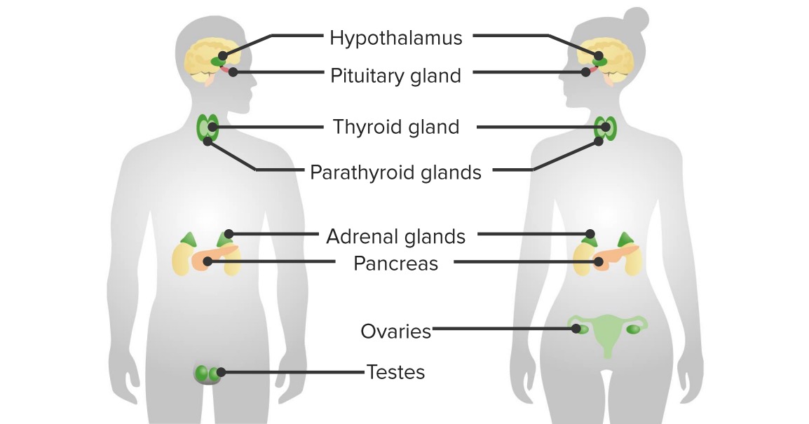
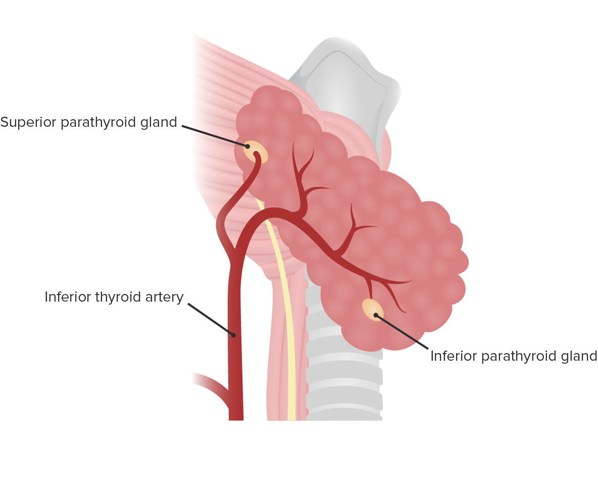
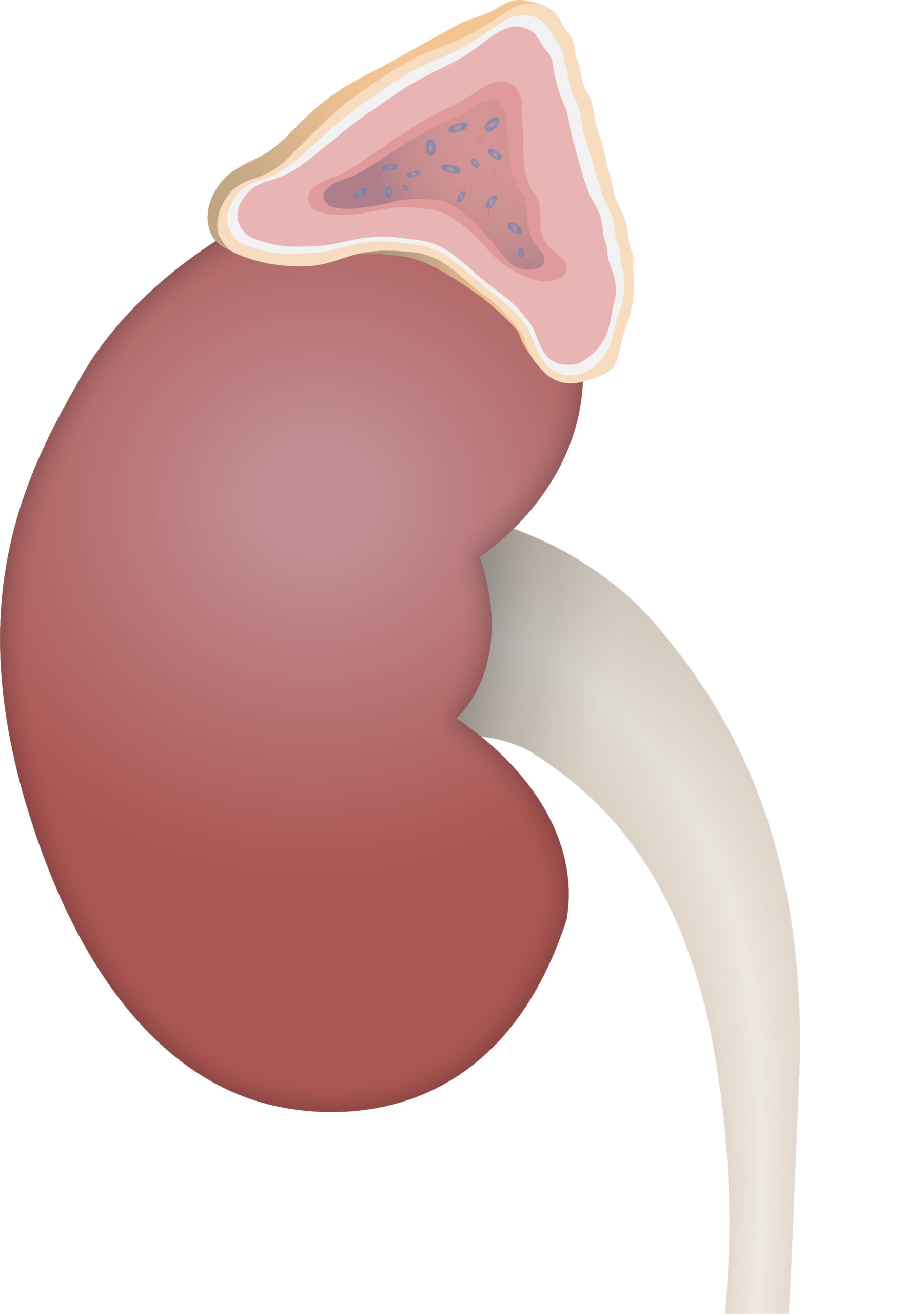
00:01 Let’s now move to the parathyroid gland. It’s a gland that doesn’t have a lot of significance histologically. It’s a pretty sort of dull looking organ, if you like, it's one of the less prettier organs of the body that we all have the pleasure of examining with a light microscope. 00:19 But it stores and secretes parathyroid hormone. And the principal cell that does that is called the principal cell or chief cell. If you look at the section on the left, you see predominantly, these cells are rather basophilic or bluish in their stain in this particular stain or dye used. If you look very carefully, you often see little clusters of sort of cells that take up a reddish stain. The reddish stains are these oxyphil stain cells. 00:54 You can see them contrasted here against the more basophilic stain chief cells that dominate the parathyroid gland. No one seems to know the function of these oxyphil cells. So I’ve put a question mark against their name. There are probably of limited significance when we consider the endocrine function of the parathyroid gland. I’d like to now describe for you the adrenal gland, often called the suprarenal gland because it sits on top of the kidney, the renal gland. The adrenal gland, again, is one of these endocrine glands like the pituitary gland that comes from two origins. It has a cortex that’s derived from mesoderm. This cortex occupies at least 90% of the gland. And it has a medulla that occupies about 10%, that is in the center of the gland, surrounded by the cortex. And this medulla is derived from neural crest cells. So have a look at these glands labelled in the center, in the section of the center, and make sure you clearly see the cortex and the medulla. One thing to notice is that the medulla has a brownish sort of stain. 02:19 And the cortex has a more pinker stain. So, different stains have been used here to help distinguish those two components of the adrenal gland, the adrenal medulla, and the adrenal cortex. 02:34 And again, like the pituitary, they probably should be considered as separate glands even though geographically, they live together. Well, when you look at the cortex, you can clearly see if you look very, very carefully that you can separate the cortex of the adrenal gland into three separate zones based on how they appear, even from this low magnification. 03:04 And I’m going to describe each of those three zones because they secrete different classes of hormonal product.
About the Lecture
The lecture Parathyroid Gland and Adrenal Gland by Geoffrey Meyer, PhD is from the course Endocrine Histology.
Included Quiz Questions
What is the origin of the adrenal cortex?
- Mesoderm
- Ectoderm
- Endoderm
- Neural tube
- Neural crest cells
Which of the following cells secrete parathyroid hormones?
- Chief cells
- Parafollicular cells
- Oxyphil cells
- Follicular cells
- Beta cells
Author of lecture Parathyroid Gland and Adrenal Gland

Geoffrey Meyer, PhD
Customer reviews
5,0 of 5 stars
| 5 Stars |
|
5 |
| 4 Stars |
|
0 |
| 3 Stars |
|
0 |
| 2 Stars |
|
0 |
| 1 Star |
|
0 |
