Playlist
Show Playlist
Hide Playlist
Ocular Emergencies

-
Slides OcularEmergencies AcuteCare.pdf
-
Download Lecture Overview
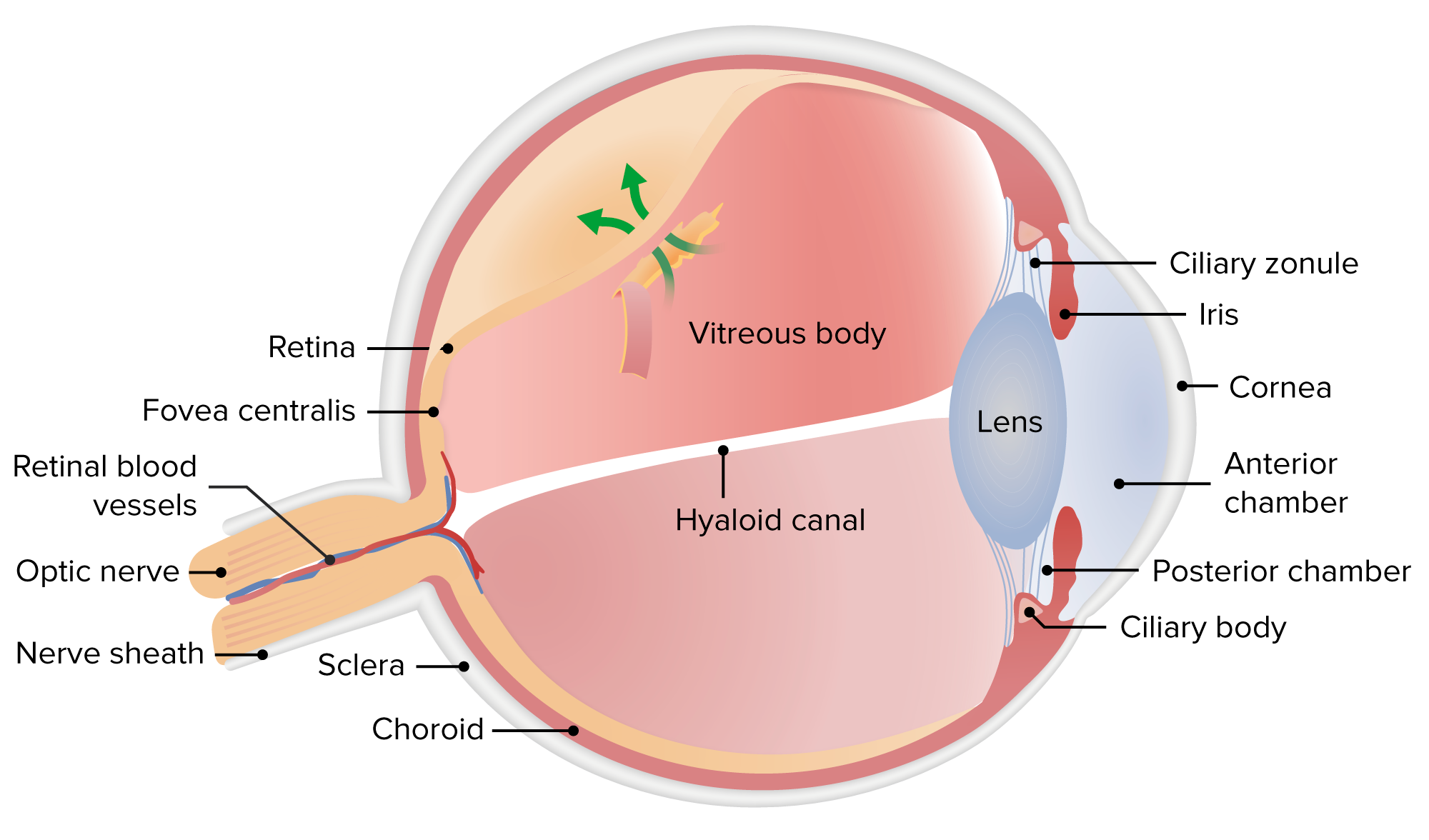
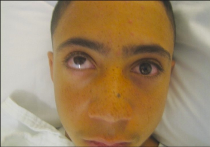
00:01 Now, primary care physicians don't see a ton of ocular emergencies as part of continuity practice, but if you ever work in an urgent care setting, you will see a lot of acute eye symptoms, and therefore, it's really important to get to know some of the diagnoses I’m about to share with you in ocular emergencies. 00:19 Even if you don't see a lot of these emergencies, there are some principles here that are really important and I think are worth sharing as well. 00:26 Let’s get underway. 00:27 And we’ll do a case. 00:28 So, I’ve got a 75-year-old male. 00:30 He had bilateral cataract surgery last year, most common surgery in the United States. 00:36 But, today, he had a problem. 00:37 Sudden onset of floaters and flashing lights in his left eye. 00:42 And so, he tried to do an exam of that eye. 00:44 And it’s difficult. 00:45 You can't really get a good look inside because of a constricted pupil. 00:50 So, just based on that limited information alone, what is the best course of management for this patient right now? Do, we, A, treat with a topical antibiotic? B, treatment with systemic antibiotics? C, immediate referral to ophthalmology? Or D, referral for neuroimaging? What do you think? The answer is C. 01:11 And I’m going to make the answer clear why in a second. 01:14 But this is a case of probable retinal detachment that needs emergent evaluation by an ophthalmologist. 01:22 So, let's talk about a retinal detachment first. 01:25 Then we’ll be moving on to globe injuries and chemical injuries to the eye. 01:30 So, this gentleman has all the risk factors for a retinal detachment except the fact that he didn’t have a previous detachment, but he is older and has a history of cataract surgery. 01:41 He has the right symptomatology. 01:43 Floaters plus flashing lights. 01:45 The visual defect can be variable. 01:47 Some patients actually can see fairly well after a retinal detachment and some people have a much more blurry field. 01:55 But unquestionably, this needs to be evaluated emergently because once a detachment occurs, their risk for ischemia goes up and may not be regained. 02:07 The good news with retinal detachment is, when the macula is involved, most patients who receive surgery do achieve a satisfactory result in terms of getting 20/40 vision or better. 02:21 And these patients should also be followed because, as I mentioned, previous detachment is a major risk factor for subsequent detachment. 02:30 So, up to a quarter will actually experience detachment on the opposite eye at some point. 02:37 And those are the points I just made. 02:39 Let’s talk about mechanical globe injury. 02:42 So, this can be a full thickness tear, either through the cornea or through the sclera. 02:48 It’s often related to high velocity, high-impact injuries like with this rocket ship. 02:52 You don't want that hitting you in the eye. 02:54 Symptoms, as you can imagine, pain, but also redness, tearing and decreased vision. 03:00 And we’ll talk about how redness is one thing, but ocular pain is a different thing. 03:07 And when I talk about ocular pain, I’m talking about a pain in the globe. 03:10 Patients with conjunctivitis feel irritation, burning, itching, but it's not necessarily a deep boring kind of pain. 03:18 A mechanical globe injury, that's going to be actual pain in the globe and always should be a red flag for an emergent condition. 03:26 Always, always. 03:28 Now, the good news with a mechanical globe injury, if it's fairly straightforward and you can diagnose it as just a conjunctival laceration and it’s less than a centimeter, they don’t need to be referred, they can receive topical antibiotics and follow-up and should be okay. 03:43 Anytime you have a mechanical globe injury, you want to avoid any type of Valsalva that could increase the intraocular pressure because that can lead to more damage. 03:55 This is another one. 03:57 Immediate ophthalmology evaluation for anything other than that superficial lesion. 04:02 And most ophthalmologists are going to recommend systemic antibiotics, oftentimes fluoroquinolones, with some kind of Staph coverage. 04:10 So, that could be a drug like levofloxacin or it could be a fluoroquinolone along with another drug, which has gram-positive coverage. 04:20 Okay. 04:20 Let’s move on to chemical injury. 04:21 We’re moving quickly today. 04:23 Just one thing that may come up on the exam. 04:25 Alkali burns, more common than acid burns. 04:28 And most of these are related to work-related accidents. 04:32 So, that's why the first step in preventing injury is appropriate eyewear and safety measures. 04:39 Symptoms include swelling and burns in the eyelids. 04:44 Your cornea will get cloudy as a result of the chemical reaction there. 04:51 Then the conjunctival injection injected as the body tries to heal itself. 04:58 And you might actually see a completely white eye due to conjunctival ischemia if the injury related to the alkali burn is particularly bad. 05:09 The management is, of course, immediately washing out the eye. 05:12 Hopefully, there's an eye station at the at the site and the sooner and the more copious irrigation is better. 05:20 But then, once they get into a medical situation, like an emergency department, topical anesthetics with a lot more irrigation. 05:27 And again, this is one for serious burns. 05:30 Better to see an eye specialist right away. 05:34 The least they are going to recommend is topical antibiotics with artificial tears and maybe even more than that because they might actually employ corticosteroids, which are always tricky to use in the eye and therefore should be done only by specialists, should not be done by primary care in a situation like a burn. 05:52 And so, my pearl to everyone, if you see these cases on a daily basis or you see one case every five years, I feel like it’s just hard to be very overly conservative with these eye issues because once you – it’s a one-time thing to preserve vision. 06:12 And once it's lost, many times it's not coming back. 06:15 So, therefore, caution is the watchword and you should be friendly with an ophthalmologist, so you can make sure if nothing else, run these issues by that person. 06:26 And if they can’t be reached, emergency department is always at your disposal. 06:31 So, let’s look at another subject, slightly different, maybe not quite as emergent, but certainly urgent, is the redeye. 06:39 Here we have a nice example of conjunctivitis. 06:41 That’s by far the most common cause of redeye. 06:44 You could see the purulent drainage on the lashes. 06:48 Can I tell just by looking at this picture whether it's viral or whether it's bacterial? It’s hard to say because, yes, it does have a purulent drainage, but that's not pathognomonic. 07:00 You get some purulence from viruses as well. 07:04 But the more copious drainage, the more severe redness, the fact that – especially when it’s unilateral and not bilateral, all those may implicate that it’s a bacterial versus a viral situation. 07:18 Now, a focal area of hyperemia, as you see here, that may be associated with episcleritis, which is associated with a variety of different autoimmune conditions. 07:30 So, that might be\ something to consider in, say, Bissett syndrome, maybe one of the signs that the patient is getting Bissett’s. 07:37 And scary for patients and their loved ones, but generally benign is subconjunctival hemorrhage. 07:44 Maybe the classic is a person has a cold, sneezes a bunch of times, then goes to bed, wakes up with this big red bright red patch in their eye under their conjunctiva. 07:58 It’s disconcerting, but usually resolves over time with no symptoms. 08:05 So, I’m going to leave you with again that clinical pearl that it's one thing to have a redeye, but it’s another to have a red and painful eye. 08:13 So, when redness comes with pain, I consider that an urgent or emergent condition because it could be a keratitis, which might be herpetic which can cause permanent vision loss, a corneal ulcer which is a serious diagnosis, iritis associated with other autoimmune disorders may exist on its own. 08:33 But, again, all of those are good reasons for referral to ophthalmology on an urgent basis. 08:40 And therefore, I think if you recognize these more serious conditions, as rare as they might be, you'll do your patients a world of good by preserving their vision. 08:51 And with that, I just wanted to say thank you and watch out for those red eyes.
About the Lecture
The lecture Ocular Emergencies by Charles Vega, MD is from the course Acute Care. It contains the following chapters:
- Ocular Emergencies
- Rentinal Detechment
- Mechanical Globe Injury
- Chemical Injury
- Differential Diagnosis of the Red Eye
Included Quiz Questions
Which of the following conditions has a higher risk of retinal detachment?
- Bilateral cataract surgery
- Bilateral glaucoma
- Hyperopia
- Macular degeneration
- More frequent use of antibiotics
Which of the following increases the risk of retinal detachment?
- Personal or family history of retinal detachment
- Personal history of retinal detachment
- Family history of retinal detachment
- A previous personal or family history of retinal detachment does not increase the risk of retinal detachment.
Which of the following types of eye injury is most likely to cause a white eye due to ischemia of the conjunctiva and scleral vessels?
- Alkaline chemical burn
- Acid chemical burn
- Corneal scratch
- Scleral tear
- Traumatic retinal detachment
Which of the following therapies is of primary importance in most chemical eye injuries?
- Irrigation
- Topical steroids
- Surgery
- Topical antibiotics
- Swabbing the fornices
Which of the following findings is LEAST likely to require emergency management in a patient with eye injury?
- Small round pupils
- Chemical eye exposure
- Periorbital swelling with proptosis
- Protruding foreign body
- Decreased anterior chamber depth
A 37-year-old presents to the clinic because of severely red conjunctiva after several episodes of emesis and retching from food poisoning the night before. He has no pain or visual disturbance. What is the next best step in management?
- Reassurance
- Topical antibiotics
- Irrigation
- Urgent ophthalmology consultation
- Topical steroids
Author of lecture Ocular Emergencies

Charles Vega, MD
Customer reviews
3,0 of 5 stars
| 5 Stars |
|
0 |
| 4 Stars |
|
0 |
| 3 Stars |
|
1 |
| 2 Stars |
|
0 |
| 1 Star |
|
0 |
Good presenter medical content could be more detailed. I would have appreciated more in-depth talk on anatomy, medications and complications
