Playlist
Show Playlist
Hide Playlist
Necrosis vs. Apoptosis

-
Slides Cellular Pathology Apoptosis.pdf
-
Reference List Pathology.pdf
-
Download Lecture Overview
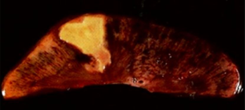
00:01 So comparing Necrosis and Apoptosis. 00:04 In necrosis, the cells are swollen. 00:07 They've taken up water because of the lack of regulation of the normal ionic environment. 00:14 So they are swollen, edematous. 00:17 In comparison, the cells in apoptosis are shrunken, they're smaller. 00:22 They're fragmenting into little apoptotic bodies. 00:25 The nucleus is variably changed in necrosis, there could be piyknosis condensation. 00:32 Complete destruction, that's karyolysis, fragmentation, karyorrhexis. 00:37 In apoptosis, it's mostly a fragmentation, so they look karyorrhectic. 00:41 The plasma membrane in necrosis, it's disrupted, completely punch full of holes. 00:47 In fact, apoptosis is completely intact. 00:50 So none of the cell contents leak out. 00:53 The only thing that's different really about that is the phosphatidylserine. 00:56 The PS has been flipped from the inside to the outside, saying, "eat me". 01:01 The Contents. 01:03 In necrosis there is auto digestion, and then there's leakage of those cellular contents to the outside world. 01:11 We actually use that when we think that there's, for example, been damaged in the liver or the heart. 01:17 We can look at enzymes leaked from the liver and the heart in the bloodstream, and we can characterize how severe the injury is. 01:26 In apoptosis, cellular contents are completely intact, so we can't. 01:30 We cannot measure apoptosis by measuring cardiac proponents, for example. 01:36 The Mediators. 01:37 So in necrosis, it's a calpains, remember those are the calcium activated cytosolic enzymes that help to degrade lipid and protein and nucleic acids. 01:49 In apoptosis, it's the caspases. 01:53 And in necrosis, inflammation all the time. 01:56 In fact, as we talk about really important component of necrosis. 02:01 In apoptosis, no, no inflammation. 02:04 It's very quiet. 02:06 Finally, just to look at the DNA fragmentation pattern. 02:09 For example, in necrosis, as we break down the nuclear material on the left hand side, we just get a smear. 02:17 There's all kinds of different sizes of nucleic acids. 02:21 So the middle band is just a control layering of different sizes of nucleic acids. 02:31 The total right hand of the three undergoing necrosis is the total DNA. 02:38 Before it gets fragmented, it's large material that doesn't flow into the gel. 02:43 And on the left hand side of that left panel, you see a complete smear, and that complete smear is because DNA is fragmented all different sizes. 02:52 It just randomly is hit. 02:54 In comparison, like apoptosis. 02:57 You see with the arrow on the right hand side that we get discreet sizes to our DNA. 03:02 It's cleaving at distinct points around his stones, so we get about 200- 220 size base pair bands all the way up. 03:14 It's called DNA laddering. 03:16 So this says that this is really well controlled. 03:19 It's not random, necrosis, that's random. 03:22 So we've talked about cells dying. 03:25 We talked about how they die, we've talked about necrosis, and we've talked about cellular suicide, which is apoptosis. 03:34 And with that, we're ready to start talking about some of the next stages of pathology.
About the Lecture
The lecture Necrosis vs. Apoptosis by Richard Mitchell, MD, PhD is from the course Cellular Injury.
Included Quiz Questions
Which of the following is characteristic of necrosis?
- Karyorrhexis
- Intact plasma membrane
- Cell shrinkage
- Caspase-mediated cell death
- Absence of inflammatory response
Which of the following is characteristic of apoptosis?
- DNA laddering on gel electrophoresis
- Leakage of the cellular content
- Calpain-mediated cell death
- Enlarged cells
- Disruption of the cell membrane
Author of lecture Necrosis vs. Apoptosis

Richard Mitchell, MD, PhD
Customer reviews
5,0 of 5 stars
| 5 Stars |
|
1 |
| 4 Stars |
|
0 |
| 3 Stars |
|
0 |
| 2 Stars |
|
0 |
| 1 Star |
|
0 |
One of the best teachers of Pathology I've ever seen.
