Playlist
Show Playlist
Hide Playlist
Glioma: Grading with MRI and Contrast Enhancement

-
Strowd CNS Tumors Glioma.pdf
-
Download Lecture Overview
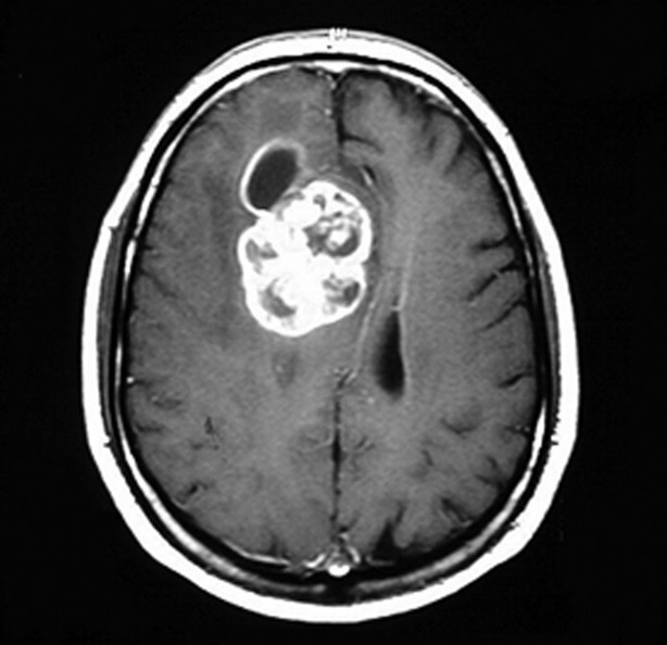
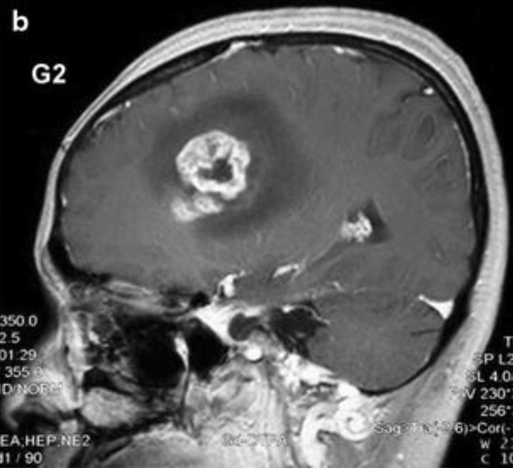
00:01 So after the patient has presented with a certain clinical syndrome, and we're worried this could be a tumor, we want to evaluate that with imaging. 00:08 And the most common imaging that's performed for patients may begin with a CT, but ends with an MRI of the brain to really characterize that lesion comprehensively. 00:20 So the second step after clinical presentation is imaging evaluation. 00:25 What types of imaging do we use? I said CT and MRI. 00:28 And MRI is really the diagnostic modality of choice, for evaluating gliomas. 00:33 Conventional MRI will show a mass like lesion in the brain parenchyma in the tissue of the brain, and this should tip us off to think about tumors that arise from the cells in the brain. 00:44 Astrocytomas, oligodendrogliomas, ependymomas, and others. 00:49 Contrast enhancement is important. 00:51 And we'll talk in the next few slides about the benefit of contrast enhancement and evaluating the aggressiveness of tumors. 00:57 Tumors that are growing more aggressively, enhanced with contrast more avidly, and tumors that are often less aggressive, or of lower grade typically do not enhance or more weakly enhance, more minimally enhanced with contrast. 01:10 Increasing contrast enhancement is seen as tumors grow from grade II, to III, to grade IV. 01:17 Let's look at the prevalence of contrast enhancement, and its benefit and value for evaluating a patient or a clinical vignette of a patient who may be presenting with a glioma. 01:28 Contrast enhancement. 01:29 Gadolinium on the scan is present in 96% of glioblastomas. 01:33 And typically, this is a ring of enhancement, as we saw with our second patient, and second case at the beginning of this lecture. 01:41 Grade III or Anaplastic gliomas, we only see contrast enhancement in about half of those patients 50 to 60%. 01:48 So presence of contrast enhancement should tip us off that we could see a higher grade, but we don't see that in all of these grade III or anaplastic gliomas. 01:57 And then we don't commonly see contrast enhancement for lower-grade gliomas. Only in about 20% of patients. 02:03 And typically, this is not that ring enhancement pattern, but a more patchy or heterogeneous pattern of contrast enhancement. 02:10 So let's look at that. 02:11 Grade I tumors are the least aggressive, the lowest-grade. 02:15 They're actually uncommon in adults and more common in children. 02:18 And we'll talk about those in the next couple of slides. 02:21 They do enhance with contrast, and this defies our rule of increased enhancement with increasing grade because we commonly see avid contrast enhancement for grade I tumors like this pilocytic astrocytoma, of the left optic nerve. 02:35 Of the infiltrating gliomas, we see that pattern of contrast enhancement. 02:40 Grade II, low-grade gliomas typically do not enhance. 02:45 Anaplastic gliomas often homogeneously enhance or have salt and pepper punctate enhancement within the inside of the lesion. 02:52 And what characterizes glioblastomas is this ring of enhancement and that should tip us off that we're looking likely at a grade IV glioma.
About the Lecture
The lecture Glioma: Grading with MRI and Contrast Enhancement by Roy Strowd, MD is from the course CNS Tumors.
Included Quiz Questions
Which of the following types of glioma has the lowest level of enhancement on MRI?
- Low-grade glioma
- Glioblastoma
- Pilocytic astrocytoma
- Anaplastic glioma
- Oligodendroglioma
Where are gliomas most commonly found in the CNS?
- Brain parenchyma
- Posterior fossa
- Brainstem
- Intraventricular space
- Foramen magnum
Author of lecture Glioma: Grading with MRI and Contrast Enhancement

Roy Strowd, MD
Customer reviews
5,0 of 5 stars
| 5 Stars |
|
5 |
| 4 Stars |
|
0 |
| 3 Stars |
|
0 |
| 2 Stars |
|
0 |
| 1 Star |
|
0 |
