Playlist
Show Playlist
Hide Playlist
Fenestration & Endothelial Transport

-
Slides 05 EpithelialTransport GeneralPhysiology.pdf
-
Download Lecture Overview
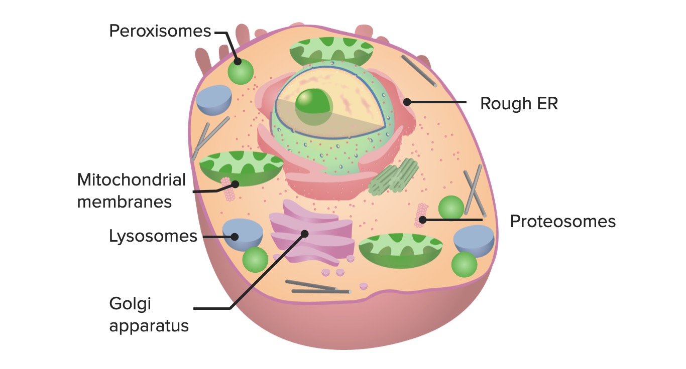
00:01 Endothelial transport is much different than epithelial transport. 00:08 It also though has tight junctions. 00:12 The tight junctions though can be very tight such as in the blood-brain barrier when there’s no movement of fluid, or in capillaries they sometimes have fenestrations, pores, or sometimes even clefts that will allow things to travel through. 00:33 So, endothelial transport is going to rely less on specific transporters than epithelial transport does. 00:43 You’re going to utilize things like pressure and osmolality to move various solutes and solvents around. 00:53 It is primarily a variable associated with the flux of the substance. 01:00 So you will filter certain things, hydrostatic pressure is highly involved, and osmotic and oncotic pressures. 01:10 Remember, osmotic pressures have to do with the ion differences and oncotic pressures have to do with protein differences to draw fluid. 01:23 This is an example of how pressure can move fluid out of a capillary bed. 01:30 The higher the amount of pressure, the more fluid travels through. 01:36 Other examples determine about how much the fenestrations are in terms of their width. 01:43 Some will allow more fluid to travel out and some will allow less. 01:47 The other issue that we need to think about with endothelium versus epithelium is that we sometimes turn the membranes a little bit differently. 01:58 The terminology used for the inside surface is the luminal surface rather than the apical membrane. 02:07 In terms of the outside is termed the basal surface rather than the basolateral membrane. 02:15 But if you keep those linked together, you’ll be better off and able to think about the differences between epithelial and endothelial surfaces. 02:26 Let’s look at how fenestrations can be regulated because normally you think of a pore either being open or being closed, but you can modulate this in certain tissues. 02:38 The lymphatic is a great example of this. 02:42 So you can have some constriction and have the pores closed. 02:47 And then, after constricting these, you can open them up. 02:54 This then will allow fluid to transport between the lymphatic circulation. 03:01 Then when you have smooth muscle constriction, you close them up. 03:06 Good examples of how you can modulate these fenestrations. 03:11 Now, many tissues you don’t have the modulatory ability because either they are open or closed, but you do have some regulation of fenestration widths. 03:22 Let’s summarize now the different ways you’re going to move a solution, a substance, or a gas across the endothelial wall. 03:35 The first thing you could do is use something called pinocytosis, which is the actual pinching off of a small vesicle that contains a solute and solvent. 03:47 It moves from the luminal surface to the basal surface and then releases it out. 03:55 Another way you could get fluid through is by fenestrations, either fluid travels through or sometimes solutes travel along with the fluid. 04:07 The fenestration width will be dependent upon what molecules can make it through. 04:12 Water will always be able to make it through, but sometimes larger substances like big proteins, maybe like albumin, have a harder time in moving through these fenestration slits so they get stuck on one side or the other. 04:27 A primary variable is the pressure at which is in the hydrostatic, which is inside the vessel to push fluid through. 04:39 And that helps with the bulk transport driven by the pressure change. 04:46 You also have diffusion that is capable of moving a solute through these fenestration slits. 04:53 So this is based upon a concentration gradient. 04:57 The bigger the concentration gradient, the more solute is allowed to travel through. 05:03 Other items, such as gases or other things that are very soluble might be able to make it through the endothelial cell all on its own, without a fenestration slit, without pinching off the particular portion of the membrane and having it travel through. 05:21 These substances are usually more lipophilic in nature and therefore can travel through the membrane on their own.
About the Lecture
The lecture Fenestration & Endothelial Transport by Thad Wilson, PhD is from the course Membrane Physiology.
Included Quiz Questions
Which of the following statements is CORRECT regarding endothelial transport?
- It relies on hydrostatic pressure to transport solutes across.
- It relies on specific transporters.
- It relies on vector transport.
- It relies on voltage differences between the two sides of the cell membrane.
- It has impermeable tight junctions throughout the body.
What is the main determinant of oncotic pressure?
- Protein concentration
- Ion differences
- Ion-derived voltage differences
- Protein derived voltage differences between the two sides of the endothelium
- Types of ions
What is the name of the surface membrane of the endothelial cell facing the blood?
- Luminal surface
- Basal surface
- Apical membrane
- Basolateral membrane
- Luminal membrane
Which of the following drives the bulk of flow across the endothelium?
- Hydrostatic pressure
- Pinocytosis
- Osmosis
- Simple diffusion
- Passage through the cell membrane
In which of the following does hydrostatic pressure have the greatest influence in the movement of fluid across the endothelium?
- Arteriolar side of the capillary
- Venous side of the capillary
- Second third of the capillary
- Last part of the capillary
- Middle of the capillary
Which of the following statements regarding lymphatic capillary fenestrations is CORRECT?
- They are open in the expansion phase.
- They are open in the compression phase.
- They are always open.
- They are open when the secondary lymph valves are open.
- They are closed when the secondary lymph valves are closed.
Author of lecture Fenestration & Endothelial Transport

Thad Wilson, PhD
Customer reviews
5,0 of 5 stars
| 5 Stars |
|
2 |
| 4 Stars |
|
0 |
| 3 Stars |
|
0 |
| 2 Stars |
|
0 |
| 1 Star |
|
0 |
Thank you for this lecture. Very good explanation, excellent structure! Dr. Wilson, you are awesome!!!
All Dr. Wilson's lectures are great. Good contents presented in a clear and interesting manner.
