Playlist
Show Playlist
Hide Playlist
Examination of the Shoulder

-
Reference List Physical Examination.pdf
-
Download Lecture Overview
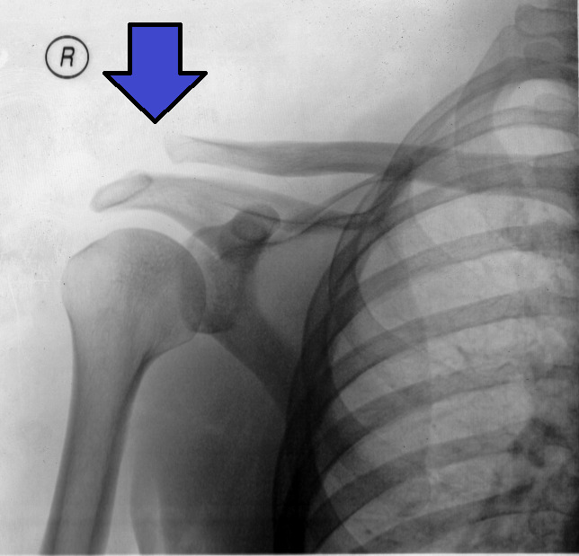
00:01 So now we're going to move on to the shoulder examination. 00:04 The shoulder is one of the most complex joints in the body, if not the most complex, because unlike the knee joint and finger joints, etcetera, it has three degrees of freedom, which means that rather than just having flexion and extension and maybe a little bit of torsion that you can have with your knee, this has flexion, extension, abduction, adduction, external rotation and internal rotation. 00:29 So it's an extremely flexible joint, which, of course, gives us a lot of room to do a lot of different things with our shoulder, but it also provides a lot of opportunities for injury. 00:38 In addition, it's the only place in the body where a muscle is passing between two bones, two bony structures, which, of course aren't very forgiving. 00:47 If there's any inflammation within that compartment. 00:51 In this case between the scapula, the acromion part of the scapula and the humeral head. 00:57 So with that in mind, Shayla has graciously allowed me to draw a tattoo of the shoulder anatomy on her arm, and so we'll take a look at some important structures. 01:06 Whenever I'm doing inspection, I first am just looking at both shoulders simultaneously. 01:11 I'm looking for any obvious asymmetry which may indicate atrophy of the muscle groups. 01:15 I'm also looking to see if there's any step off where the acromioclavicular joints are to see if there's a recent injury to the AC joints. 01:24 Atrophy may show up on the supraspinatus areas if there's loss of musculature there or you'll see some flattening in the infraspinatus areas. 01:33 I'll also always add, if somebody has any shoulder pains, particularly back shoulder pain, that's a common place for shingles, Zoster, you don't want to miss Zoster. 01:42 Make sure you have the patient take off their shirt and put a gown on so you can take a look at the skin in the area. 01:48 With that, we'll move on to palpation. 01:50 So I like to do a full circle to get all the structures without missing any. 01:55 So I'll typically start with the cervical spine. 01:58 Now, patients who have neck pain, oftentimes it's radiating axial axial pain can radiate from the neck to the posterior shoulder. 02:08 And anytime somebody complains a posterior shoulder pain, I'm usually thinking about neck disease, not shoulder disease. 02:14 So that's why I start over here on the cervical spine to look for tenderness there, the paraspinal musculature. 02:20 Then I'm moving over here to the supraspinatus. 02:23 Then I can feel the spine of her scapula, which I'll show here in my model. 02:27 Here's the spine. 02:27 So I'm going above it for supraspinatus and below it for the infraspinatus. 02:32 looking for any tenderness or again, loss of muscle bulk in that area. 02:37 Then I'm going to move along here to find the AC joint, which is that nice, palpable groove that ends the clavicle and where it attaches to the acromion. 02:48 It's typically around 4 centimeters proximal or medial from the end of the acromion, which I've shown here. 02:56 In my model here, the acromion is a nice squared off section of bone there, which is a part of your scapula, your shoulder blade, and you should be able to very clearly feel this sulcus between the end of the acromion and the top of the humeral head. 03:10 And so I'm pushing on right here to identify that location. 03:14 This is an important location for when ultimately you may be doing a acromio- forgive me, a subacromial injection of steroids or lidocaine for the purposes of diagnosis or treatment. 03:25 I can also actually palpate this is representing the head of the humerus, the supraspinatus, the subscapularisis, the infraspinatus and the teres minor. 03:35 I can actually palpate the supraspinatus tendon if I bring her elbow backwards. 03:41 I'm actually bringing this humeral head forward. 03:44 I can actually now palpate approximately where her supraspinatus muscle is. 03:49 And so if there's soreness in that area, I'd be more concerned about a supraspinatus injury. 03:54 I'll bring her her humeral head back. 03:56 You can sort of see what happens there when I bring her elbow back. 03:59 You see that bony prominence that pops forward. 04:01 That's the top of her humeral head and this is where her supraspinatus is located. 04:06 Now, I'll move that back to where it was. 04:08 I've already done the AC joint, so continuing to wrap around. 04:11 I'm moving along the clavicle here, ending at the sternoclavicular notch right here. 04:17 One last structure that's worthy of mention because it's in this area is the biceps tendon, the long head of the biceps tendon. 04:24 I love my model here because it really shows this quite well. 04:27 This is the bicipital groove on the humerus. 04:31 And this little string here represents the long head of the biceps tendon, which runs along that groove, goes deep through this groove and ultimately is going to insert on the glenoid labrum deep inside this joint. 04:46 And so while we think of this muscular tenderness areas inserting perhaps out here, it's actually really quite deep inside the groove there. 04:54 But if I rotate the humerus internally and externally, I'll actually be able to palpate that cable, that tendon underneath my fingers. 05:04 So I'm going to put my fingers right about here and just rotating her arm in and out, I can feel this tendinous structure passing right underneath my fingertips. 05:13 And since the biceps tendon can be ruptured or injured, it's good to be able to identify that on the physical exam. 05:21 So having done inspection and palpation, we'll go ahead and do range of motion. 05:25 There's a quick way and a longer way, we'll do the quick way first. 05:28 Shayla, if you wouldn't mind, just touching the back of your head and now touch the small of your back. 05:34 That essentially runs through the entirety of range of motion, and if a patient can do that painlessly, I'm almost done with the shoulder exam because whatever is causing them pain, it's probably not the shoulder if they can do those maneuvers painlessly. 05:46 Again, I mentioned that if somebody says they have posterior shoulder pain, it's probably not the shoulder. 05:50 It's probably a neck problem. 05:51 So putting that aside, I said there's a long way to do range of motion, so we should do that as well. 05:56 So we're going to demonstrate shoulder flexion, which is bringing your arms straight from your body, shoulder extension. 06:03 Great, then putting your arms like this will have you reach, I'm sorry, just lifting your shoulders up. 06:10 That's a ABduction, abduction and then bring your arms across your body. 06:15 That's ADduction, adduction and now putting your arms out again. 06:19 We'll have you rotate up for external rotation and then down for internal rotation. 06:26 So this is a good place to pause and just highlight that patients who have adhesive capsulitis, also known as frozen shoulder, they're going to have a lot of trouble with range of motion and may not be able to get more than 30 or 40 degrees off from the ribcage. 06:41 So that's an easy, quick way to see if adhesive capsulitis is at play, particularly if that was active range of motion where she was doing the work, but if I passively can't get very far with range of motion, then that would be a good a good indication for adhesive capsulitis. 06:55 Active range of motion ensures that her muscles are doing the work. 06:59 And so you're stressing the rotator cuff and all the muscles. 07:02 But passive range of motion, she shouldn't be using any muscles or ligaments at all or any muscles or tendons at all. 07:07 This is just me doing the work, so when I have significant limitations in that regard. 07:11 I'm thinking about adhesive capsulitis.
About the Lecture
The lecture Examination of the Shoulder by Stephen Holt, MD, MS is from the course Examination of the Upper Extremities.
Included Quiz Questions
Where can the examiner palpate the supraspinatus tendon during a physical exam?
- At the top of the humeral head
- At the acromioclavicular joint
- At the top of the scapula
- Near the T3 vertebra
- It is not possible to palpate the supraspinatus tendon on an exam.
Which range of motion is tested by having the patient raise their arm forward in front of them?
- Flexion
- Extension
- Abduction
- Internal rotation
- External rotation
What is the medical terminology for "frozen shoulder?"
- Adhesive capsulitis
- Biceps tendinitis
- Rotator cuff tendinitis
- Bursitis
- Osteoarthritis
Where does the long head of the biceps tendon originate in the shoulder?
- At the supraglenoid tubercle above the glenoid cavity of the scapula
- At the intertubercular sulcus of the humerus
- On the radial head
- At the apex of the coracoid process of the scapula
- On the acromion of the scapula
Author of lecture Examination of the Shoulder

Stephen Holt, MD, MS
Customer reviews
5,0 of 5 stars
| 5 Stars |
|
2 |
| 4 Stars |
|
0 |
| 3 Stars |
|
0 |
| 2 Stars |
|
0 |
| 1 Star |
|
0 |
I have watched a couple other reviews of the shoulder examination and this was the best by far!
1 customer review without text
1 user review without text
