Playlist
Show Playlist
Hide Playlist
Diagnosis: Other Pulmonary Function Tests – Lung Disease
-
Slides 06 Respiratory Medicine Basics Brown.pdf
-
Download Lecture Overview
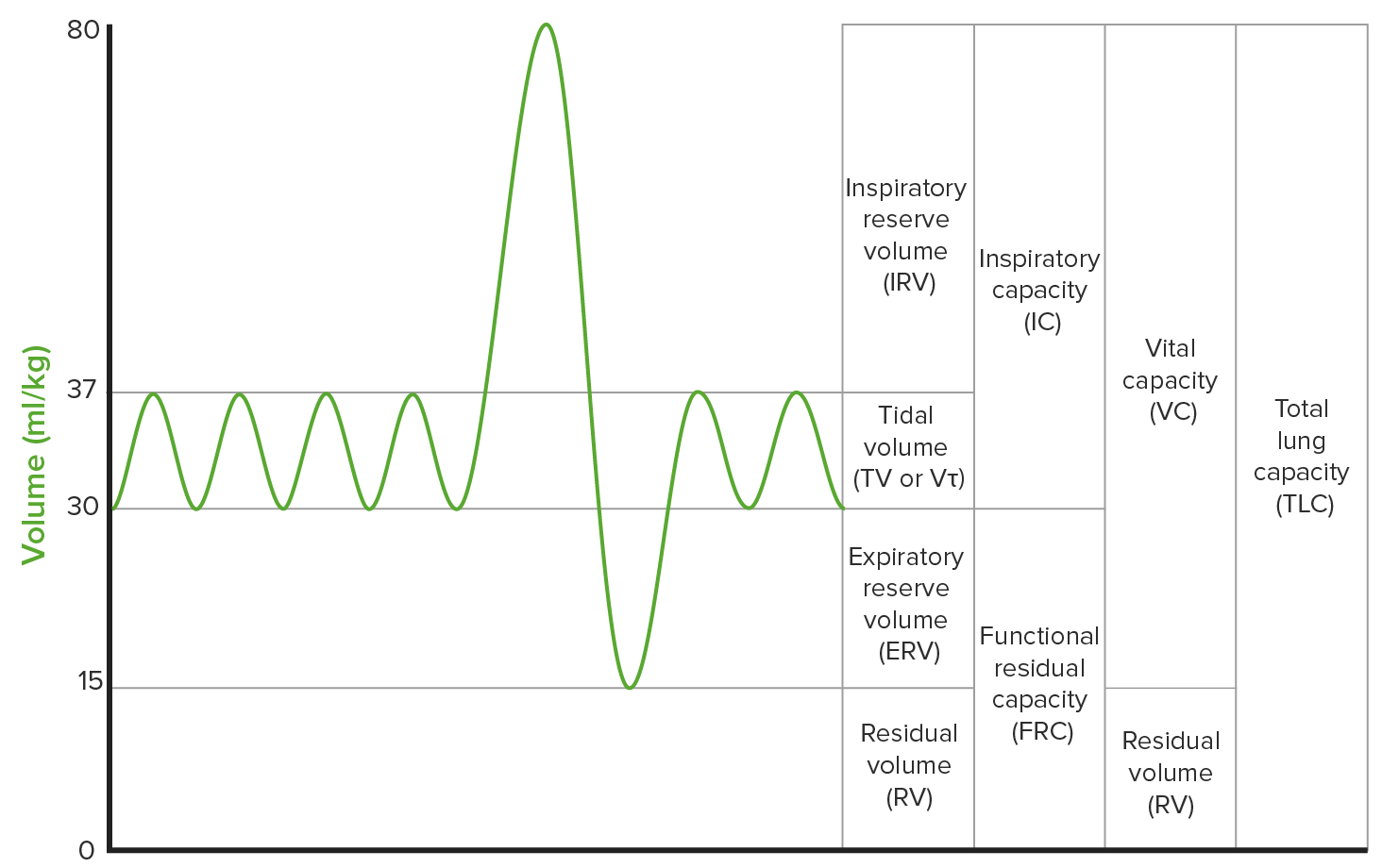
00:00 Right. Lung-function tests: the volumes. 00:03 So the capacity of the lung can also be measured. How big are the lungs? And this is really mainly important to identify people who have shrunken lungs – that happens in pulmonary fibrosis. So they have reduced total lung capacity. But it’s also useful in obstructive airways disease. Because obstructive airways disease – whether it’s COPD or ongoing severe asthma – causes an increase in the residual volume. And as the volume of the lung is dictated by the residual volume as well as you vital capacity, then the total lung capacity will go up in these patients. 00:39 So you get this paradoxical situation where patients with airways disease – such as COPD – will have low vital capacity but high lung volumes. And that’s largely because they’ve got residual volume increases. They have air trapping in their lungs and that makes their lungs bigger than they should be. 00:59 So what do we mean by air trapping? Well, essentially as you breathe out, you generate a positive pressure. And that squeezes the air out of the lungs. But it also squeezes the tubes taking the air out of the lungs – the bronchial tree. And if the bronchi are compressed by that positive pressure, then air flow’s going to be limited. And in patients who have airways disease, the problem here is that, on inspiration, they can breathe in relatively freely but, on expiration, the obstruction is made worse by this positive pressure. And you get a degree of air trapping with every breath. And that slowly but surely inflates the lungs and leaves them operating with a high residual volume. And this is particularly pronounced in emphysema because the alveolar destruction that occurs in emphysema allows the airways to collapse when faced with this positive pressure on expiration. That’s called dynamic airways collapse. 01:57 So we can use flow volume loops to give a little bit more information about how the patient’s air flow is changing during the inspiratory and expiration cycles. They’re quite complex. They give distinctive patterns with different diseases. And so they’re especially useful in emphysema and in patients with upper airways obstruction. 02:24 I’ll show you the pictures that we might get in those circumstances. So, on the left, we have somebody with emphysema. So in expiration, we do get this dynamic airways collapse. And how that is presented in this is that the flow on expiration is initially rapid and then, suddenly, there’s a fall. And that’s due to the dynamic airways collapse. And that’s followed by a long tail or slow, low air flow expiration. If you have an obstruction to the major airways – so a tracheal tumour for example – then you get this distinct squared-off appearance to the flow volume loop. And that can be very helpful in identifying these patients who are otherwise hard to identify. 03:05 So the lung-function tests we’ve described so far describe the volumes of the lung and how much air you’re able to shift during inspiration and expiration and the rate with which you shift that. 03:16 What they don’t measure is how easy it is for oxygen to get from the alveoli into the blood. And to measure that we use the transfer factor. 03:24 This is measured by inhaling a low concentration of carbon monoxide and then measuring how much carbon monoxide is exhaled. And the difference between the two is a factor that reflects how efficient oxygen transfer will be across the alveolar membrane into the pulmonary capillaries. 03:48 There are two factors we measure: one is the DLCO – which is the absolute factor. But the problem with that is that, if you remove somebody’s left lung and left them with just one lung, you’re going to halve the transfer factor because you’ve halved the surface area which they have for oxygen diffusion or carbon monoxide diffusion. So you need to adjust the transfer factor for the alveolar volume. And that’s called KCO. And that’s the transfer factor divided by alveolar volume. And that reflects how efficient oxygen transfer will be per units of lung. And using that parameter, you can identify patients who actually seem to have lung disease as opposed to just decreased lung volume. 04:28 There are a variety of factors that affect transfer factor but these fall into the following categories. One is the alveolar surface area. So if you reduce the alveolar surface area then you’re going to reduce the transfer factor. And that can happen after a lung resection, as we just discussed, but it also occurs in emphysema characteristically where the alveoli destruction decreases the surface area available for gas exchange. 04:53 If you increase the alveolar barrier thickness – and that occurs in interstitial lung disease – then you will reduce the ability of oxygen to cross that barrier. So that is also another cause of a low transfer factor. 05:08 But transfer factors critically depend on blood supply to the lung. And if you reduce the blood supply to the lung, then it will go down. And that occurs in pulmonary emboli, pulmonary hypertension and also right-to-left shunts. 05:21 It’s also critically dependent on the ability of that oxygen to pick up oxygen and that requires haemoglobin. So the transfer factor is low in patients who are anaemic. 05:31 Cardiac function also is relevant of him. So mitral valve disease, pulmonary oedema etc. will also affect the transfer factor. 05:42 So we can actually measure how much oxygen is present in the blood. And we do two measurements for that: one is the oxygen saturation which measures how saturated the haemoglobin is with oxygen. Low saturation = low oxygen availability. High saturation = good oxygen availability. 05:58 In general, people should have saturations of 95 to 99%. Because of the steepness of the oxygen saturation curve, as you become more hypoxic and the saturation falls to 90%, you get a lot of fluctuation in the values here. So, although in patients who have 95%+ the saturation remains relatively stable, when you get down below 90% they often fluctuate 3 or 4%. It’s a very useful test because it’s non-invasive and you can use it actually continuously to monitor how patients are doing. And it’s also useful not just in acute circumstances but also for the monitoring of patients who may be developing respiratory failure due to chronic lung disease. It’s inaccurate if the patient has poor peripheral perfusion – if they’ve got cold fingers – and there’s thick nail varnish on as it measures through the nailbed itself. 06:55 The other measure of oxygen are blood gases. And what that measures is how much oxygen is dissolved in the blood. But in addition to that, it gives you other parameters which are very important: it gives you the carbon dioxide level dissolved in the blood, it gives you the pH and it gives you bicarbonate and the base excess. So it’s very useful in identifying acid-base problems as well and problems with excretion of CO2 – hypoventilation. 07:23 So we use blood gases to see whether somebody has type-I or type-II respiratory failure. 07:28 That is either hypoxia with no increase in the carbon dioxide or somebody with hypercapnia. 07:33 And if they have type-II respiratory failure, we need to know whether they have acidosis with that or not. And it’s also used when acid-base disorders may be present.
About the Lecture
The lecture Diagnosis: Other Pulmonary Function Tests – Lung Disease by Jeremy Brown, PhD, MRCP(UK), MBBS is from the course Introduction to the Respiratory System.
Included Quiz Questions
Which of the following statements about pulmonary function tests is TRUE?
- Peak flow rate is measured in L / minute.
- Peak flow rate is measured in L / second.
- FEV1 is measured in L / minute.
- FVC is measured in L / second.
Which of the following sums of volumes equals total lung capacity (TLC)?
- Forced vital capacity (FVC) and residual volume (RV)
- FEV1 and residual volume (RV)
- Inspiration reserve volume and tidal volume
- Expiratory reserve volume and tidal volume
- Tidal volume and FEV1
Regarding pulmonary function tests, which of the following changes would be expected in patients with COPD?
- An increased residual volume (RV)
- A decreased inspiratory reserve volume
- An increased tidal volume
- A reduced total lung capacity (TLC)
- No changes would be observed.
Which of the following flow volume loop patterns would be expected in a patient with a tracheal obstruction?
- "Squared off" pattern
- Rounded pattern
- Triangular pattern
- "Mountain" pattern
- Flattened inspiratory loop pattern
Customer reviews
5,0 of 5 stars
| 5 Stars |
|
5 |
| 4 Stars |
|
0 |
| 3 Stars |
|
0 |
| 2 Stars |
|
0 |
| 1 Star |
|
0 |


