Playlist
Show Playlist
Hide Playlist
Diagnosis if a Baby is Blue and Hyperoxia Test


-
Slides Cyanotic Heart Disease.pdf
-
Reference List Pediatric Nursing.pdf
-
Download Lecture Overview
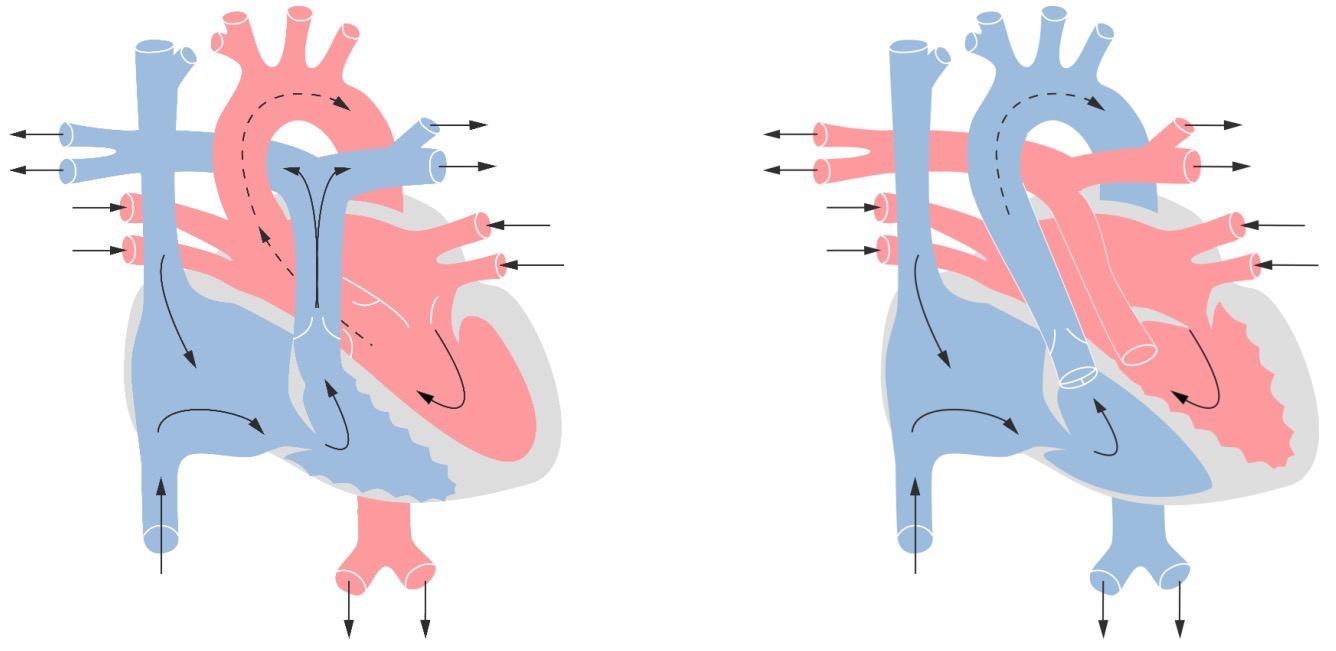
00:01 Hi, I'm Brian Alverson. 00:04 In this lecture, we're going to focus on cyanotic heart disease in children. 00:08 When you see a baby who's blue when they're just born, we often leap to the diagnosis of cyanotic heart disease maybe because it's in the name - cyanotic. 00:19 But there's a lot more that might be going on with that baby that you have to consider. 00:24 This lecture will focus on cyanotic heart disease, but I want you to remember the other things that might be causing a baby to be blue. 00:32 Examples would be a problem with their blood cells, such as methemoglobinemia. 00:37 It's possible that they have a problem in their lung. 00:40 They might have respiratory distress syndrome or VQ mismatch. 00:43 Something else going on with the blood is having difficulty getting oxygenated through the lungs, and the lung is actually the problem. 00:52 Alternatively, they may have cyanotic heart disease. 00:55 They may also have obstructive heart disease, and we think of these two in different categories. 01:01 Generally, cyanotic heart disease is blood that is being shunted from the right side to the left side and is bypassing the lungs. 01:10 There are certain types of congenital heart disease that are classified as cyanotic heart disease. 01:15 We're going to go through those one at a time. 01:18 But when you see that baby when they're blue, you have to do a very thorough examination to try and figure out what exactly is the problem. 01:26 It's important to take a very good history, and in particular, to note the age of onset of that child's cyanosis. 01:35 Were they blue when they came out of the uterus and has remained blue the whole time, or has something changed in the interim? It's important to know about the birth history. 01:46 Babies who are very large, for example, are in increased risk for persistent pulmonary hypertension of the newborn, which is not cyanotic heart disease. 01:55 And that would be a clue, as an example, of something that you would ask about to get a sense of what might be the underlying history, or historical problem that might be driving this baby to be blue. 02:07 Another example, what if there is relatively little amniotic fluid, does a child have a syndrome? Is there something else going on that's part of the situation that might be more than just cyanotic heart disease, but a whole syndromic problem? Prenatal history is important, too. 02:23 Did the mother have a history of a small amount of amniotic fluid? Were there infections in utero? Things like that. 02:31 It's important to do a physical exam as well and a thorough one. 02:34 Differences between the arms and the legs in terms of cyanosis can be a critical clue because the patent ductus arteriosus arises between where the blood supply comes off the aorta to the right arm and when it comes off to the rest of the body. 02:49 So if a patient is cyanotic in one place but not the other, it might give you clues as to the etiology of a problem. 02:56 Diseases that do this are transposition of the great arteries and persistent pulmonary hypertension of the newborn. 03:04 The lung exam is critical as well. 03:07 A patient with crackles, or a very wet lung exam, may have a pulmonary problem, or maybe a congestive heart failure. 03:16 Likewise, the cardiac exam is, of course, critical. 03:20 Typically, low-pitched murmurs are going through larger holes. 03:25 Those are often benign murmurs of infancy, and about half of children are born with a benign murmur. 03:33 However, very loud murmurs, very high-pitched murmurs, murmurs that are radiating from an unusual location, may give you a clue as to what the underlying problem is. 03:44 As you start listening to infants, it's very hard to listen to murmurs and it takes a lot of practice. 03:51 What I would suggest is, as you listen to your first baby with a murmur, take a careful listen, try and describe what you hear, and describe it to your attending, and then you can get some feedback on that, but it does take a lot of practice. 04:04 When you have a baby with a suspicious history or a suspicious physical exam where you're concerned about cyanotic heart disease, you do get some labs, and getting those labs is critical to figuring out what exactly is going on. 04:18 The first lab we have to get, and perhaps the most important screening lab, is the arterial blood gas or the ABG. 04:25 The arterial blood gas is obtained through doing phlebotomy of typically the wrist artery. 04:34 Upon getting that lab, you interpret the pH and the carbon dioxide to look for acidosis and alkalosis, and other metabolic problems, but key is getting the oxygen level. 04:46 In a patient where there's low levels of oxygen - so we're concerned about cyanotic heart disease - we will often do a hyperoxia test, and we'll talk about that in a few minutes. 04:56 Additionally, while you're drawing that blood, you'll want to get a CBC because remember that it could be this is something like hereditary spherocytosis, or a problem with the red blood cells. 05:08 The child might be anemic, which might be important to understand in the setting of a child with congenital heart disease, or that might be driving the problem. 05:17 Additionally, the testing to really truly diagnose the problem is the echocardiogram. 05:23 We'll often quickly get a chest x-ray or an EKG as a clue to what's going on. 05:29 The echo sometimes takes a little while before you can get it because you have to consult the cardiologist. 05:35 So all of these things you've done, you suspect cyantic heart disease, well, what do you do next? Let's get back to that hyperoxia test for a minute. 05:46 In the hyperoxia test, we're going to take the infant and we're going to place them in 100 percent oxygen for 10 minutes. 05:54 We're essentially going to try and hypersaturate the blood with oxygen. 05:59 The goal is, in a normal baby, to make sure that every single red blood cell's ability to bind to oxygen - all of those irons are all bound. 06:09 So we'll draw an ABG after they've been in the oxygen and we'll check the level of the oxygen in that blood. 06:17 If the level is over 200 mmHg, cardiac disease is extremely unlikely. 06:24 So you've really ruled out that that's likely the problem and you should be thinking more along the lines of pulmonary disease. 06:31 If the level is between 50 and 150, that's a typical range for a mixing lesion. 06:40 A mixing lesion is one where the right-sided blood and the left-sided blood is mixing before it goes out to the body. 06:46 Examples of those include truncus arteriosus and tricuspid atresia. 06:51 If the level is very low and less than 50, it's likely that infant has two circuits - and that would be classic for a transposition of the great vessels. 07:03 And we'll talk about all of these diseases next.
About the Lecture
The lecture Diagnosis if a Baby is Blue and Hyperoxia Test by Brian Alverson, MD is from the course Pediatric Cardiology.
Included Quiz Questions
Which of the following congenital heart diseases is most likely the cause of PaO₂ < 40 mm Hg after a hyperoxia test in an infant?
- Transposition of the great vessels
- Tetralogy of Fallot
- Tricuspid atresia
- Truncus arteriosus
- Totally anomalous pulmonary venous return
Which of the following causes blue baby syndrome?
- Methemoglobinemia
- Hereditary spherocytosis
- ABO incompatibility
- Ventricular septal defect
- Patent ductus arteriosus
Which of the following does not cause blue baby syndrome?
- ABO incompatibility
- Lung disease
- Cyanotic heart disease
- Methemoglobinemia
- VQ mismatch
Author of lecture Diagnosis if a Baby is Blue and Hyperoxia Test

Brian Alverson, MD
Customer reviews
5,0 of 5 stars
| 5 Stars |
|
5 |
| 4 Stars |
|
0 |
| 3 Stars |
|
0 |
| 2 Stars |
|
0 |
| 1 Star |
|
0 |
Excellent lecture as it opens up the differential diagnosis of cyanosis in a baby.
its very helpful and informative many students can prepare for exam throught it
tnx sir for this nice video..help me lotttttt.tnx again.
veryyyyyyy informative thanks lots!! it was very intersting i learnt so much
