Playlist
Show Playlist
Hide Playlist
General Terminology

-
Slides Dermatology Inflammatory Skin Diseases.pdf
-
Reference List Pathology.pdf
-
Download Lecture Overview
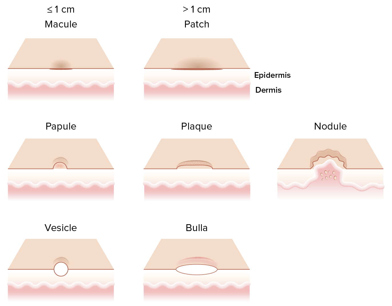
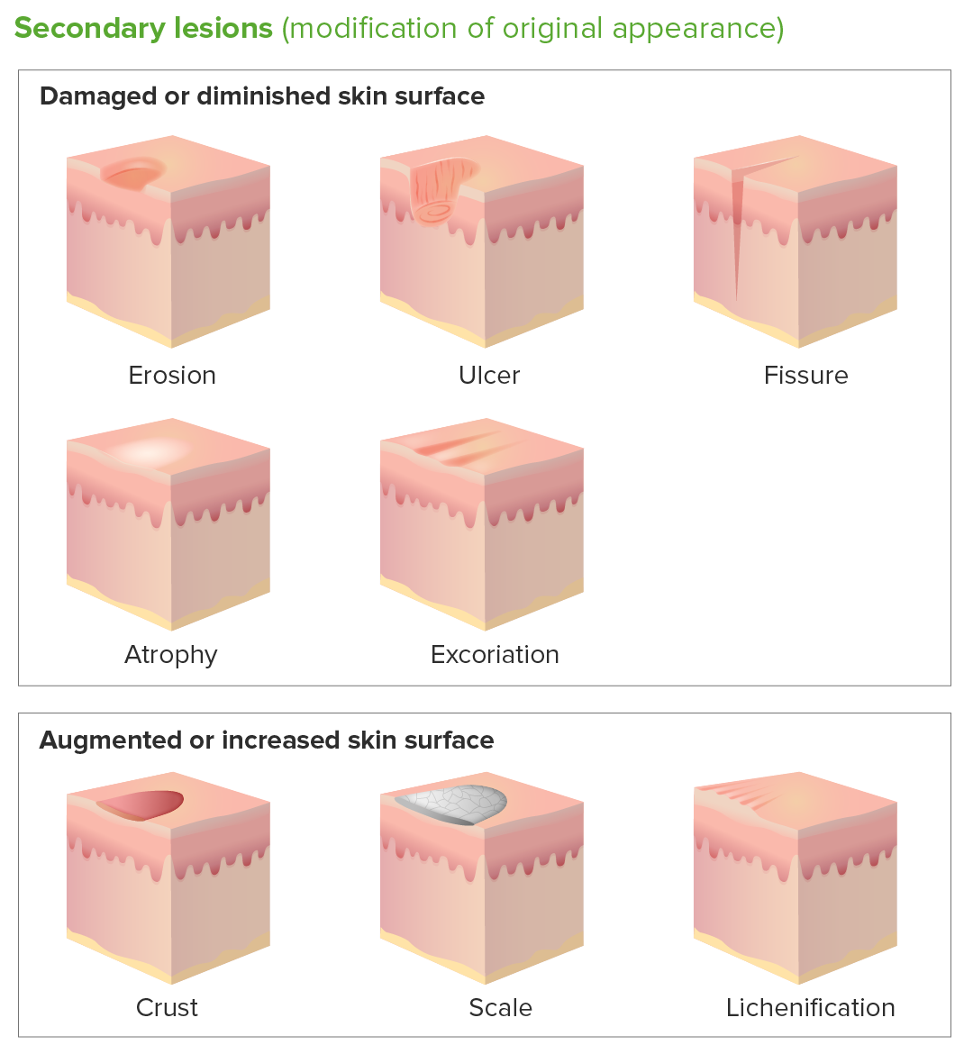
00:01 Welcome to dermatology. 00:02 Here, we’ll take a look at inflammatory disorders. 00:06 Our topic, first, we’ll begin with general terminology. 00:09 It will behoove you to spend a little bit of time to make sure that you have a proper understanding of the following descriptions. 00:16 You will see these descriptions either being described by your attending or in a clinical vignette, and this will then clue you in as to what kind of dermatologic condition you are referring to. 00:27 For example, if it’s a macule, you cannot feel this. 00:30 So therefore, you place your hand on the macule. 00:33 For example, café au lait spot. 00:35 It’s a macule, it’s mocha-colored. 00:37 By definition, less than one centimeter that is not palpable. 00:41 That is the most important topic. 00:44 Patch is a lesion that’s a little bit greater than 1 centimeter, and once again, that is not palpable. 00:50 Both of these are not palpable, but the size with the macule was a little bit smaller. 00:57 Once you get into papules, these are elevations. 01:00 And so therefore, it’s possible that you’re able to feel these, but the elevation here is less than one centimeter. 01:07 Or you might have a patient that has a condition we’ll talk about called psoriasis. 01:11 And psoriasis, this is a plaque, and this would be flat-topped elevation. 01:16 You’ve heard of salmon-colored plaque with psoriasis. 01:19 And here, the elevation would be greater than 1 centimeter. 01:23 A nodule, with nodule, I want you to think of something like a rheumatoid nodule or subcutaneous nodule that you might find with the criteria, Jones criteria, for rheumatic fever. 01:33 And so therefore, nodule will be round and with an elevation of less than 2 centimeters. 01:38 Whereas, if it’s a tumor, then remember, tumor, all it means is swelling. 01:43 And if it’s a particular tumor or swelling that has not developed due to, perhaps, injury or trauma, then you’re looking at an elevation of greater than 2 centimeters. 01:53 Then we get into what’s known as fluid-filled. 01:55 Now, what kind of fluid is then filling up your particular lesion? You’ve heard of herpes. 02:01 You’ve heard of chickenpox or varicella. 02:04 This is referring to your vesicle. 02:06 It’s a clear fluid blister, blister, blister. 02:09 You’ve heard of painful blisters that may occur with a condition, a porphyria pathway disease known as porphyria cutanea tarda, where that type of vesicle would then be painful in your patient upon exposure to UV light. 02:23 And here, the blister is less than 1 centimeter. 02:26 A pustule, I said pustule on purpose because here, you’re going to have fluid that’s filling up this blister, which is made up of pus, and we see pus less than 1 centimeter, and this is what you’re referring to. 02:39 Bulla. 02:40 A bulla, when we get into what’s known as your vesiculobullous or in other words, your bullous type of conditions, pemphigoid vulgaris or bulla pemphigoid. 02:51 A bulla is going to be fluid-filled as well but a little bit larger, greater than one centimeter in diameter. 02:57 Then we have erosions. 02:59 With an erosion, it’s a loss of part of the epidermis, part, erosion. 03:04 Whereas, if it’s an ulcer, that you’re going to then notice on the skin, then please understand the full thickness of loss of the epidermis. 03:13 The fine difference between erosion and ulcer becomes important to you in terms of description.
About the Lecture
The lecture General Terminology by Carlo Raj, MD is from the course Dermatopathology: Foundations.
Included Quiz Questions
What type of skin lesion is greater than 1 cm in size and not palpable?
- Patch
- Macule
- Pustule
- Plaque
- Papule
What type of skin lesions are most typical of psoriasis?
- Plaque
- Patch
- Papule
- Vesicle
- Erosion
What do you call a fluid-filled lesion with a diameter greater than 1 cm?
- Bulla
- Vesicle
- Pustule
- Nodule
- Tumor
Author of lecture General Terminology

Carlo Raj, MD
Customer reviews
3,8 of 5 stars
| 5 Stars |
|
1 |
| 4 Stars |
|
1 |
| 3 Stars |
|
2 |
| 2 Stars |
|
0 |
| 1 Star |
|
0 |
Good lecture, liked the general overview and structure. Happy that i found it.
good and simple explanation however it would be more useful if there are examples for each lesion
it's clear and simple, i like dr. raj so far.but there are other lesions that should be covered ( mainly secondary) and pictures should be added, it's a a dermatology course if we don't see we won't understand. i would suggest to add those things in the articles (which are amazing btw) if you want to keep the video simple
I like his way to explain. He did not mention if these are 1ry or 2ry lesion. There is no pic.
