Playlist
Show Playlist
Hide Playlist
Clinical Assessment of Stroke

-
Slides Stroke and intracranial hemorrhage.pdf
-
Download Lecture Overview
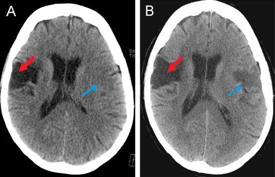
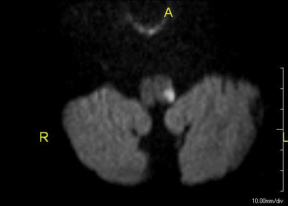
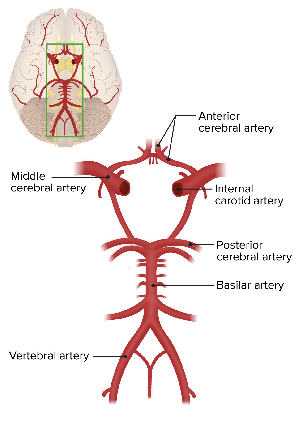
00:01 So, let's talk about how we diagnose stroke and how we think about the localization and categorization of ischemic stroke. 00:10 Importantly, when we're evaluating stroke patients, we have to know our vascular anatomy. 00:15 It's not enough to know just the anatomy of the brain, we need to know the arterial supply to each of those areas. 00:22 Strokes comes from a problem with the arteries and we're looking for problems that localize to those specific vascular territories or arterial beds. 00:31 The first way to think about the arterial system to the brain is the anterior and posterior circulation. 00:38 At any point in time, the brain receives about 20% of cardiac output, that's a ton of blood that's going up to the brain. 00:45 The majority of that, about 80% of that blood, is coming through to carotid arteries, they run on the anterior portion of the neck and go up right up to the middle of the brain. 00:56 They provide the vast majority of brain with blood supply and we'll look at them closer on the next few slides. 01:03 The posterior circulation is fed by two vertebral arteries. 01:07 The vertebral arteries come off both the aortic arch and the subclavian artery, and go up and meet near the base of the brain and form the basilar artery. 01:17 The basilar artery is one of the most important arteries in the brain. 01:21 It supplies the entire brain stem with blood. 01:24 Blockage of the basilar artery can cause catastrophic dysfunction of the brain stem and death, and so the posterior circulation, while it carries the minority of blood to the brain, goes to critical structures and that's something we need to be careful about and consider closely when evaluating these patients. 01:41 Here, in the schematic, you can see both the common carotid arteries travelling up in the neck, branching into an internal carotid artery which has no branches and it will branch into an external carotid artery which has a number of facial branches. 01:55 The external carotid provides arterial perfusion to the face. 01:59 We also see the two vertebrals coursing behind the carotid arteries and connecting into a single basilar artery up at the surface or base of the brain. 02:09 If we go into the brain and look at that arterial supply from top down in this axial section, we can see again the anterior and posterior circulation. 02:19 The two internal carotid arteries travel up into the skull through the skull and enter the brain compartment where the internal carotid artery divides into a middle cerebral artery which you see here, and that supplies the temporal lobe and parts of the frontal and parietal lobe with blood and an anterior cerebral artery which goes to the anterior portion of the frontal lobes and then courses back over the frontal lobe. 02:45 That, those two arteries comprised the anterior circulation in the cranial vault. 02:51 From the posterior standpoint side, we see the posterior cerebral artery. 02:55 So the basilar artery courses up on the brain stem and then divides into two posterior cerebral arteries, they course around the brain in the medial temporal lobe region and then provide blood supply to the two occipital lobes. 03:09 So, again, we can see the division between the anterior circulation, the MCA, ACA and the posterior circulation giving rise to the two PCA's as you can see here on this MR angiography image. 03:22 Importantly, the anterior and posterior circulations are connected and what connects them is the Circle of Willis, a series of communicating arteries that hooks posterior circulation to anterior circulation. 03:37 This allows collateralization in the brain, so that one vascular territory isn't required to give blood flow to all areas of the brain, and when we think about an ischemic stroke, blockage to one artery can be taken over by another, if there's sufficient collateralization. 03:56 And so we look for an intact Circle of Willis, a normal circle, that allows the posterior and anterior circulations to connect, and that can portend a more favorable prognosis in patients who are presenting with ischemic stroke. 04:09 So, let's walk through the Circle of Willis. 04:11 Here you can see the two internal carotid arteries. 04:14 They would be coming up through the plane of the slide in the patient's head. 04:19 They give rise to two middle cerebral arteries, as you can see here, and also, two anterior cerebral arteries that are coursing up to the frontal parts of the brain. 04:30 Importantly, there's a connection between the right and the left side of the brain. 04:35 In the anterior circulation that's called the anterior communicating artery, and it usually connects both ACA's. 04:42 Next, if we go to the posterior circulation, we see the two vertebrals coursing into the basilar artery and giving off two PCAs or posterior cerebral arteries to the occipital lobes. 04:55 There's a very important connection between the posterior circulation and the anterior circulation and that's is through two posterior communicating arteries that you can see here. 05:04 This is what's connecting those two circulations in the brain. 05:09 As we look at what gives rise to the posterior circulation, we see two vertebral arteries. 05:14 They actually provide blood flow and blood supply to the spinal cord through the anterior spinal artery as you see here, and give off a number of branches to the cerebellum and cerebellar fibers, that includes the posterior inferior cerebellar artery, which provides lateral circumferential arterial supply to the inferior cerebellum; the anterior inferior cerebellar artery which supplies blood supply to the middle and superior parts of the cerebellum, and the superior cerebellar artery which supplies lateral circumferential blood supply to the superior cerebellum. 05:49 Importantly, Pontine perforating arteries right off the basilar provide all of the midline structures of the brainstem with blood supply, and a large clot in the basilar that obstructs flow to all or many of these Pontine perforators will be catastrophic. 06:06 Lastly, we see a few other interesting and important arteries, the ophthalmic artery. 06:11 Blood clots in the carotid can get dislodged, go up into the internal carotid artery and the first branch is the ophthalmic artery. 06:20 Blood clots in that area cause Amaurosis Fugax or transient monocular vision loss which is why that artery is important to recognize. 06:28 And then we see the anterior choroidal artery which comes right off the MCA or middle cerebral artery and provides blood supply to some of the subcortical gray matter structures in the thalamus.
About the Lecture
The lecture Clinical Assessment of Stroke by Roy Strowd, MD is from the course Stroke and Intracranial Hemorrhage.
Included Quiz Questions
Where do the posterior cerebral arteries originate?
- The posterior cerebral arteries are branches of the basilar artery.
- The posterior cerebral arteries are branches of the vertebral arteries.
- The posterior cerebral arteries are branches of the middle cerebral arteries.
- The posterior cerebral arteries are branches of the cerebellar arteries.
- The posterior cerebral arteries are branches of the carotid arteries.
What artery supplies the temporal and parietal lobes of the brain?
- Middle cerebral artery
- Anterior cerebral artery
- Posterior cerebral artery
- Vertebral arteries
- Basilar artery
Where do the posterior inferior cerebellar arteries (PICA) originate?
- Vertebral arteries
- Basilar artery
- Circle of Willis
- Posterior cerebral arteries
- Anterior inferior cerebellar arteries
Author of lecture Clinical Assessment of Stroke

Roy Strowd, MD
Customer reviews
5,0 of 5 stars
| 5 Stars |
|
5 |
| 4 Stars |
|
0 |
| 3 Stars |
|
0 |
| 2 Stars |
|
0 |
| 1 Star |
|
0 |
