Playlist
Show Playlist
Hide Playlist
Carpal Tunnel – Anatomy of the Hand

-
Slides 07 UpperLimbAnatomy Pickering.pdf
-
Download Lecture Overview
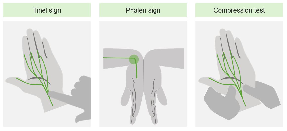
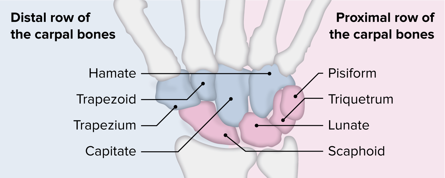
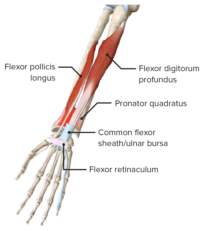
00:01 So now let’s turn to the carpal tunnel. Very similar to the arrangement we had on the dorsum of the hand, at the wrist joint where we had the extensor tendons passing between the extensor retinaculum and the carpal bones. But this time, we’re looking at the flexor retinaculum. 00:21 And deep to the flexor retinaculum are a series of tendons from those long extrinsic muscles in the forearm, that are passing through this carpal tunnel towards the hand. And we can see this here. We can see the flexor retinaculum in this section, and we can see we’ve got the tendons here surrounded by a flexor sheath. And we can just talk through these tendons. 00:45 So the long tendons of extrinsic muscles enter the hand through the carpal tunnel. This carpal tunnel is formed by the flexor retinaculum, and the concave palmar aspect of the carpal bones. So the way the carpal bones are arranged is that along their palmar surface, they are actually concave. And then with the flexor retinaculum passing over, they then create this tunnel, includes the flexor tendons of flexor digitorum profundus, flexor digitorum superficialis, and flexor pollicis longus. So the tendons of these forearm muscles, these extrinsic muscles pass through the carpal tunnel. And we can see this here. In this diagram with the carpal tunnel opened up, we can see the tendons of flexor digitorum profundus here, and these are passing towards the palm. 01:45 If we look here, we can see the cut tendons of flexor digitorum superficialis and this also would have entered through the carpal tunnel. We can see that here. We have the most superficial tendons of flexor digitorum superficialis. And then deeper between those tendons and the row of carpal bones here, we can see flexor digitorum profundus. 02:08 We can also see flexor carpi radialis tendon here, and we can see flexor pollicis longus tendon here. So we can see these tendons passing through the carpal tunnel. We can also see that we have the median nerve. We can see the median nerve is deep to the flexor retinaculum. 02:36 So if you were to have swelling within the sheaths, overuse of the tendons perhaps, irritation of the tendons could lead to inflammation and swelling, and that could push the tendon against the tight flexor retinaculum, and you could develop carpal tunnel syndrome. 02:54 We also have passing superficial to the flexor retinaculum. We have the ulnar artery and nerve. And this is within a region known as the ulnar canal. So also running towards the hand, we know passing towards the hand has to be the ulnar nerve because it supplies those hypothenar muscles. But importantly, the ulnar artery and its accompanying nerve do not pass into the hand via the carpal tunnel. We can see here that they lie above the carpal tunnel, superficial to the flexor retinaculum. So these do not in fact run within the carpal tunnel.
About the Lecture
The lecture Carpal Tunnel – Anatomy of the Hand by James Pickering, PhD is from the course Upper Limb Anatomy [Archive].
Included Quiz Questions
Which structures pass through the carpal tunnel? Select all that apply.
- Tendons of flexor digitorum profundus
- Ulnar artery
- Tendons of flexor digitorum superficialis
- Tendon of flexor pollicis longus
- Median nerve
Author of lecture Carpal Tunnel – Anatomy of the Hand

James Pickering, PhD
Customer reviews
1,0 of 5 stars
| 5 Stars |
|
0 |
| 4 Stars |
|
0 |
| 3 Stars |
|
0 |
| 2 Stars |
|
0 |
| 1 Star |
|
1 |
very minimal interactive video. I could have read all that.
