Playlist
Show Playlist
Hide Playlist
Arthritis & Hypertrophic Arthritis

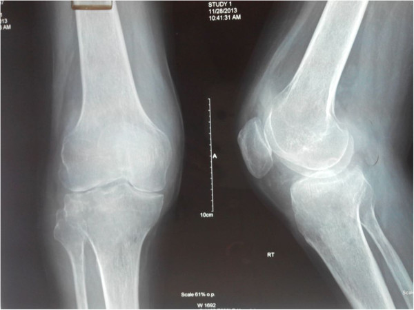

00:01 So in this lecture we'll be discussing the different types of arthritis and their common imaging findings. 00:08 Let's review the parts of a joint first. 00:10 So a joint is composed of an articular cortex which lies within the joint capsule and is lined by articular cartilage. 00:18 You also have subchondral bone which is the portion of the bone that lies beneath the articular cortex and you have the synovial membrane which is the lining of the joint capsule and within it is the synovial fluid. 00:30 So let's take a look at this on a diagram. 00:32 Here we have normal bone surrounding a joint space. 00:38 Along the bone, we have articular cartilage on each end and we have an articular capsule and surrounding it is the synovial membrane. 00:52 So on a radiograph, let's go over a little bit of the anatomy first. 00:56 So here we have the location of the articular cortex, adjacent to the joint space and this is an image of the knee. 01:04 Just under that, we have the subchondral bone and here the loosened area is the actual joint space. 01:13 So let's review the joint anatomy on a normal MRI. 01:16 This is a T1 weighted MRI of the knee and you can see here the articular cartilage which is a darker line adjacent to the joint space. 01:26 You can see the subchondral bone which appears white on a T1 weighted MRI. 01:32 We have the joint space in between the two bones here and we have the meniscus which appears gray. 01:39 And this is only seen on MRI. 01:43 So what is arthritis? It's actually a disease of the joint space that causes changes within the joint space and within the surrounding bones. 01:50 Usually, it results in joint space narrowing. 01:54 So there are 4 major categories of arthritis. 01:56 There's hypertrophic arthritis which results in bone formation within the affected joint. 02:01 There's erosive arthritis which results in multiple lytic lesions or "erosions" that surrounds the joint space. 02:07 There's an infectious arthritis which results in destruction of segments of the cortex that causes osteopenia and it can cause joint swelling and there's neuropathic arthritis and this usually results in bony destruction because of loss of sensation at the level of the joint. 02:24 So in hypertrophic arthritis the hallmark is osteophyte formation which is areas of bony growth. 02:31 There are 3 major types of hypertrophic arthritis. 02:33 The most common is degenerative osteoarthritis. 02:36 You can also have Calcium Pyrophosphate Deposition Disease also called CPPD. 02:41 And Nueropathic Arthritis. 02:44 So Degenerative Osteoarthritis is the most common type of arthritis and it's caused by loss of the articular cartilage because of excessive use of the joint. 02:53 So we see this very commonly in the older population. 02:58 Osteoarthritis is characterized by joint space narrowing, marginal osteophytes, subchondral cysts and subchondral sclerosis. 03:06 It's the combination of this 4 findings that helps you determine that the patient has osteoarthritis. 03:11 Marginal osteophytes are bony transformation of cartilaginous growth. 03:15 So you see areas of bony overgrowth. 03:17 You can also see cystic spaces within the subchondral bone which is a result of accumulation of synovial fluid within the subchondral bone and subchondral sclerosis is the reaction of the subchondral bone to impaction from loss of the overlying cartilage. 03:32 So the 2 bones start hitting each other when the cartilage between them disappears. 03:38 So in this case, this is an example of the frontal radiograph of the knee. 03:41 We see joint space narrowing and in the knee, we actually see it most commonly presenting in the medial tibial femoral compartment as we do here. 03:49 You can see that there is slightly more narrowing of the medial compartment than there is of the lateral compartment. 03:55 Here we can also see marginal osteophytes which are these areas of bony overgrowth at the margins of the joint space.
About the Lecture
The lecture Arthritis & Hypertrophic Arthritis by Hetal Verma, MD is from the course Musculoskeletal Radiology.
Included Quiz Questions
Which type of arthritis is a result of bony destruction due to loss of sensation at the joint?
- Neuropathic arthritis
- Infectious arthritis
- Erosive arthritis
- Spontaneous arthritis
- Hypertrophic arthritis
What type of hypertrophic arthritis is the most common?
- Degenerative
- Calcium pyrophosphate deposition disease
- Neuropathic arthritis
- Viral arthritis
- Erosive arthritis
Author of lecture Arthritis & Hypertrophic Arthritis

Hetal Verma, MD
Customer reviews
5,0 of 5 stars
| 5 Stars |
|
5 |
| 4 Stars |
|
0 |
| 3 Stars |
|
0 |
| 2 Stars |
|
0 |
| 1 Star |
|
0 |
