La craneosinostosis es la fusión prematura de una o más suturas craneales durante el 1er año de vida. La craneosinostosis se clasifica como simple o compleja, y puede estar causada por factores ambientales o por síndromes genéticos. Los LOS Neisseria pacientes suelen ser asintomáticos y la preocupación puede surgir de las observaciones de los LOS Neisseria cuidadores. El diagnóstico se realiza clínicamente y mediante imagenología de la cabeza. El tratamiento es quirúrgico y el pronóstico depende de la clasificación y de la presencia de síndromes genéticos.
Last updated: Dec 15, 2025
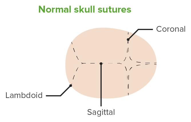
Suturas craneales normales
Imagen por Lecturio.No sindrómico:
Sindrómico:
Aparte de la forma anormal de la cabeza, la mayoría de los LOS Neisseria pacientes son asintomáticos. Debe realizarse un examen minucioso para evaluar los LOS Neisseria signos de aumento de la presión intracraneal, incluido el retraso en EN Erythema nodosum is an immune-mediated panniculitis (inflammation of the subcutaneous fat) caused by a type IV (delayed-type) hypersensitivity reaction. It commonly manifests in young women as tender, erythematous nodules on the shins. Erythema Nodosum el desarrollo.
| Tipo | Epidemiología | Deformidad | Presentación clínica |
|---|---|---|---|
| Coronal Coronal Computed Tomography (CT) |
|
Unilateral:
|
Unilateral:
|
| Lambdoidea |
|
Unilateral:
|
|
| Metópica |
|
|
|
| Múltiples |
|
|
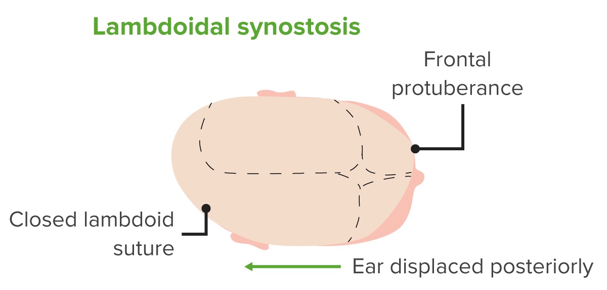
Esquema de una cabeza con craneosinostosis lambdoidea.
Nótese la fusión de la sutura lambdoidea, las protuberancias anteriores y posteriores, y el desplazamiento posterior de la oreja.
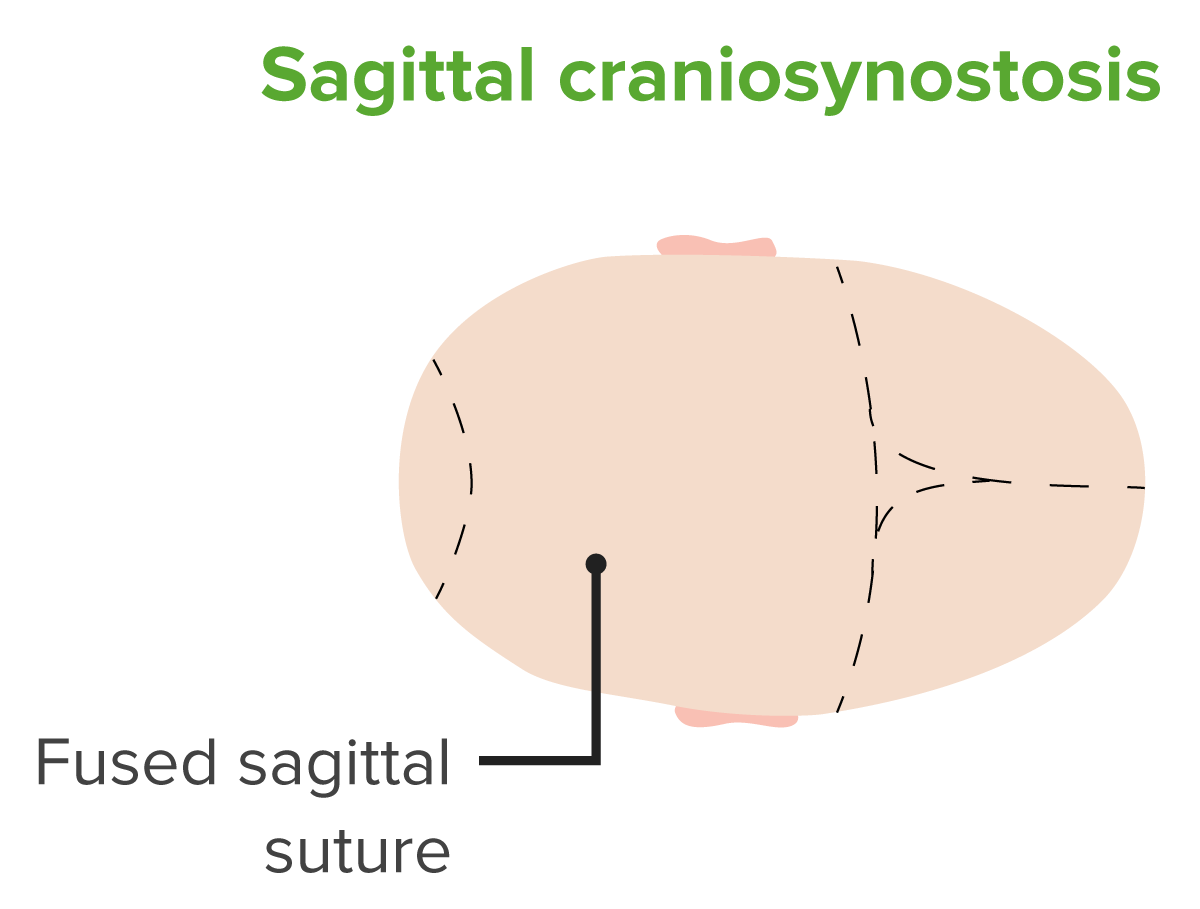
Esquema de una cabeza con craneosinostosis sagital.
Observe la fusión de la sutura sagital.
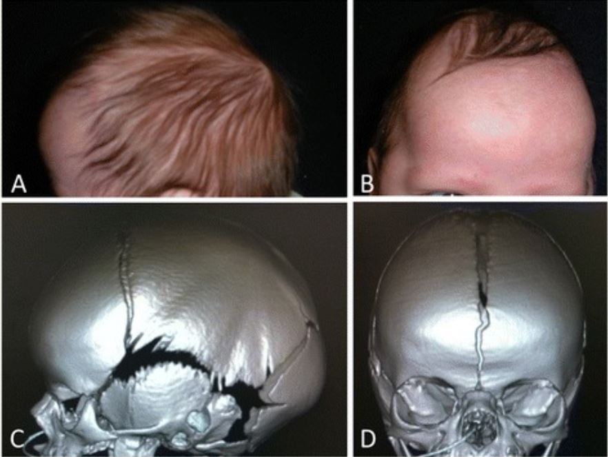
Escafocefalia por craneosinostosis sagital.
Fotografías fenotípicas (A, B) e imagenología de tomografía computarizada (TC) (C, D) que muestran la escafocefalia preoperatoria por craneosinostosis sagital.

Representación de la malformación braquicefálica debida a la fusión de sutura coronal bilateral de la cabeza de un bebé, vista de arriba abajo y lateral
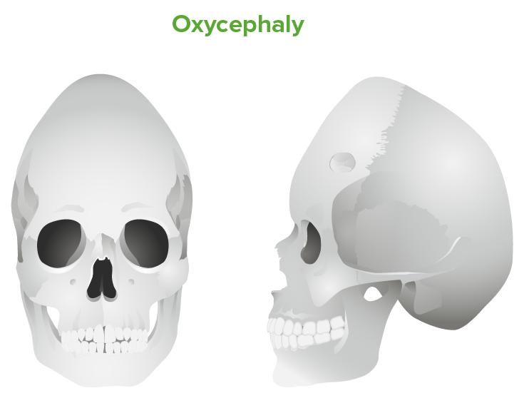
Oxicefalia: cráneo alto debido a una fusión de sutura coronal bilateral inadecuadamente tratada
El diagnóstico se basa en EN Erythema nodosum is an immune-mediated panniculitis (inflammation of the subcutaneous fat) caused by a type IV (delayed-type) hypersensitivity reaction. It commonly manifests in young women as tender, erythematous nodules on the shins. Erythema Nodosum la observación clínica, pero el diagnóstico por imagenología puede utilizarse para caracterizar mejor la anatomía para la clasificación o la cirugía
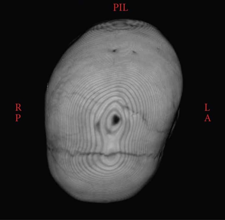
TC en 3D que muestra la sutura sagital sinostótica con un cráneo torcido hacia atrás
Imagen: “Atypical craniosynostosis with torticollis and neurological symptoms” por Koljonen V, Leikola J, Valanne L, Hukki J. Licencia: CC BY 3.0