Playlist
Show Playlist
Hide Playlist
Warts (Cutaneous and Genital) in Darker Skin

-
Slides Warts Cutaneous Genital in Darker Skin.pdf
-
Download Lecture Overview
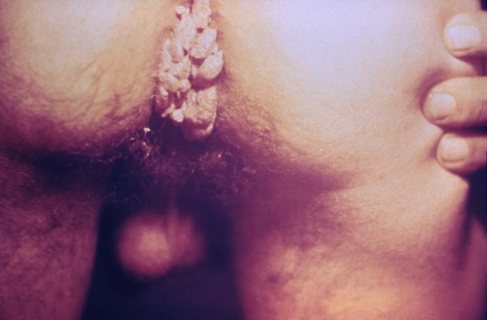
00:01 Welcome to our lecture on verruca or warts. 00:06 These are benign conditions where there is proliferation of skin and mucosa caused by the human papillomavirus, or HPV. It affects approximately 7% to 12% of the population worldwide. 00:24 It is twice more frequently seen in whites than in blacks or Asians. 00:29 The male to female ratio is the same. 00:33 It can occur at any age, but is unusual in infancy or early childhood. How do we classify HPV virus infection? There's more than 100 types of HPV that have been identified. 00:50 The cutaneous types range from serotype 1, 2, 3, 4, 7, 10, 27, 57, etc. 00:58 The genital types usually 6 and 11, but more than 90% of all genital warts are non-oncogenic . 01:08 The ones that are oncogenic are the serotype 16 and 18. Let's take a quick look at the pathophysiology of human papillomavirus. 01:21 HPV is usually transmitted via direct person-to-person contact, such as unprotected sexual intercourse, sharing contaminated needles or syringes, and sharing personal items, etc. in order for HPV to infect, there needs to be disruption of the skin or mucosal surface. 01:44 These are usually typically minor skin abrasions or microtrauma during sexual intercourse. 01:52 HPV invades the basal keratinocytes. 01:57 The viral genome gets transported into the cell nucleus. 02:02 Then proliferation of the infected keratinocytes also causes a replication of the viral genome. This means that cells derived from the infected basal keratinocyte also contain the viral DNA in their nucleus. Once non-infected cells leave the basal layer, they lose their ability to further proliferate and begin their terminal differentiation. However, on the contrary, the HPV infected cells keep proliferating because the virus maintains the cell replication cycle. 02:38 This uncontrolled proliferation of the keratinocytes results in the formation of warts. Let's take a look at the clinical manifestations. 02:49 There are five clinical presentations of what's the common warts or verruca vulgaris, plantar warts or verruca planter, flat or plain warts or verruca plana, filiform warts and genital warts, or what we call condyloma acuminata. Now let's take a look at each type of verruca and its differential diagnosis. 03:16 Going to start with Verruca vulgaris or Common warts, vulgaris means common. 03:22 These present as cauliflower-like papules with a rough papillomatous and hyperkeratotic surface. 03:31 It can also present as solitary lesions or multiple lesions, as you can see on the left and on the right hand side. 03:41 Some of the predilection sites include hands, fingers, and palms, the knees. 03:48 The lesion usually resolves spontaneously up to 6 to 9 months without treatment, but usually patients will request treatment or intervention, so the differential diagnosis includes, amongst others, molluscum contagiosum, which tends to be umbilicated. The second one is squamous cell carcinoma. 04:10 We should always be considered when wart like papules or plaques have irregular growth, ulceration, and/ or resistant to Therapy. Verruca plana, the second one, or plantar warts one, gets single, rough hyperkeratotic papule or thickened cobblestone plaque with a presence of small black dots which which are due to thrombosed capillaries. 04:39 And this you see if you try and shave the lesion. 04:43 Some of the predilection sites are the feet, the toes, mid metatarsal areas and the weight bearing areas of the heels. The differential diagnosis is the planter callus or corn. 05:00 You may get obscuring of the normal skin lines but it lacks the thrombosed capillaries. And this you can only see if you take a scalpel and you shave the upper part of the what or callus. 05:13 And in a verruca plana you will see the thrombosed vessels which you don't see in a cone. The plane warts of verruca plana, one gets multiple small, flat topped, skin colored papules, and they tend to involve the face, the hands, and sometimes the legs. 05:36 The differential diagnosis includes acquired epidermodysplasia verruciformis, and this is usually associated with HIV infection. The fourth type, the filiform ones, is a cluster of fine fronds emerging from a narrow pedicle base, and this is usually found on the face, and you can see the typical filiform lesion on that picture. 06:07 With anogenital warts, or what we call Condylomata acuminata, one sees lobulated papules 2 to 5mm in size and it tends to be multifocal. 06:23 One can see variable color from that of the skin. 06:27 You may also see erythema or hyperpigmentation depending on the skin type. The complications of HPV virus infection include periungual warts, which can cause nail dystrophy and destruction, and these tend to be challenging to treat and take longer. 06:51 Genital warts complications include giant condylomata acuminata. You may also get genital urinary malignancies in both males and females. 07:05 With vertical transmission, you may get recurrent respiratory papillomatosis. Right. 07:13 So how do we diagnose this condition? Clinical examination and history are usually enough to make a diagnosis. 07:20 We sometimes use dermoscopy which shows typical features which will not cover here. A skin biopsy is needed in some cases to differentiate from malignancy. 07:35 How do we treat this condition? It may not be necessary, as spontaneous resolution may occur, particularly in immunocompetent children. 07:46 However, most of the time parents will want you to treat their patients. 07:51 Here is a list of options that can be used to treat HPV virus infection. Depending on what's available to you, we can also use Intralesional intervention as listed over here. 08:08 But usually it's a dermatologist who uses this kind of treatment. 08:13 Cryotherapy, which is liquid nitrogen, repeated every 1 to 2 weeks. Electrocautery can also be used. 08:22 In darker skin prototypes, always beware of hypo or hyperpigmentation post-treatment, and always discuss this potential side effect with patients so that they can anticipate it and expect it post-treatment.
About the Lecture
The lecture Warts (Cutaneous and Genital) in Darker Skin by Ncoza Dlova is from the course Viral Skin Infections in Patients with Darker Skin.
Included Quiz Questions
What is the approximate worldwide prevalence of warts (verruca) in the general population?
- 7% to 12%
- 1% to 3%
- 15% to 25%
- 30% to 40%
- 50% to 60%
Which type of wart presents as a cluster of fine fronds emerging from a narrow pedicle base and is typically found on the face?
- Filiform warts
- Verruca vulgaris
- Verruca plantaris
- Verruca plana
- Condyloma acuminata
What is the mechanism by which HPV causes wart formation?
- HPV maintains the cell replication cycle in infected keratinocytes, causing uncontrolled proliferation
- HPV triggers an inflammatory response that causes localized swelling and hyperkeratosis
- HPV directly destroys basal keratinocytes, leading to compensatory hyperplasia
- HPV stimulates excess melanin production, resulting in pigmented hyperplastic lesions
- HPV blocks cell death pathways, causing accumulation of senescent keratinocytes
What key feature helps differentiate plantar warts (verruca plantaris) from plantar calluses or corns?
- Presence of small black dots representing thrombosed capillaries in plantar warts
- Yellow coloration that is characteristic of plantar warts but not calluses
- Sharp, stabbing pain with plantar warts versus dull, aching pain with calluses
- Presence of a central core in calluses that is absent in plantar warts
- Symmetrical distribution of plantar warts versus asymmetrical pattern of calluses
Author of lecture Warts (Cutaneous and Genital) in Darker Skin

Ncoza Dlova
Customer reviews
5,0 of 5 stars
| 5 Stars |
|
5 |
| 4 Stars |
|
0 |
| 3 Stars |
|
0 |
| 2 Stars |
|
0 |
| 1 Star |
|
0 |
