Playlist
Show Playlist
Hide Playlist
Uterus – Female Reproductive System

-
Slides 09 Human Organ Systems Meyer.pdf
-
Reference List Histology.pdf
-
Download Lecture Overview
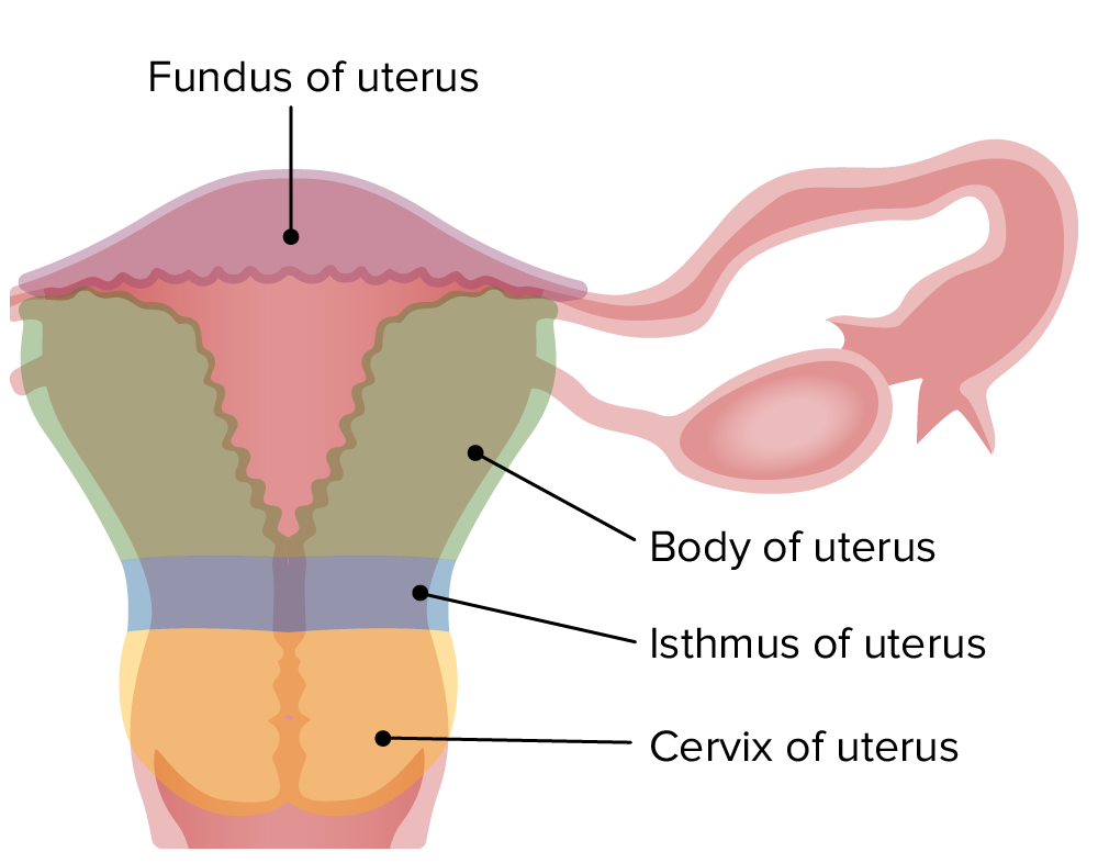
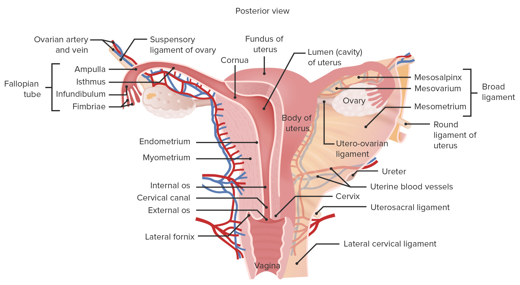
00:01 that we spoke about earlier. I want to move on now to discuss the uterus. And again on this slide, this diagram shows you where the uterus is located. It’s a relatively large organ associated with the female reproductive system. It has two components that I’ll now describe. 00:18 The uterus undergoes drastic changes throughout the menstrual cycle. Let’s just look at this slide, and on the top left-hand side, there is a low magnification picture of the uterus. And then there is a closer up picture on the left bottom of the slide illustrating an example of the proliferative endometrium. The uterus has two components. It has a myometrium and an endometrium. And you can see on the far right-hand side a larger section showing the secretory endometrium. And I’m going to talk about these two phases of the menstrual cycle, and pay particular attention to the changes that occur during the proliferative stage of the menstrual cycle, and the secretory phase of the menstrual cycle. I mentioned earlier that the uterus has two parts to it. It has the endometrium which you see here, and I’ve spoken about briefly, it also has a myometrium, a very thick muscular wall. 01:31 And that myometrium consists of smooth muscle cells predominantly. And those smooth muscle cells are around 50 microns in length, and they’re all joined together by gap junctions and they all contract at the end of pregnancy to expel the fetus. But during pregnancy, those smooth muscle cells can grow drastically in size, mostly by hypertrophy. They can grow from 50 microns to about 500 microns. So there is this enormous growth of the myometrium, the muscle layer of the uterus during pregnancy. The endometrium is the component of the uterus that changes with every menstrual cycle. I want to first describe the secretory phase, the secretory endometrium because that is a better basis for then looking at the other stages. On the left-hand side, you can see the secretory endometrium labelled. This is the internal lining of the uterus. The external lining is the thick muscle layer you can see on the top left-hand slide. So we’re going to describe the changes to do in the secretory endometrium. First of all, when you look at the section, it has got a very bluish stain to it. It’s a very active tissue. All the cells there are highly active doing various jobs that I’ll describe as we go through this lecture. You can see some little wiggly white lines running through the section of the endometrium. These represent very coiled glandular tissue. This secretory endometrium is prepared for possible implantation of a fertilized egg. So it has to have a number of features. It must have glands that are very active, and that’s represented by the coiled nature you see there. It must have many cells there that are providing secretion, particularly glycogen, to provide nutrition for the possibility of the implanting blastocyst. It also must be warm and moist, so there’s a very large blood supply to the endometrium even though you don’t really see it that clearly in the histological sections because a lot of the capillaries collapse down. 04:10 This is the ideal environment to have to be the womb of the developing child, a nice nutritious warm environment for implantation of the fertilized egg. On the right-hand side, it shows diagrammatically the sorts of histological components that I want to emphasize. First of all, down the bottom of the slide, is the myometrium. Besides being muscle, as I explained before, it also carries in it some large blood vessels, branches of the uterine artery carrying blood to the endometrium, and that blood circulates into the endometrium and returns back to the system via the uterine vein. So that’s the myometrium. Then look above and look at the endometrium. It’s divided into two components. One is the functional layer, and the other is the basal layer. And the basal layer is very important because that’s the layer that remains after menstruation. And that is the layer that has the ability to then proliferate and repair all the lost endometrial surface. The component of the endometrium that’s lost during menstruation is the functional layer that’s illustrated here. Now, just have a look at the endometrial glands, those long yellow shaded tubes that you could see extending from the surface of the endometrium down into the basal layer. Well, under the influence of progesterone, those glands become secretory. They become very coiled like you see in the histological section. And they accumulate glycogen. They become very very secretory to provide nutrition as I mentioned earlier for the possibility of an implanting blastocyst. And those glands have got to that stage of being secretory because under the influence of estrogens, during the follicular phase of the ovarian cycle, has repaired the uterus, the endometrial component of the uterus that’s shed during menstruation. 06:36 And I’ll talk about that in a moment. The other feature I would like you to understand at this stage looking at this particular diagram is the arteries you see. You have a very basal straight artery, and you also have a spiral endometrial artery, an artery that coils its way to the surface of the endometrium, and then gives rise to tiny little branches and supplies all the blood to all those cells and glands in the functional part of the endometrium that are so important. And as I said earlier, those capillaries then drain into a venous plexus and then drain out through the uterine vein. So they are the important components within the endometrium at the stage where ovulation has just occurred, and the endometrium, as I explained, is now ready for the possibility of an implanting blastocyst. It’s now grown from about one millimeter to six millimeters in thickness. So it grows very, very significantly under the influence of estrogens. And now, once that growth has occurred and that thickness of the endometrium has been achieved, then progesterone acts on the glands to become very secretory, and the blood supply to be increased to that region. So they are the main structural and functional changes occur during this secretory phase of the endometrium. 08:14 And it’s often called the progestational period because it’s under the influence now of progesterone. Have a look now at a description, and also some detailed evidence
About the Lecture
The lecture Uterus – Female Reproductive System by Geoffrey Meyer, PhD is from the course Reproductive Histology.
Included Quiz Questions
Which ONE of the following is CORRECT?
- The myometrium undergoes dramatic changes during pregnancy.
- The myometrium of the uterus changes with each menstrual cycle.
- The proliferative and secretory endometrium contains smooth muscle cells joined by gap junctions.
- Smooth muscle cells in the endometrium don't undergo changes during pregnancy
- Skeletal muscle makes up the bulk of the myometrium.
Which of the following is INCORRECT?
- Progesterone repairs the endometrium that is shed during menstruation.
- Progesterone promotes the coiling of endometrial glands.
- Progesterone promotes the production of a glycogen-rich secretion.
- Menstruation may be accompanied by a series of uterine contractions.
Which of the following statements regarding the myometrium is MOST ACCURATE?
- It is located between the endometrium and the serosa.
- It is mostly composed of striated muscle.
- It is contains the majority of the uterine glandular tissue.
- It sheds during the second phase of the menstrual cycle.
- It is largely absent in nonpregnant women.
Which of the following statements regarding the blood supply to the layers of the uterus is MOST ACCURATE?
- The myometrium provides the blood supply to the endometrium.
- The endometrium provides the blood supply to the myometrium.
- The circulating blood to the endometrium does not return to the myometrium.
- The endometrium arterial supply is independent from that of the myometrium.
Author of lecture Uterus – Female Reproductive System

Geoffrey Meyer, PhD
Customer reviews
5,0 of 5 stars
| 5 Stars |
|
5 |
| 4 Stars |
|
0 |
| 3 Stars |
|
0 |
| 2 Stars |
|
0 |
| 1 Star |
|
0 |
