Playlist
Show Playlist
Hide Playlist
Tooth

-
Slides Digestive system oral cavity.pdf
-
Reference List Histology.pdf
-
Download Lecture Overview
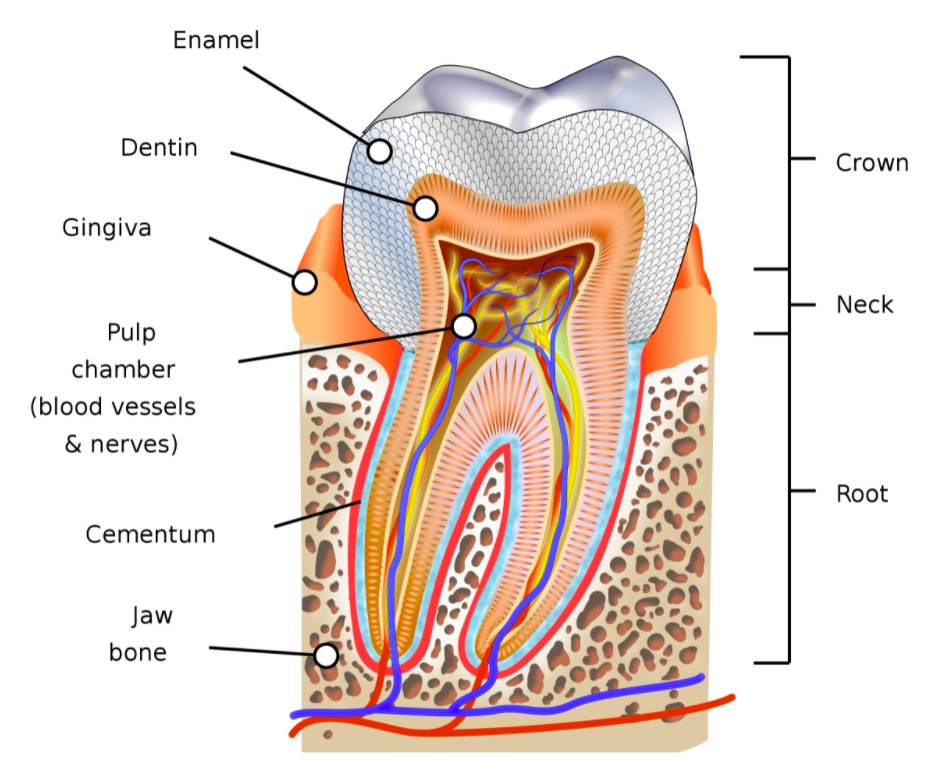
00:01 Let's now look at the tooth. On the left-hand side is a diagram of a tooth sitting in its socket in alveolar bone. And various parts of the tooth are labelled. 00:15 The enamel is on the surface of the tooth. The clinical crown is that part of the enamel you can actually see projecting from the gingival surface or the gingiva, that piece of the oral mucosa that covers the bone housing the tooth. 00:35 The anatomical crown is all of the enamel. Part of which is actually embedded just below the gingiva. You can see the enamel on the surface, then the dentin, and then in the middle, the pulp cavity. And the pulp cavity is continuous with the root canal of the tooth where blood vessels, nerves, etc, can enter into the pulp cavity and provide nutrition, and also carry sensations away from the tooth. And the tooth sits in the alveolar bone and it's held in its place, in its socket, by the periodontal ligament that I'll refer to in a moment. Sometimes you see lines through the enamel, and also through the dentin. These different striae or lines represent incremental growth of both the enamel and also the dentin. And I'm not going to mention them more in this lecture. You'll see them when you look across at the ground section of the tooth in the middle, and also when you look at histological sections of decalcified tooth. As I said, they represent just incremental growth of the tooth. 01:52 Sometimes, they're actually evidence, historical evidence of things that may have occurred in our lives during the tooth development, such as changing our diet or getting various environmental products circulating our body such as lead. 02:10 All these sorts of historical events can often be embedded in the tooth in the enamel, particularly, and also in the dentin in this striae. 02:22 They represent changes in the direction or frequency of these lines. And for that reason, this evidence can be often used for forensics. In the middle is the ground section of tooth where the tooth is being cut and then ground down to be very, very thin, and then a lot is shown through it to create the different patterns and different series of areas of the tooth that we can identify. On the right-hand side is a section of the tooth that has been decalcified. You see the large red component. That's the dentin. In the middle is the pulp cavity. The enamel is absent, as I explained before because it's mineralized and the decalcification process takes away the enamel. In this section, I want to just briefly explain
About the Lecture
The lecture Tooth by Geoffrey Meyer, PhD is from the course Gastrointestinal Histology.
Included Quiz Questions
Which of the following options correctly describes the structures from the inner to the outer tooth?
- Pulp - dentin - enamel
- Enamel - pulp - dentin
- Pulp - enamel - dentin
- Enamel - dentin - pulp
- Dentin - pulp - enamel
Which of the following is NOT found within the root canal?
- Cementum
- Arteries
- Veins
- Nerves
- Connective tissue
Which of the following tooth structures is primarily affected by decalcification?
- Enamel
- Pulp cavity
- Root
- Dentin
Which ONE of the following is INCORRECT?
- The clinical crown is the non-visible portion of the tooth that is covered by gingiva.
- The pulp cavity is continuous with the root canal.
- The tooth is supported in its bony socket in the alveolar bone by the periodontal ligament.
- The striae of Retzius are incremental growth lines or bands seen in tooth enamel.
- Periodontal ligaments connect the alveolar bone to the cementum.
Author of lecture Tooth

Geoffrey Meyer, PhD
Customer reviews
5,0 of 5 stars
| 5 Stars |
|
5 |
| 4 Stars |
|
0 |
| 3 Stars |
|
0 |
| 2 Stars |
|
0 |
| 1 Star |
|
0 |
