Playlist
Show Playlist
Hide Playlist
T-Cell Development – Lymphocyte Development

-
Slides Adaptive Immune System.pdf
-
Reference List Immune System.pdf
-
Download Lecture Overview
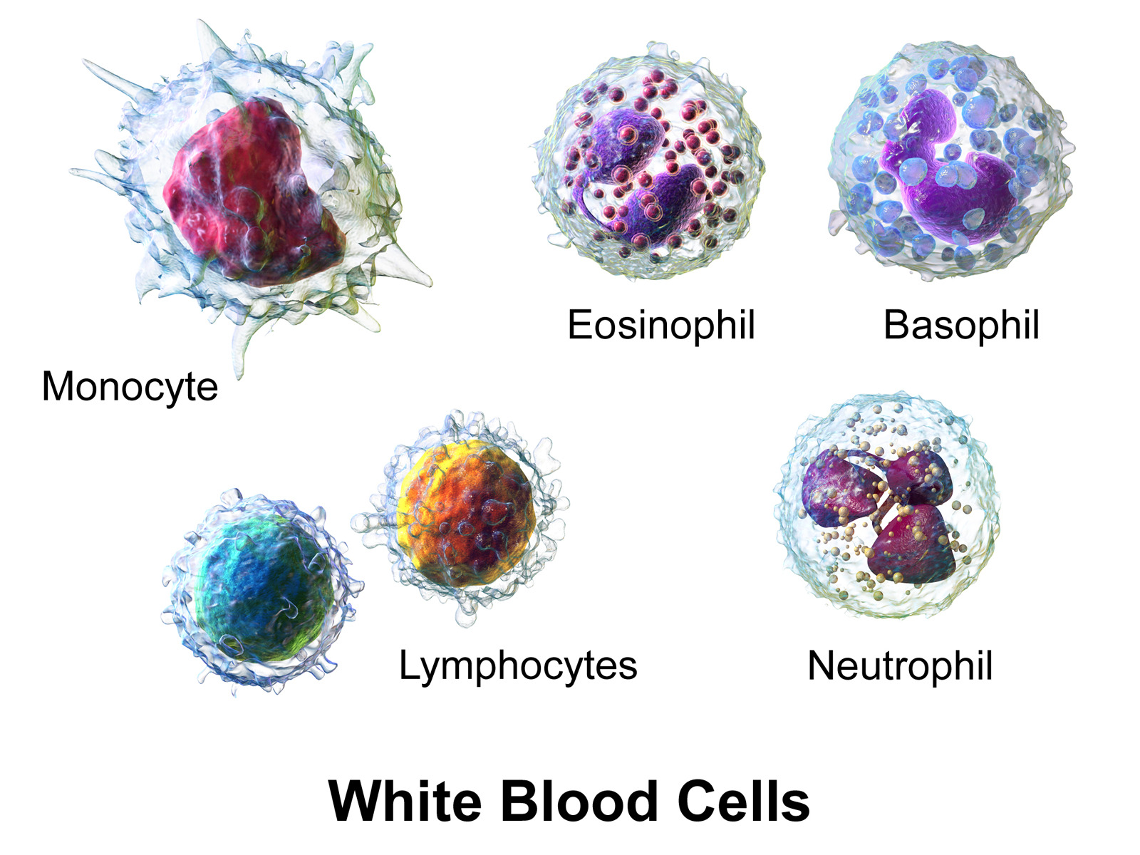
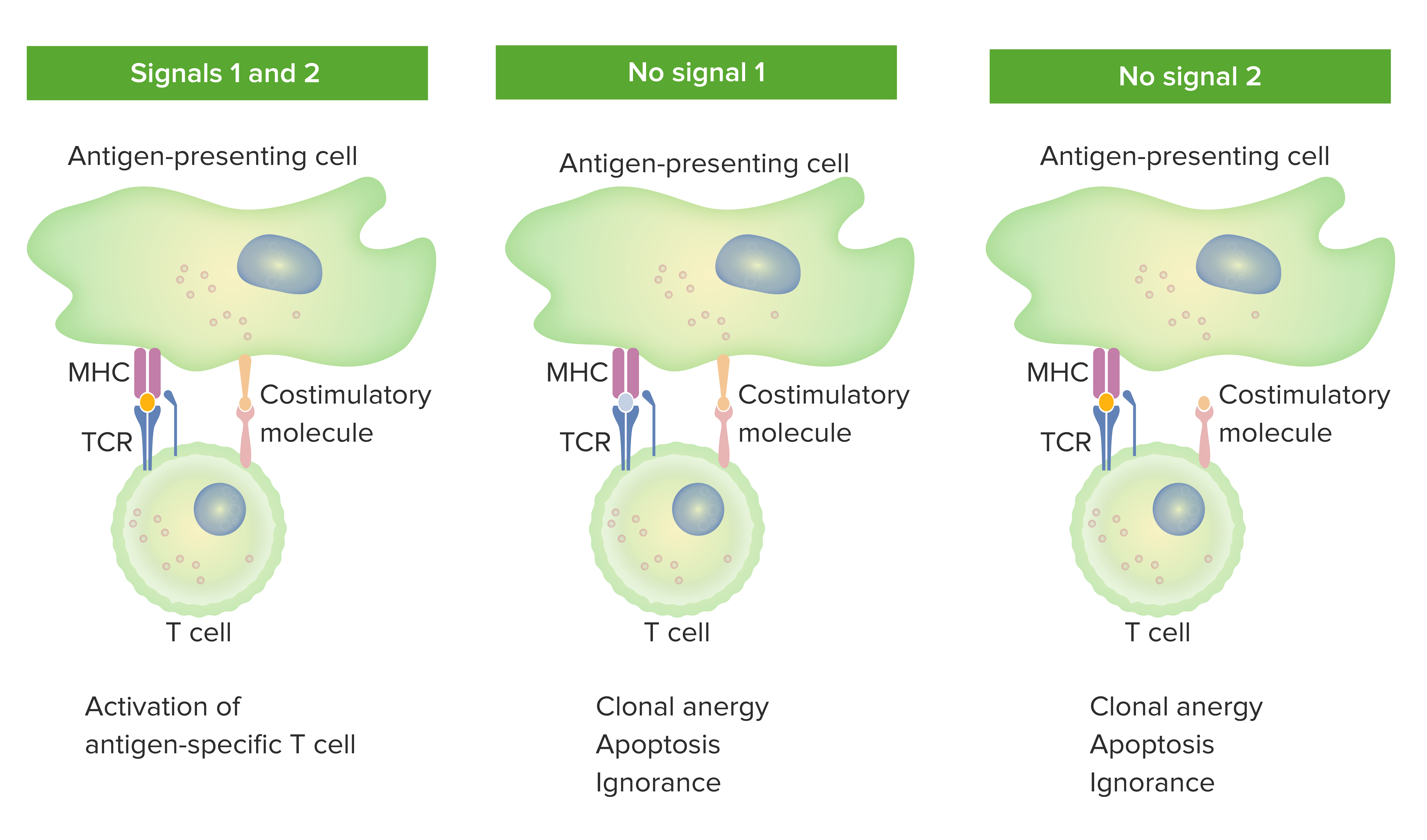
00:01 Unlike B-cells which directly develop in the bone marrow; remember B, bone marrow. 00:07 T-cells develop in the thymus. 00:11 They arise from hematopoietic stem cells in the bone marrow, like just virtually every other cell of the immune system. But they need to travel from the bone marrow to the thymus in order to become mature. And that’s where they recombine their T-cell receptor genes and so forth. 00:29 So T-cell precursors enter the thymus, and then they develop into the different types of T-cell that exists. 00:37 And these are predominantly of three different types: helper T-cells, cytotoxic T-cells which are often also referred to as cytotoxic T-lymphocytes or CTLs. 00:49 They’re exactly the same thing, cytotoxic T-cell or cytotoxic T-lymphocyte. 00:55 And naturally occurring regulatory T-cells or Tregs. 01:03 Following their development in the thymus, these various types of T-cells move to the secondary lymphoid tissues - the lymph nodes, the spleen, the mucosa-associated lymphoid tissue. 01:16 So the thymus is where T-cells develop. 01:23 Let’s have a look at exactly what is going on in the thymus as regards T-cell development. 01:30 We can see in this section of thymus that there are two clearly different areas that are stained differently. 01:38 The darker area is the cortex and this is densely packed with developing T-cells. 01:46 Whereas the lighter staining area is the medulla, and this contains more mature T-cells. 01:53 Upon arriving in the thymus, the T-cells undergo a process that’s referred to as thymic education. 02:00 You can think of the thymus as being like a school for T-cells. 02:04 It’s where they learn to recognize our own variants of the MHC molecules and where any reactivity against self antigens is eliminated. 02:15 They pass through the thymus, going through the cortex and the medulla. 02:21 Initially in the cortex they interact with thymic epithelial cells, and then as they pass through the medulla they particularly interact with dendritic cells and macrophages. 02:34 And as we’ll see very shortly, these processes of thymic education depend upon these interactions with other cells in the thymus. 02:47 So here we have a T-cell precursor, a cell that is going to become a T-cell, and some of these T-cell precursors will recombine the T-cell receptor gamma (γ) and delta (δ) chains. 03:01 And they will express on their cell surface, a γδ T-cell receptor. 03:07 We refer to these T-cells as γδ T-cells. 03:11 They recognize antigen directly or sometimes antigens, for example glycolipid antigens presented by an MHC-like molecule; it’s not an MHC molecule but it’s very similar to an MHC molecule of the CD1 family. 03:28 Particularly CD1d is utilized for this purpose. 03:33 These T-cells, γδ T-cells are very much a minority of the T-cell population. 03:39 The majority of T-cells that are developed in the thymus become alpha (α) beta (β) T-cells. 03:45 These αβ T-cells initially put on their cell surface a preliminary form of the T-cell receptor, a pre-T cell receptor. 03:56 This consists of a conventional T-cell receptor β chain together with a surrogate α chain, pre-Tα. 04:06 It’s a little bit like the pseudo light chain or surrogate light chain that the pre-B cell receptor has on the surface of B-cells. 04:14 Following recombination of the T-cell receptor α chain genes, then the pre-Tα is replaced by the fully mature normal version of the T-cell receptor α chain. 04:26 And we have the T-cell receptor comprised of an α chain and a β chain and refer to these T-cells as αβ T-cells. 04:36 And they are, if you like, the conventional T-cells and they recognize peptide shown to the T-cell receptor by MHC molecules. 04:45 Just like the B-cell receptor on the surface of B-lymphocytes is associated with other molecules, Ig-α and Ig-β, the same is true of the T-cell receptor on the surface of a T-cell. 05:00 So here we can see the αβ T-cell receptor on the surface of the T-lymphocyte held there by a transmembrane segment. 05:09 The T-cell receptor α chain and β chain have very short cytoplasmic tails. 05:16 But associated with the T-cell receptor are molecules that are referred to as CD3 molecules. 05:24 And these come in three different versions. 05:28 We have CD3 epsilon (ε), CD3γ and CD3δ. 05:34 And here we can see a pair of CD3 molecules, CD3ε and CD3γ. 05:41 You will note that these molecules have longer cytoplasmic tails than the T-cell receptor itself. 05:49 And most commonly, the association is with four different CD3 molecules as you can see here. 05:57 A pair that is ε and γ, and another pair that is ε and δ. 06:05 Associated with these CD3 molecules, are also two identical molecules called zeta chains. 06:13 So this is a homodimer; a dimer - two molecules, homo - the same. 06:17 A homodimer of two zeta chains. 06:20 And the zeta chains have even longer cytoplasmic tails. 06:25 Both the CD3 molecules and the zeta chains importantly contain these ITAMs (Immunoreceptor Tyrosine-Based Activation Motifs) that are so necessary in initiating the signaling once the T-cell recognizes its antigen.
About the Lecture
The lecture T-Cell Development – Lymphocyte Development by Peter Delves, PhD is from the course Adaptive Immune System.
Included Quiz Questions
Which of the following cluster of differentiation (CD) molecules is responsible for presenting lipid antigens to gamma delta (γδ) T cells?
- CD1
- CD2
- CD3
- CD4
- CD5
Which of the following cells is produced outside the thymus?
- Multipotent progenitor cell
- Helper T cell
- Cytotoxic T cell
- Regulatory T cell
Which of the following options are the main components of the CD3 molecule associated with the alpha/beta T-cell receptor complex?
- CD3 epsilon/gamma and CD3 epsilon/delta
- CD3 alpha/beta and CD3 epsilon/gamma
- CD3 alpha/beta and CD3 epsilon/delta
- CD3 epsilon/gamma and CD3 gamma/delta
- CD3 gamma/alpha and CD3 delta/beta
Which of the following is NOT true regarding T-cell development in the thymus?
- They interact with multipotent progenitor cells in the medulla.
- They interact with thymic epithelial cells in the cortex.
- They recognize self-major histocompatibility complex molecules.
- They recognize self-antigens.
- They interact with macrophages within the medulla.
Author of lecture T-Cell Development – Lymphocyte Development

Peter Delves, PhD
Customer reviews
5,0 of 5 stars
| 5 Stars |
|
1 |
| 4 Stars |
|
0 |
| 3 Stars |
|
0 |
| 2 Stars |
|
0 |
| 1 Star |
|
0 |
Clear and concise information presented in a systemic manner. Good balance between clinical and scientific knowledge.
