Playlist
Show Playlist
Hide Playlist
Sympathetic Nervous System (SNS): Output and Thoracolumbar Outflow

-
Slides 1 NervousSystem BrainAndNervousSystem.pdf
-
Reference List Anatomy.pdf
-
Download Lecture Overview
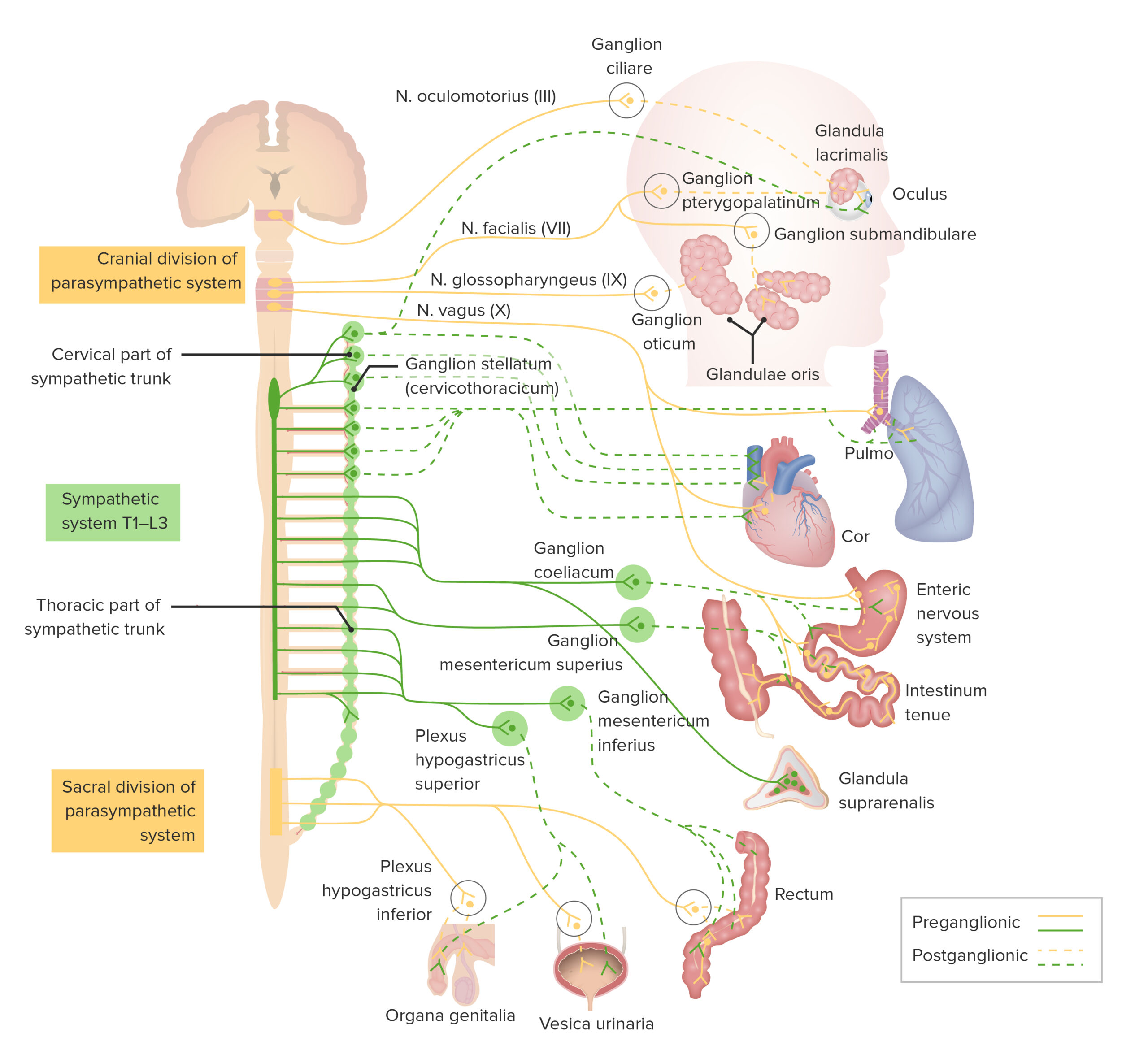
00:01 The sympathetic system is referred to alternatively as the thoracolumbar system because the preganglionic nerve cell bodies reside in these segments of the spinal cord specifically T1 down to L2 or even as well as L3 and the near output will be to the viscera through preganglionic fibers and communicating with ganglia. The segments key 1 through key 5, these nerve cell bodies reside in the intermediate gray horns. They will send axons out to the viscera in the head, neck, and thorax region and so this will be the level of the spinal cord innervating those particular structures. T5 down through L2/3 will supply viscera in the abdominal area and the pelvis via splanchnic nerves. Those can be seen in this illustration, so we'll zoom in. The greater splanchnic nerve is shown in through here and then its preganglionic fibers will synapse with ganglia that are associated with the branches of the aorta so here is a celiac trunk and you can see some ganglia associated with it. Another splanchnic nerve is the lesser splanchnic nerve. There is also the least splanchnic nerve coming in and then more inferiorly we have the lumbar splanchnics which are shown in through here. And then we have pelvic sympathetic splanchnic nerves shown here in green. This should not be confused with the pelvic splanchnic nerves associated with the parasympathetic division which are coming off S2, 3, and 4 and those are shown in purple. Now the mystery of the autonomic nervous system resides in the sympathetic outflow. How do you distribute the sympathetics outwards to the periphery or to maintain our distribution internally to viscera? We're going to have to break this down by the pattern of distribution. And so when we think about sympathetic outflow, we need to think about how that outflow is distributed to the body wall as well as to the limbs for example. And so this pattern that you see here has over here to the far right, the spinal cord will reside over here, we have the anterior nerve root of the spinal cord and it is distributing motor fibers out toward the periphery and the fibers you see here in red are going to be sympathetics that we're going to kind of highlight and understand their route. 03:17 We have this structure here which is a white ramus communicans. We have a sympathetic ganglion and then sympathetic trunk and this sympathetic chain of ganglia is distributed along the vertebral column. We also have this communication called the gray ramus communicans and then this more lateral component here is the spinal nerve that's going out toward the musculature and the skin of the body wall and the extremities. So our first consideration for this sympathetic outflow to body wall and limbs is that preganglionic neurons will reside in the lateral gray horns, again T1-T12 and L1 down to L2 and/or 3. From here, axons then travel to the sympathetic trunk via the white ramus communicans because these are myelinated, imparts that white name to the white ramus communicans. So here we see some of these preganglionic fibers coming through the anterior root. They then enter the white ramus communicans and then we see them either synapsing with a postganglionic neuron at that level with the sympathetic ganglion. However, the preganglionic neuron may go up or may go down and synapse at a more inferior level. So for practical purposes, these preganglionic neuron is going to synapse at the same level with a ganglion that it entered. From here, the postganglionic neuron then will leave the sympathetic ganglion via the gray ramus communicans. So now we're going to go from here where the synapse occurred outwards as the postganglionic nerve fiber outwards to the gray ramus and then it will enter the spinal nerve and then can travel through the dorsal ramus to the spinal nerve or the anterior ramus to the spinal nerve to be distributed to the target structures of the limb or the body wall. 05:40 Sympathetic outflow to the cervical and thoracic viscera should be considered separately from the distribution of the body wall and limbs. Here again, we have the same illustration and so we are going to begin with that preganglionic neuron. The axons are traveling outwards through the anterior nerve root through the white ramus communicans and then they're going to synapse within the ganglion at/or above the level which they entered. So, innervation to cervical and thoracic viscera, the preganglionic neuron will synapse at the level of the ganglia which it entered or will send them synapse at a higher level. The postganglionic neurons then will leave that ganglion where they synapse and then will travel outwards to the viscera. In many cases, they will follow arteries but in some cases they will travel more directly to that structure. So in this case, this preganglionic neuron is synapsing at a higher level and then it leaves that synaptic ganglion to be distributed to a thoracic visceral structure or a structure more superior to that. Now we need to consider separately yet again sympathetic outflow to the abdominal pelvic viscera. Here, the preganglionic nerve axons will pass through the sympathetic trunk. However, when they do so, they will not synapse. Instead they will continue and help to form those splanchnic nerves that we identified earlier. So, this could continue on without synapsing in the sympathetic trunk, go in to the greater splanchnic nerve to go in to the lesser splanchnic nerve, could enter the least splanchnic nerve or this could continue onwards in the lumbar splanchnics or even continue onward within the pelvic splanchnics. So the take home message here is they do not synapse at all within the sympathetic trunk, they go out more peripherally. They will then synapse with prevertebral ganglia situated at major arteries that branched from the aorta. So, they can extend out in branch with the celiac ganglion associated with the celiac trunk or these preganglionic neurons may synapse with the superior mesenteric ganglion or renal ganglion or even more inferiorly with the inferior mesenteric ganglion as some examples. Once they synapse with those peripheral ganglia at major arterial branching points from the aorta, then the postganglionic neurons from then will travel to reach their visceral targets, perhaps the stomach, perhaps the small intestine but that gives you an idea of how they extend and are distributed.
About the Lecture
The lecture Sympathetic Nervous System (SNS): Output and Thoracolumbar Outflow by Craig Canby, PhD is from the course Autonomic Nervous System (ANS). It contains the following chapters:
- Sympathetic System
- Symphatetic Outflow
Included Quiz Questions
Axons of preganglionic neurons in the lateral gray horns of T1–T12 and L1–L3 travel to the sympathetic trunk via which of the following structures?
- White ramus communicans
- Gray ramus communicans
- Dorsal column
- Central canal
- Intermediate horn
Axons of postganglionic neurons leave the sympathetic ganglion via which of the following structures?
- Gray ramus communicans
- White ramus communicans
- Cauda equina
- Ventral root
- Vertebral body
At what level do the preganglionic sympathetic axons, providing innervation to cervical and thoracic viscera, synapse within the ganglion?
- The level they entered or 1 level above
- The level they entered
- One level below where they entered
- One level above where they entered
Which of the following is NOT an example of a prevertebral ganglion?
- External iliac ganglion
- Celiac ganglion
- Superior mesenteric ganglion
- Renal ganglion
- Inferior mesenteric ganglion
Which of the following statements regarding the sympathetic preganglionic neuron axons providing innervation to the abdominopelvic viscera is most accurate?
- They pass through the sympathetic trunk without synapsing and synapse with the prevertebral ganglia.
- They synapse at the sympathetic trunk and do not enter the prevertebral ganglia.
- They provide innervation to the target organs without a synapse.
- They synapse at both the prevertebral and sympathetic ganglia.
- They do not enter the sympathetic trunk and synapse with the prevertebral ganglia.
Author of lecture Sympathetic Nervous System (SNS): Output and Thoracolumbar Outflow

Craig Canby, PhD
Customer reviews
3,3 of 5 stars
| 5 Stars |
|
3 |
| 4 Stars |
|
0 |
| 3 Stars |
|
1 |
| 2 Stars |
|
2 |
| 1 Star |
|
1 |
Easy to lose general view of what we are talking about with the pathways of S.S.. More focus on details that in understanding what we are talking about
not clear enough, should be improved. slides can be more schematic or include mnenomics or at least make it more interesting by adding more facts
Simple and concise explanation. Covered all necessary aspects without elaborating too much on details.
Great, I loved the slides and how he explained them! I'm happy to take this lecture
