Playlist
Show Playlist
Hide Playlist
Supporting Structures of the Tooth

-
Slides Digestive system oral cavity.pdf
-
Reference List Histology.pdf
-
Download Lecture Overview
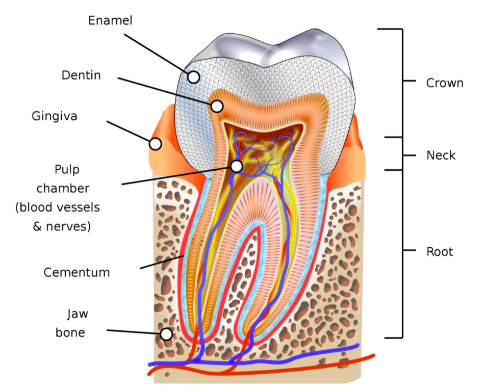
00:00 cells, and neurofibers that carry sensations away from the tooth. The tooth is embedded in the alveolar bone in a socket by the periodontal ligament. 00:07 Periodontal ligament, unlike many ligaments, is very very cellular. 00:13 In fact, it's even vascular. It has got a number of different functions. It's often called a periodontal membrane. But the name membrane or ligament doesn't really give a credit for all the functions that it does. It holds the tooth in its socket. You can see Sharpey's fibres that embed into the alveolar bone, holding that tooth in its socket. And that periodontal ligament plays a role in the eruption of teeth. It plays a role in remodeling the bone, the alveolar bone when different stresses are put on the tooth. Under certain dietary situations, it can start to break down. So you might get a wiggly tooth in its socket and the tooth might even fall out. It majors proprioception forces on the tooth. It has got an enormous range of functions. Therefore, it's not just a ligament or a membrane, at least in name and structure it is, but unlike other ligaments and membranes, it has this enormous other array of functions. On the right hand side, you can see a section taken through where the enamel and the gingiva join together. The enamel space is labelled there because, as I pointed out before, the enamel is missing, because it's highly mineralized and this is a decalcified section. The enamel is now acellular. Those ameloblasts that I spoke about earlier laying down the enamel are lost once the tooth erupts. So you can't repair enamel if it's damaged. There are no cells there to repair it. 02:08 And some bacteria can actually secrete enzymes that can dissolve that very strong calcified material and create a dental caries. But have a look at the gingiva, on the left-hand component or left-hand side of that right-hand section, on the gingiva, there is typical oral or masticatory mucosa. The gingiva is at a very small part of the oral cavity that lies against the tooth. And then on the enamel side, you've got the epithelial attachment labelled. The epithelial attachment refers to the basal lamina or the basement membrane of the epithelial membrane or the cellular epithelial surface you see there on the inner aspect of the gingiva sitting up against where the enamel would be. 03:08 That epithelial attachment, as I say, is the basement membrane of that epithelium. 03:15 And it's stuck to the enamel by very numerous numbers of hemidesmosomes. And then just above that enamel or that epithelial attachment is a little space called the gingival sulcus that sometimes gets embedded with food debris and can often cause a number of problems. So let me just summarize the sorts of structures we've looked at
About the Lecture
The lecture Supporting Structures of the Tooth by Geoffrey Meyer, PhD is from the course Gastrointestinal Histology.
Included Quiz Questions
Which of the following statements about the function of the periodontal ligament (PDL) is INCORRECT?
- The PDL attaches the enamel of the tooth to the surrounding alveolar bone.
- The PDL maintains the vitality of the surrounding cells.
- The PDL innervation involves mechanoreception.
- The PDL is a part of the periodontium.
- The PDL can influence the remodeling of bone.
Author of lecture Supporting Structures of the Tooth

Geoffrey Meyer, PhD
Customer reviews
5,0 of 5 stars
| 5 Stars |
|
5 |
| 4 Stars |
|
0 |
| 3 Stars |
|
0 |
| 2 Stars |
|
0 |
| 1 Star |
|
0 |
