Playlist
Show Playlist
Hide Playlist
Superficial Layer of Posterior Compartment – Anatomy of the Leg
-
Slides 06 Lower Limb Anatomy.pdf
-
Download Lecture Overview
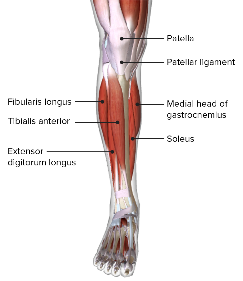
00:01 the posterior compartment are also supplied by the tibial nerve. So if we look at the anatomy of these, then we can see in greater detail the nice fleshy gastrocnemius. We can see the lateral head here. And then from the medial aspect of the lateral head, we can see plantaris. And here, we’ve got the medial head. Remember, it’s this lateral surface of the medial head, and the medial surface of the lateral head that forms the inferior boundary of the popliteal fossa. We can then look at this diagram and see the fleshy soleus and we can see the long tendon of plantaris. So the posterior compartment evolved in plantarflexion allowing you to stand on tiptoes. The superficial layer, gastrocnemius, soleus, and plantaris, are innervated by the tibial nerve and supplied by the posterior tibial artery and some fibular vessels which run within the deep layer. The two heads of gastrocnemius and soleus, together, are known as the triceps surae. This is responsible for elevating the heel, and therefore, depressing the forefoot. And this is what happens when you stand on your tiptoes. You elevate the heel and you depress the forefoot, the front part of your foot. These three muscles really, the two heads of gastrocnemius and the soleus, known as the triceps surae form the calcaneal tendon. And within the calcaneal tendon, the fibres of gastrocnemius and soleus rotate, so that gastrocnemius is actually lateral and soleus is medial within the calcaneal tendon. And this increases the elastic property of the tendon, and therefore, its ability to recoil really important features in being able to plantarflex. Plantaris is a small muscle with a long tendon. It has a very small muscle belly, and the tendon, as we can see, runs in between soleus and gastrocnemius. If we look at the deep layer of the posterior compartment, and we can see here the muscles have been reflected. So here, we can see we’ve got soleus that’s been reflected, popliteus has been reflected. Then we can see these muscles passing down here. We have tibialis posterior here in a slightly deep dissection. 02:17 Most superficially, we can see flexor digitorum longus here. And coming out from quite deep, we have flexor hallucis longus here. And these are giving their tendons that pass posterior to the medial malleolus. We can see that in this diagram here. So if we look at the deep layer, including popliteus, flexor digitorum longus, flexor hallucis longus, and tibialis posterior, they were all innervated by the tibial nerve and they’re supplied by the posterior tibial artery and fibular vessels. We can see them all lined up here, fibularis, flexor digitorum longus, tibialis posterior, and flexor hallucis longus. We can see them here. And their tendons, as we can see them passing down here, are running posterior to the medial malleolus. We can see they’re also enclosed via this flexor retinaculum, which is running from the medial malleolus to the calcaneus. We can see these tendons passing to the sole of the foot. The three long tendons or flexor digitorum longus, flexor hallucis longus, and tibialis posterior pass posterior to the medial malleolus, from medial to lateral. So here, if we have a look, we can see we have the medial aspect of the foot and the lower leg here. Here, we’ve got the lateral aspect. And if we go from medial to lateral, so if we go from medial to lateral, we see we have the tendon of tibialis posterior. 04:01 We’d see the tendon of tibialis posterior here. Then moving laterally, we have the tendon of flexor digitorum longus. And then if we move laterally again, we have flexor hallucis longus. So from medial to lateral, we have tibialis posterior, flexor digitorum longus, and flexor hallucis longus. And this is at the level of the medial malleolus. So it’s posterior to the medial malleolus. Here, we can see it at the ankle joint, we can see medial malleolus here, then we have tibialis posterior, we have flexor digitorum longus, and then we have flexor hallucis longus. And these are now passing to enter into the foot. 04:45 In this region, we also have the posterior tibial artery and the tibial nerve passing into the sole of the foot. Flexor hallucis is particularly important as it provides the final push for elevation of the foot, after plantarflexion has been initiated by the triceps surae. 05:05 So the triceps are important in elevating the heel, depressing the forefoot. But the final push off is by the great toe carried out by flexor hallucis longus. It’s a very important muscle. So in this lecture, we started off by looking at the deep fascia of the leg and the intermuscular septae in the cross-section of the leg. We then looked at the anterior compartment. We looked at the muscles like tibialis anterior, extensor hallucis longus, extensor digitorum longus, and fibularis tertius. We looked at the extensor retinacula. We then looked at their function and innervation. We then moved onto the lateral compartment, looking at fibularis longus and brevis, their relationship to the lateral malleolus and the fibular retinacula, and also their function and innervation. And then finally, we looked at the posterior compartment, the muscles in the deep and superficial layers, and the relationship of those deep muscles to the medial malleolus. We looked at the triceps surae and the formation of the calcaneal tendon. We finally, and throughout the lecture, looked at their function and innervation.
About the Lecture
The lecture Superficial Layer of Posterior Compartment – Anatomy of the Leg by James Pickering, PhD is from the course Lower Limb Anatomy [Archive].
Included Quiz Questions
Which statement describes the soleus muscle of the posterior leg?
- It is attached to the tendocalcaneus.
- It is the most superficial muscle in the leg.
- It has the tibial vessels and nerves lying between it and the gastrocnemius.
- It is an extensor at the knee joint.
- It is lateral to the gastrocnemius within the calcaneal tendon.
Which nerve supplies the popliteus muscle?
- Tibial nerve
- Popliteal nerve
- Peroneal nerve
- Common peroneal nerve
- Deep femoral nerve
Which nerve innervates the plantaris muscle?
- Tibial nerve
- Radial nerve
- Superficial fibular nerve
- Deep fibular nerve
- Superior gluteal nerve
Which muscle provides the final push-off of the foot after plantarflexion initiation?
- Flexor hallucis longus
- Triceps
- Gastrocnemius
- Popliteus
- Soleus
Which muscle is part of the lateral compartment of the leg?
- Fibularis longus
- Gastrocnemius
- Popliteus
- Soleus
- Plantaris
Customer reviews
5,0 of 5 stars
| 5 Stars |
|
1 |
| 4 Stars |
|
0 |
| 3 Stars |
|
0 |
| 2 Stars |
|
0 |
| 1 Star |
|
0 |
because i learnt alot from it which i could not easily understand while reading


