Playlist
Show Playlist
Hide Playlist
Stomach

-
Slides Digestive system espohagus and stomach.pdf
-
Reference List Histology.pdf
-
Download Lecture Overview
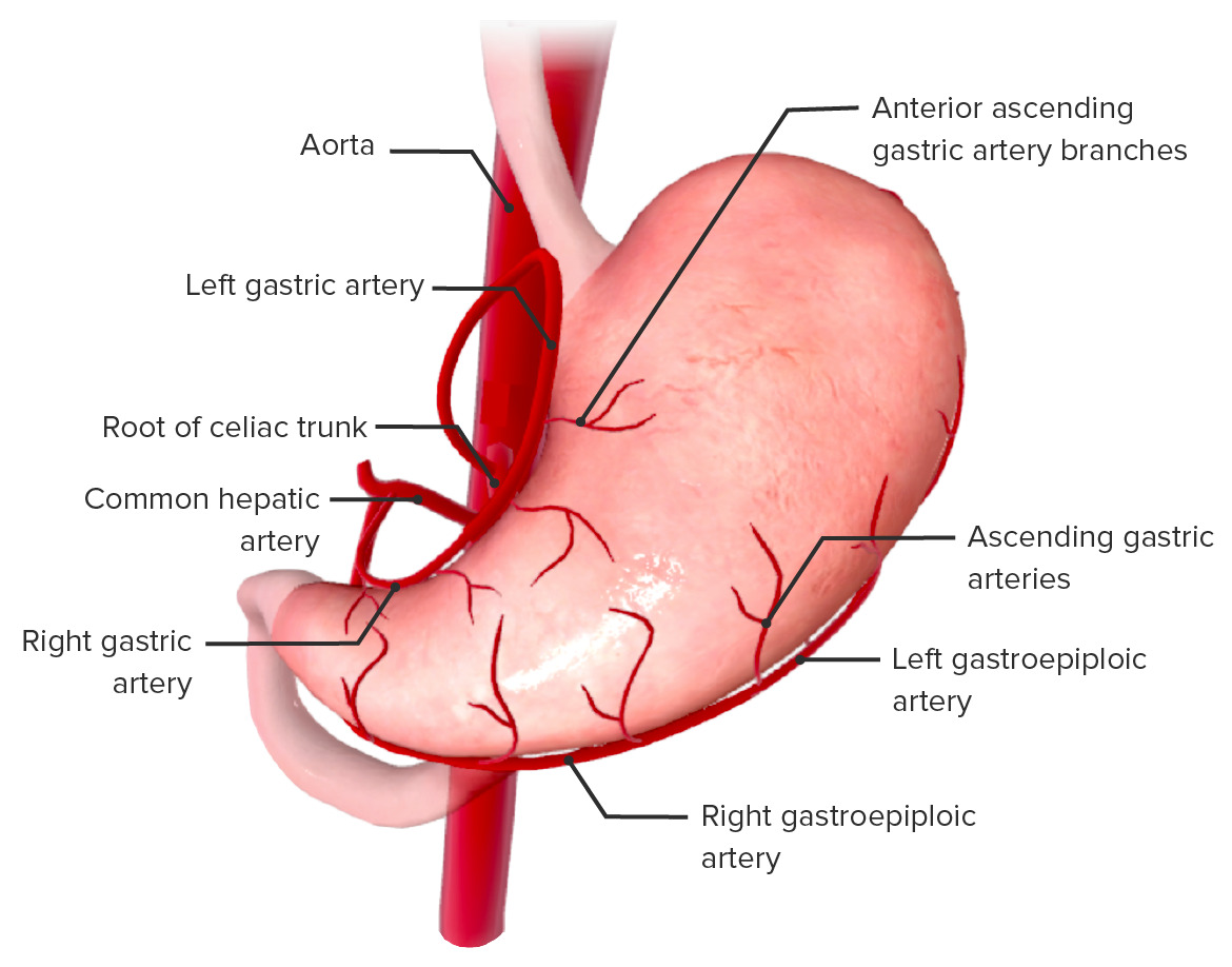
00:00 The stomach is an organ that receives food from the esophagus, and then mechanically breaks it down, but also chemically breaks it down. 00:14 There are anatomic regions that we assign to the stomach. On the left-hand side, you can see two images. 00:23 One is the stomach, whole stomach, a real section or a real image of the human stomach. In the middle of the stomach is being cut down its length to show the internal structure. And on the right-hand side is the beginnings of the histology of the stomach mucosa that I want to stress in this lecture. The cardia of the stomach is the top part, sort of towards the heart, if you like, where the esophagus enters into the stomach. And then there's the pyloric region at the end of the stomach, the end where the stomach content is going to pass into the duodenum. And at those two regions, the muscularis externa becomes very, very pronounced and forms two sphincters. These sphincters are important because they separate contents of the organs of the digestive tract from other organs, from other content in the lumen. In this case, it stops acid refluxing back into the esophagus. So the sphincter closes after we swallow, and after we complete our meal, to prevent that reflux. In the pyloric region, the sphincter is close until the chyme or the food in the stomach is being broken down sufficiently to almost be a solution. And then that sphincter will release that content to move further on in the gut, in this case, into the duodenum. 02:05 So sphincters are very important. Of course, we have another sphincter at the very end of the gut to make sure that we only eliminate digested products and waste products and the voluntary control when we receive this appropriate stimulus. Now focus your attention on the middle section, on the middle anatomical part of the stomach that has been cut open. That is the main part of the stomach. It contains mostly the most deepest gastric glands that do all the work in the stomach. And if we look in the more open door section view of the stomach, you can see it's folded into different regions. It has a number of elevations or folds called rugae. This is a collapsed stomach. Those rugae are real folds of the mucosa and submucosa, and we'll point that out in a moment on the right-hand histological section. Of course when the stomach fills with food, it will expand and open just like an open bag, and that means that those rugae will disappear. 03:21 So these rugae are just folds in a relaxed stomach that allow the stomach then to expand when it's filled with food. Move over to the right-hand side, and in the green square, you can see a histological section taken through one of these rugae. Imagine that rugae flattening out when the stomach fills. What happens will be that the submucosa supporting, or in the core of that rugae, will actually flatten out and it would be very, very hard to see. It will thin out. That explains again the importance of the submucosa allowing the mobility of that mucosa. You can just see a little small part of the muscularis mucosa being the boundary between the big folded mucosa on the surface of the rugae and the underlying submucosal core. Then look at the actual surface, the mucosa itself. It's very thick and it's very different to what you saw previously in the esophagus, which was just a simple stratified squamous wear and tear epithelium. 04:40 This mucosa, as we'll see in a moment, contains the gastric glands, and they open into the glandular tissue below by pits. If you look at the surface of the stomach that just has a lot of little holes or pits in it, those pits open into all the glandular tissue below in the mucosa that we'll see in a moment.
About the Lecture
The lecture Stomach by Geoffrey Meyer, PhD is from the course Gastrointestinal Histology.
Included Quiz Questions
Which of the following options lists 3 components of the stomach in the correct order, going from proximal to distal?
- Cardia, body, pylorus
- Pylorus, fundus, cardia
- Cardia, pylorus, fundus
- Fundus, pylorus, cardia
- Fundus, antrum, body
What is the anatomic term for the prominent folds on the luminal side of the gastric wall?
- Rugae
- Pits
- Crypts
- Plicae
- Sphincter
Which of the following is NOT correct about the anatomy of the stomach?
- The cardiac sphincter lies between the stomach and duodenum.
- The main sections include cardia, fundus, body, and pylorus.
- The largest section of the stomach is generally the body.
- The section of the stomach immediately adjacent to the esophagus is the cardia.
- The pylorus is the section of the stomach that empties contents into the duodenum.
Author of lecture Stomach

Geoffrey Meyer, PhD
Customer reviews
5,0 of 5 stars
| 5 Stars |
|
1 |
| 4 Stars |
|
0 |
| 3 Stars |
|
0 |
| 2 Stars |
|
0 |
| 1 Star |
|
0 |
Español: Fácil de aprender. Conciso y directo. Buena infografía. English: Easy to learn. Concise and direct. Good infographic.
