Playlist
Show Playlist
Hide Playlist
Small Intestine: Digestion and Absorption Processes

-
Slides 15 Human Organ Systems Meyer.pdf
-
Reference List Histology.pdf
-
Download Lecture Overview
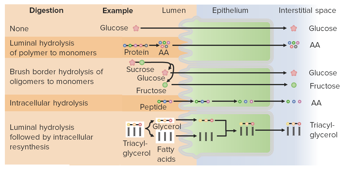
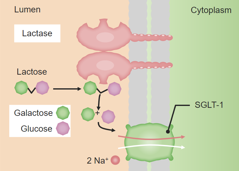
00:01 On the right-hand side of this slide is an electron micrograph taken of this brush border. It consists of numerous microvilli. A microvillus is labelled. Each microvillus has a central core of actin filaments. And those actin filaments interact with other actin filaments running parallel to the apex of the cell. 00:27 There's also myosin filaments running along the apex of the cell. And when this contracts, because it's intermingled with the actin filaments, it causes the microvilli to slightly spread apart. And this allows a greater surface area of the microvilli for absorption. 00:50 Their main role is absorption, and that's the main role of these enterocytes in the small intestine. The diagram on the left-hand side isn't meant to be too detailed and too descriptive for a histology course. So don't panic about the content. The reason why I've put it here is to just emphasize how important these microvilli are in the absorption of proteins, and also later on, the absorption of lipids. If you look at the diagram, you can see it explains the breakdown of proteins in the lumen of the intestine by pancreatic enzymes. 01:31 And those proteins, when broken down, are then taken into the surface of the microvilli by attaching to a certain carrier of proteins and other structures. And then they get internalized into the cell, at the apex of the cell, and they move through the cell to where they finally dealt with inside the cell cytoplasm. So again, let me stress not to worry about the details of the physiology described here, but it's just to emphasize how important these microvilli are in the absorption of proteins from the lumen of intestine. 02:12 On this slide, you can see an illustration on the right-hand side of how important these microvilli are in absorbing lipids. This description of the process is labelled one to five of this absorption process, and the numbers coincide with various stages through the cell on the right-hand side. Again, I don't want you to get too involved with the details here. 02:42 It's physiology. But I just want to stress how important again these microvilli are. 02:50 The lipids are broken down in the lumen of the intestine by bile salts, and also pancreatic lipase. They are then diffused towards the microvilli where they're taken in through the microvilli. You can see them as little yellow representations in this diagram. 03:14 And they get internalized also in the apex of the cell. And that's labelled 2 on the diagram. 03:21 They then move towards the Golgi apparatus where they're turned in or transformed into chylomicrons. 03:32 And also in the Golgi apparatus, again labelled 3 here, they're coated with a membrane. 03:39 And then they can diffuse to the basolateral surfaces of the enterocytes. And the membrane that binds these chylomicrons and the content then fuses with those lateral borders of the cell, and so the chylomicron is released into the intercellular space. They can't leak back into the lumen again because of the junctional complexes towards the apex of the cell, as explained in a lecture on epithelia that I gave in this histology course. 04:15 So, the only place for them to go is to go into the basolateral component and then diffuse into the interstitium behind the cells where the capillaries are and where the lacteals are. 04:30 Down the bottom of the diagram, you can see a lacteal in the villus of the duodenum. 04:39 These lacteals are blind-ended vessels that absorb all the excessive interstitial fluid, and they eventually form a hall network called lymphatic vessels. And they finally drain back into the venous system up towards the neck and shoulder region. This is where these chylomicrons and where lipids make their way to be absorbed back into the body. They go into these lymphatic vessels, these lacteals, and find their way into the vascular system through this way. Here again you see diagram on the left, and on the right, it just illustrates again these microvilli, but also the junctional complexes. 05:30 Hard to see in the electron micrograph but they're at the apex of the cell. So, all the movement of these digested lipids passes down the lateral borders as I described in the previous slide. This slide shows again the diagram illustrate the absorption of lipids across the enterocyte on the left-hand side. But it also shows in the right-hand side a light microscope picture taken through a villus. You can see the brush or striated border on the enterocytes, and you can see a lacteal shown in the middle of the villus, in the lamina propria. These lacteals are blind-ended tubes that collect all the interstitial fluid or excessive interstitial fluid and return that fluid along lymphatic channels, lymphatic vessels to finally deposit that content, that lymph into veins up in the shoulder or neck region. These lymphatic vessels, therefore, transport the lipid that's absorbed across the microvilli into the cell and then pass out of the cell in the basolateral borders, and then find their way through the interstitium into these lacteals or lymphatic vessels. That's how lipid is absorbed and returned back into the vascular system.
About the Lecture
The lecture Small Intestine: Digestion and Absorption Processes by Geoffrey Meyer, PhD is from the course Gastrointestinal Histology.
Included Quiz Questions
The intestinal brush border is mainly composed of which of the following?
- Microvilli
- Stereovilli
- Cilia
- Flagella
- Gut-associated lymphoid tissue
Enzymes from which of the following organs is mainly responsible for the breakdown of protein in the small intestine?
- Exocrine pancreas
- Gallbladder
- Liver
- Endocrine pancreas
- Thyroid
Bile primarily contributes to the digestion of which of the following?
- Lipids
- Proteins
- Carbohydrates
- Vitamin C
- Vitamin B12
Which of the following is a lymphatic capillary that absorbs dietary fats in the villi of the small intestine?
- Lacteal
- Stereocillia
- Lingula
- Sinusoid
- Thoracic duct
Which of the following is the primary form of lipid transport from the intestines to other locations in the body?
- Chylomicrons
- Low-density lipoprotein
- High-density lipoprotein
- Phospholipids
- Low-density lipoprotein and high-density lipoprotein
Author of lecture Small Intestine: Digestion and Absorption Processes

Geoffrey Meyer, PhD
Customer reviews
5,0 of 5 stars
| 5 Stars |
|
1 |
| 4 Stars |
|
0 |
| 3 Stars |
|
0 |
| 2 Stars |
|
0 |
| 1 Star |
|
0 |
A Well-done explanation of Lipid Absorption ! Thank You, Dr. Meyer !
