Playlist
Show Playlist
Hide Playlist
Second-degree AV Blocks

-
Slides Heart Blocks.pdf
-
Reference List Electrocardiogram (ECG) Interpretation.pdf
-
Download Lecture Overview
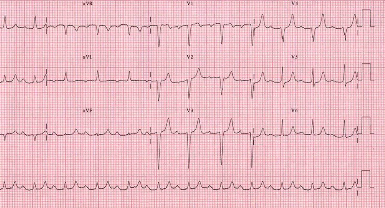
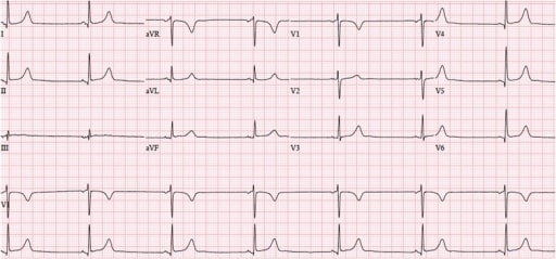
00:01 Let's move on and talk about second-degree AV block. 00:03 Second-degree AV block is more serious in that there are dropped beats. 00:08 There are two types of second-degree AV block. There's type I and type II. 00:13 Type I is often called Wenckebach block or Mobitz type I AV block. 00:18 And you'll see there's a Mobitz type II AV block, both of these are second-degree AV block. 00:23 And Wenckebach and Mobitz were people - physicians who studied electrical activity in the early days of the electrocardiogram. 00:31 What happens with type I second-degree AV block or Wenckebach or Mobitz one, it's characterized by a progressive increase in the PR interval and eventually there's a missing QRS. 00:45 The interval gets so long that the impulse doesn't get through the ventricle and you drop a beat and the sequence repeats itself. 00:54 Now usually it's 3 to 5 beats and a dropped beat. 00:57 For example; three beats and the fourth beat is missing and again, three beats and the fourth beat is missing and so forth. 01:02 Could be up to five beats and the sixth one is missing and you'll see increasing PR interval's, I'm gonna show you that. And of course, the last P-wave is not followed by a QRS. 01:12 So, type I second-degree AV block occurs when the impulse travels down from the sinus node through the atrium and arrives at the AV node. 01:23 And there's a pause before it gets through into the main His-Purkinje system and enter the ventricle, and of course the left bundle and the right bundle, and the Purkinje fibers and the ventricle. 01:36 So, the block occurs higher up in the AV node and therefore the impulse doesn't get through to the ventricles, to the right and left bundle and so forth. 01:45 So, the block of the electrical transmission occurs just above or in the upper portion of the AV node. 01:52 And you can see the little green box showing where that is. 01:58 Now, here's an example; notice, this is a Wenckebach block. 02:02 Three impulses get through with lengthening PR interval and the fourth one is blocked. 02:08 Let's take it one sequence at a time. 02:12 The first the one with the first green box shows you a normal P-wave with a normal PR interval. 02:18 Then there's a QRS and a T. 02:20 The next P is normal-looking, but look, the PR interval has lengthened; the second green box followed by a QRS and T. 02:28 Now look at the third. Oh, the PR interval is even longer QRS and then T, then comes a P-wave. 02:35 Uh-oh, we're missing the QRS and then eventually it resets itself and starts again. 02:41 So, this is 3 to 4, that is three impulses get through to four atrial impulses. 02:50 And you can see the increasing PR intervals. 02:53 So, this is type I second-degree AV block and as it says here, the fourth PR interval is too prolonged to capture the ventricle and therefore it's missing. 03:02 And the next P-wave starts the whole sequence over again. 03:07 Now, here is another example of it and if you stand back and look at this from a distance, you see what's known as group beating. Notice, look at the bottom line or at the top line. 03:23 But the bottom line shows you the P wave nicely. You see four beats and a dropped beat. 03:28 Four beats and a dropped beat and so forth. 03:32 And so, when you hold this out at arm’s length, you see this groups with pauses. 03:36 Groups, abnormal beats and a pause, groups, abnormal beats and a pause. 03:40 And here of course we show you where the missing QRS is. 03:44 The second type of type II AV block is a serious type. 03:49 It's also called high-grade AV block or Mobitz type II, AV block. 03:55 It is lower down in the conduction system, not usually below the AV node. 04:03 And it consists of P-wave that are repeatedly not followed by a QRS telling you that there's higher grade block below the AV node. 04:14 It can be 3:1 in which you have three normal beats followed by a blocked beat after the fourth P-wave. 04:23 Or it could be a single blocked beat that is repeated multiple times. 04:32 And the PR intervals of course on the normal beats are normal and then all of a sudden, there's a P-wave with no QRS and that's an example where the fourth beat was blocked, so you had a P-wave with no QRS. 04:46 With 2:1 AV block, it may be difficult to tell whether you're seeing type I or type II. 04:52 And there the electrophysiologist can make measurements often in the electrophysiology laboratory and if it's type II AV block, they often follow that with pacemaker insertion. 05:04 So, let's look at an example. Here is an example of type II second-degree AV block. 05:10 You'll notice the first complex PQRS and T: normal. Second complex, PQRS and T: normal. 05:18 Third complex, PQRS and T: fourth P-wave -- no QRS. And then it takes over normally again; PQRS and T.0 So, this could have been a very - a 2:1 type I block, but it's very worrisome and we would have to make sure that it's not a type II. Why? Because this kind rapidly goes into complete heart block, 3rd degree heart block that could be life-threatening. 05:48 So, it - the P-wave is completely blocked in or below the AV node. 05:54 It can be frequent with many dropped beats and usually is. 05:57 So, when you get a long rhythm strip, you see what's going on. 06:00 And again, it implies serious disease in the conduction system, usually requiring pacemaker therapy. 06:07 Here's another example. You'll notice here, we see it twice. 06:11 There's a normal beat, the first beat is a PQRS and T. 06:16 Then there's a P-wave with the green arrow, no QRS. 06:20 Then we have a normal beat then another P-wave; green arrow, no QRS. 06:25 So, here's two complexes that are lacking conduction to the ventricle. 06:29 And when you take a longer rhythm strip and you see multiples of these, the patient gets almost always a pacemaker because they're very close to 3rd degree heart block. 06:39 Just a little information about it. 06:43 Type I AV block - second-degree AV block can occur in normal, healthy individuals. 06:48 It's common in well trained athletes and in young people. 06:52 It's due to increased vagus nerve activity which goes with training and usually it's benign. 06:58 And when I'm at the University, every year I see some of our super athletes who are training for the Olympics or for a national competition? And they're sent over, "Oh, this person has an irregular heartbeat." And often they have type I second-degree AV block and sinus bradycardia because of the training effect. 07:14 And pacing is definitely not needed. There - we reassure them, this is normal. 07:19 You put a patient like that on the treadmill, their heart rate accelerates normally, so it's not really disease, it's a training effect. 07:26 Now, type II second-degree AV block is the nasty one. 07:30 The block of electrical transmission occurs just below or in a lower portion of the AV node. 07:36 Here, we see the system again. 07:38 So again, both type II's are occurring in and around the AV node but type I in the upper portion or just above. Type II, in the lower portion or just below. 07:48 And type II is more serious because it often can lead on to 3rd degree or complete heart block as we'll talk about. 07:54 And of course, you see the impulse once it gets through the His-Purkinje system, it gets transmitted throughout the ventricle through the left and right bundles and into the ventricular muscle.
About the Lecture
The lecture Second-degree AV Blocks by Joseph Alpert, MD is from the course Electrocardiogram (ECG) Interpretation. It contains the following chapters:
- Type 1 Second Degree AV-Block
- Typ 2 Second Degree AV-Block
Included Quiz Questions
What best describes a second-degree Mobitz type I block (Wenckebach Phenomenon)?
- Progressive lengthening of the PR interval until a beat is dropped
- Consistently prolonged PR interval (> 200 ms)
- Dropped beats that are not preceded by a change in the length of the PR interval
- P waves and QRS complexes that are not rhythmically associated
- Abnormally fast accessory conduction pathway from the atria to ventricles that bypasses the AV node
Where is the block of the electrical transmission in second-degree Mobitz 1 AV block?
- In the upper portion of the AV node
- In the upper portion of the SA node
- In the Purkinje fibers
- In the bundle of His
- In the bundle of Kent
Which of the following best describes a second-degree Mobitz type 2 AV block?
- Dropped beats that are not preceded by a change in the length of the PR interval
- Progressive lengthening of the PR interval until a beat is dropped
- Consistently prolonged PR interval (> 200 ms)
- P waves and QRS complexes that are not rhythmically associated
- Abnormally fast accessory conduction pathway from the atria to ventricles that bypasses the AV node
Which of the following is true regarding a Mobitz type 1 AV block?
- It is characterized by progressive PR lengthening, then dropped QRS.
- It is characterized by a progressive increase of a QR interval.
- It typically requires pacemaker therapy.
- It can rapidly progress to third-degree heart block.
- It often leads to complete dissociation between the atria and ventricles.
Author of lecture Second-degree AV Blocks

Joseph Alpert, MD
Customer reviews
4,0 of 5 stars
| 5 Stars |
|
1 |
| 4 Stars |
|
0 |
| 3 Stars |
|
1 |
| 2 Stars |
|
0 |
| 1 Star |
|
0 |
Good lecture overall. Downloadable slide materials do not contain all slides and need to be updated (I note this issue was raised by another commenter 3 years ago and responded to indicating it was fixed at that time although the issue persists currently), a few mis-spoken moments need either an audio re-record or an on-screen note (3:1 type 1 2nd degree block first described as 3:4, then as 2:1)
it was very very very excellent and easy to understand
