Playlist
Show Playlist
Hide Playlist
Saggital Section of the Eye – Anatomy Review
-
Slides Structures of the eye.pdf
-
Reference List Pathology.pdf
-
Download Lecture Overview
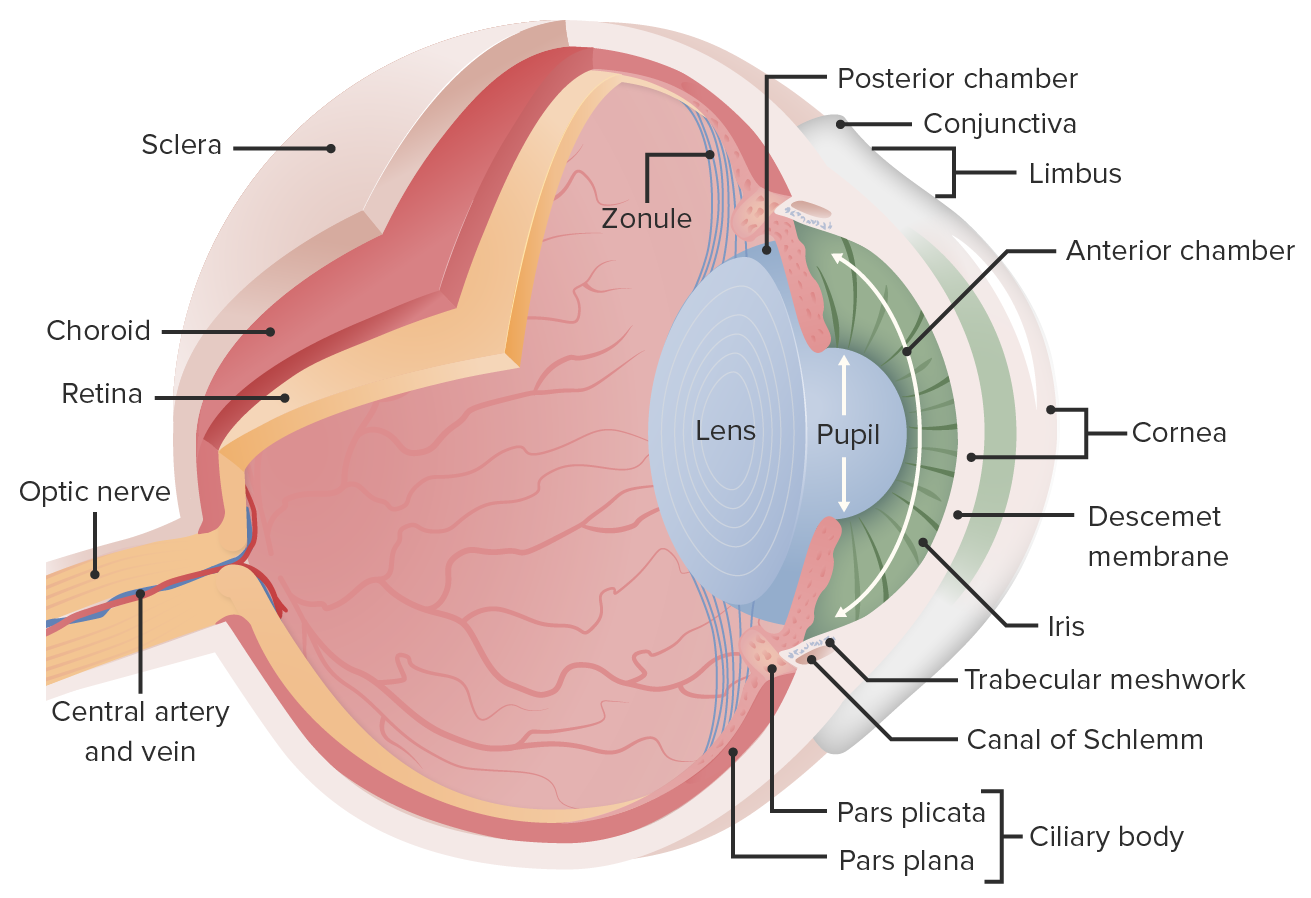
00:01 Okay, many, many, many structures that we're going to talk about here. 00:05 And we will revisit these in some detail as we go along. 00:09 This is a lateral cut through the eye. 00:11 And I just want you to - have you get a sense of all the different structures. 00:14 So, on the right-hand side, starting from the top is the levator palpebrae. 00:20 So, it's going to superioris and that's going to be responsible for opening the eyelids, not for squeezing the eyelids, remember, that's the orbicularis muscle, that's going to squeeze them close. But to keep them open, hopefully, they're keeping them open now as you're watching this, is the levator palpebrae superioris. 00:42 Okay, next one down is the sclera. The sclera is the white of the eye and it's all the way around the eye, and we've talked about it. 00:48 It is dense connective tissue. We'll see images of it near the end of this session. 00:52 And it allows us to have insertion of all the various muscles around the eye, the extraocular muscles. 01:00 The next one down is the bulbar conjunctiva. 01:03 So remember, conjunctiva is a highly vascularized membrane that sits not only on the inside of the eyelid, that's the palpebral conjunctiva. 01:14 But also over the front of the eye, on the white of the eye of the sclera will not be over in front of the iris and lens, but will extend down to that level. 01:28 That conjunctiva is going to be important for keeping the front of the eye reasonably well lubricated. 01:36 Again, it's also being helped by products from the lacrimal gland. 01:41 Next one down is the iris, we'll see more of the histology of the iris in the middle in a bit. 01:48 But the iris is the colored portion of the eye. 01:50 Next is going to be the lens, and you can see how it sits and there's zonular fibers that connect the lens up to the ciliary body, that is going to be continuous with our choroid, and we'll talk more about that. 02:09 In front of the lens is the anterior chamber. 02:13 So that's the anterior chamber of the eye, kind of a general area between the cornea and the lens. 02:19 And then, there is a much smaller posterior chamber that sits between the iris and the lens. 02:28 So, the posterior chambers pretty small. The anterior chambers is, yeah, so much. 02:32 We're going to talk about also there being a vitreous chamber in a bit. 02:36 Okay, so that's going from top to bottom on the right-hand side. 02:41 On the left-hand side, we're going to look at some of the other structures. 02:44 So from the top, the orbicularis oculi muscle, that's the palpebral part. 02:49 So that's the part that's going to be over the eyelid, eyelid or the palpebrae and the slip between your eyelids, that's the palpebral fissure. We've talked about that. 02:58 Underneath that it's the superior tarsus, the tarsus is just connective tissue for the most part that provides integrity, strength, and a kind of a framework upon which the orbicularis oculi muscles sit. 03:14 But it does have within it some meibomian glands, the tarsal or meibomian glands, and these become important for things like blepharitis and for chalazions, and we'll talk about that. 03:29 There's sebaceous glands that sit and will provide sebaceous fluid lubrication to keep your eyelashes, the cilia, the eyelashes nice and supple. 03:40 Those sebaceous glands are Moll's glands is one of them. 03:45 They can become infected. They can become obstructed. 03:48 And they will cause things like hordeolums or styes. 03:53 Okay, and finally, there is the cornea. So, the cornea sits on the front of the eye, it is a clear structure, we'll describe it more in detail. 04:06 It forms the most anterior portion of the anterior chamber. 04:11 Okay, so chambers of the eye, general structures again, just make sure we're all on the same page. 04:18 So the anterior chamber of the eye is everything that's in front of the lens, in front of the iris and behind the cornea. Got it? Okay. 04:26 Posterior chamber really small. It's between kind of the ciliary body and our iris. 04:33 Okay, so that's the posterior chamber. 04:35 And then, there's this whole vitreous chamber. 04:38 Okay, so that's not the posterior chamber. It's the vitreous chamber. 04:42 Posterior chamber is more specifically, what's highlighted in green. 04:45 And then, we can also be a little bit more broad, we can talk about the anterior segment of the eye and the posterior segment of the eye. 04:54 And that's more or less at the midpoint of the lens. 04:57 So everything that's kind of mid-lens and forward is the anterior segment, everything that's midpoint of the lens and back, posterior segment. 05:05 Okay, so that gives us all the vocabulary so that we can talk about the various pathologies in subsequent sessions. 05:14 And with that, we'll close.
About the Lecture
The lecture Saggital Section of the Eye – Anatomy Review by Richard Mitchell, MD, PhD is from the course Introduction to Ophthalmology.
Included Quiz Questions
What muscle helps in opening the eyelids?
- Levator palpebrae superioris
- Orbicularis oculi
- Superior oblique
- Inferior rectus
- Medial rectus
What is the function of the superior tarsus?
- Provides support to the upper eyelid
- Elevates the upper eyelid
- Supports the lacrimal gland
- Houses the lacrimal caruncle
- Provides lubrication to the eye
What is the location of the posterior chamber?
- Between the iris and the lens
- In front of the lens
- Between the lens and the retina
- Between the cornea and the midpoint of the lens
- In the posterior segment of the eye
Customer reviews
5,0 of 5 stars
| 5 Stars |
|
5 |
| 4 Stars |
|
0 |
| 3 Stars |
|
0 |
| 2 Stars |
|
0 |
| 1 Star |
|
0 |


