Playlist
Show Playlist
Hide Playlist
Ribosome & Endoplasmic Reticulum

-
Basic Histology 02.pdf
-
Reference List Histology.pdf
-
Download Lecture Overview
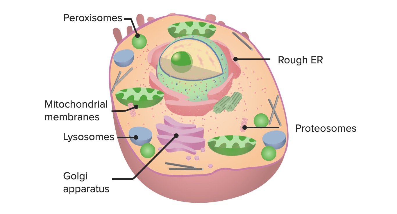
00:01 Ribosomes are protein complexes and they consist of RNA synthesized in the nucleolus. 00:12 Remember, the nucleolus produces ribosomal RNA that passes out through the nuclear pore into the cytoplasm of the cell, and these ribosomes are in two different forms. 00:27 Firstly, they could be loosely lying within the cytoplasm. Sometimes, they're combined with a messenger RNA fiber to produce a chain of ribosomes and they're called polyribosomes. 00:44 When you see individual ribosomes which are about 20 nm in diameter or you see polyribosomes in the cytoplasm of the cell freely associated, that means they're involved with making proteins that the cell uses for itself, proteins that are manufactured for usage by the cell whereas sometimes, ribosomes become attached to the endoplasmic reticulum, which I'll speak about in a moment. 01:24 When you see ribosomes attached to the membrane of the endoplasmic reticulum, that means that the proteins synthesized by those ribosomes in collaboration with the endoplasmic reticulum is making protein designed to be exported from the cell so there's quite a difference. 01:47 Loosely bound, they produce proteins for use in the cell. 01:52 Associated with the endoplasmic reticulum, they're producing proteins to export from the cell. 02:00 And if you look very carefully at this electron micrograph, what you see there are very clear channels or cisternae or passageways which have a homogeneous grayish stain to them and then you see these studded structures. They're ribosomes and they happen to be, in this case, associated with the membranes of the endoplasmic reticulum. 02:28 A bit hard to understand in this slide until you've had a bit more information about protein secretion further on in the course. 02:40 Now here's a description of the endoplasmic reticulum. 02:44 You can see on the image that there are really two different forms of the endoplasmic reticulum. 02:52 The endoplasmic reticulum, it's a vast network throughout the cell. 02:58 It consists of sacs, cisterns or passageways interconnecting tubules, and there are two forms. 03:10 Firstly, one particular form is called rough endoplasmic reticulum or granular endoplasmic reticulum, and it's called rough or granular endoplasmic reticulum because as you see in the diagram, it has little tiny ribosomes associated with it, and in that case, that part of the endoplasmic reticulum, the rough endoplasmic reticulum with associated ribosomes is making protein for export, whereas the smooth endoplasmic reticulum which is more on the peripheral part of the image shown here has no ribosomes associated with it at all. 03:58 Its job is to make lipids, steroids, metabolize glycogen and also degrade toxic substances, so it's a very busy membranous system as is the rough endoplasmic reticulum. 04:15 Now, if you look at cells like a plasma cell, making antibodies proteins or hepatocytes making blood proteins or salivary glands making protein digestive enzymes or fibroblasts making protein connective tissue fibers or other cells making enormous amounts of protein, then when you look at the ultrastructure of those cells, it's going to be dominated by rough or granular endoplasmic reticulum, whereas in cells that make lots of steroid, for instance, the testes in the male, the ovary in the female, the adrenal cortex, then you're going to have lots of smooth or agranular endoplasmic reticulum. 05:15 In the hepatocyte, you're going to have both because the hepatocytes not only makes lots of blood proteins and therefore requires lots of rough endoplasmic reticulum, it also detoxifies or degrades substances so it needs lots of smooth endoplasmic reticulum. 05:38 Here is a picture showing you again some large motor neurons. 05:44 You can see the nucleus and the nucleolus in most of these, and you see a lot dark-stained material inside the cytoplasm, perhaps more so in this image here. 05:57 All that clumpy dark purplish-stained material in this large motor neuron is rough endoplasmic reticulum. 06:08 In fact, it almost blocks out the view of the nucleus and cytoplasm in this particular image, and that large amount of rough endoplasmic reticulum in this neuron is making neurotransmitter proteins that serve to bring about the transmission of the impulse from the neuron on through skeletal muscle. 06:32 And when we see these large clumps of rough endoplasmic reticulum or granular endoplasmic reticulum in these large motor neurons, we refer to those as Nissl bodies or Nissl substance. 06:48 And here is a couple of electron micrographs, perhaps focused more on the one on the right-hand side, and you can just make out plates or channels of the endoplasmic reticulum particularly on the left and the right-hand side of the nucleus in the middle.
About the Lecture
The lecture Ribosome & Endoplasmic Reticulum by Geoffrey Meyer, PhD is from the course The Mammalian Cell. It contains the following chapters:
- Ribosome
- Endoplasmic Reticulum (ER)
Included Quiz Questions
Which ONE of the following statements about ribosomes is INCORRECT?
- Ribosomes link nucleic acids together.
- They may be attached to the rough endoplasmic reticulum.
- They may occur singly in the cytoplasm.
- They are about 20 nanometers in diameter.
- They may be found as groups within the cytoplasm.
What is the primary function of the agranular (smooth) endoplasmic reticulum?
- Synthesis of steroids and lipids
- Protein synthesis
- Enzyme degradation
- Formation of phagolysosomes
- Synthesis of adenosine triphosphate
Author of lecture Ribosome & Endoplasmic Reticulum

Geoffrey Meyer, PhD
Customer reviews
4,0 of 5 stars
| 5 Stars |
|
1 |
| 4 Stars |
|
0 |
| 3 Stars |
|
1 |
| 2 Stars |
|
0 |
| 1 Star |
|
0 |
it was very informative,thank you so much proff.big up
The flow of the video is fairly slow, however, it's pretty detailed and informative.
