Playlist
Show Playlist
Hide Playlist
Rectum and Anal Canal

-
Slides 15 Human Organ Systems Meyer.pdf
-
Reference List Histology.pdf
-
Download Lecture Overview
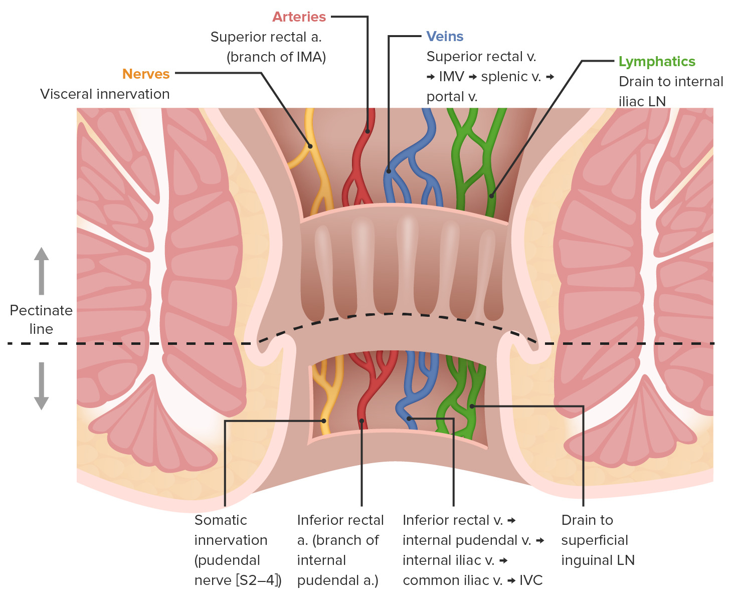
00:01 Finally, let's have a look at the rectum and anal canal briefly. Here is a diagram explaining the structure, the anatomical structure of the anorectal canal. And I briefly just want to touch on a couple of features. 00:21 Those of you who studied anatomy of this region will know a lot more details. 00:26 The mucosa in the anal canal is folded into longitudinal directed folds called columns. 00:38 And between these columns, there are joints, valves. And between the columns are structures called sinuses. 00:51 These longitudinal anal columns and the sinuses prevent leakage from the rectum into the anal canal and beyond. 01:02 There's lots of mucus-secreting glands there. 01:05 And when the anal canal distends when passing the feces, those mucus glands secrete all the mucus to help lubricate the elimination of these feces. And the epithelium changes. 01:23 Up in the rectal region, there is typical colon large intestine epithelium glands dominated by goblet cells. 01:35 And as we move from the rectum to the anorectal junction, those goblet cells and those glands are going to start to diminish until finally, in the anal canal and then beyond, the epithelium will change from the colon type epithelium to a stratified squamous epithelium. 01:56 And then it blends with the perianal skin. 02:00 There's also, on this slide, an illustration showing you the internal hemorrhoidal plexus. 02:08 This internal hemorrhoidal plexus creates problems sometimes with people. Look now at the slide. 02:20 This shows you the transition of the epithelium of the colon and rectum on the left hand side, towards on the right hand side, a diminishing of these glands and an abrupt change to be stratified squamous epithelium on the far right hand side of the right hand section. 02:40 On the left hand section, you can see evidence of these internal hemorrhoidal plexi. 02:48 You can see blood vessels containing blood cells. It's not a good section. 02:53 It has been shuttered during the cutting process, and so you see some evidence of artifact here. 03:00 But then in histology, not every section is perfect as we would like. 03:05 And so I think it's important that you often get used to examining sections that just not as perfect as perhaps one would like them to be. 03:14 In summary then, the small intestine is designed to chemically digest food and then absorb it. 03:24 There are ways in which the small intestine is designed so that the surface area is increased for that absorption. 03:32 The small intestine also has secretory cells. Mainly, paneth cells secreting antimicrobial substances, and those endocrine cells, the argentaffin cells that secrete endocrine products that are going to affect nearby cells, but also cells in other locations. And the large intestine contains those crypts which are dominated by cells that really are involved in watery absorption, but also, they have goblet cells to help lubricate the feces. 04:07 The large intestine or colon is also a storage organ for undigested products, and they're excreted as feces. 04:17 And also, remember that there are components along the gut wall, particularly in the mucosa that are designed to detect any foreign antigens and pathogens, and then alert the immune system about this. 04:35 So I hope you now understand a lot of the histology of the small intestine and the large intestine. 04:44 And thank you very much for listening to this lecture.
About the Lecture
The lecture Rectum and Anal Canal by Geoffrey Meyer, PhD is from the course Gastrointestinal Histology.
Included Quiz Questions
The upper and lower segments of the anal canal are separated by which of the following?
- Pectinate line
- Taenia coli
- Rugae
- Lacteal
- Linea alba
What is the function of goblet cells in the rectum?
- Mucus secretion
- Water absorption
- Dilution of faeces
- Antibody production
- Physical barrier
What is the hemorrhoidal plexus?
- A group of veins that surrounds the rectum
- A group of veins that surrounds the anus
- A group of nerves that surrounds the rectum
- A group of nerves that surrounds the anus
- A group of arteries that surrounds the rectum
Author of lecture Rectum and Anal Canal

Geoffrey Meyer, PhD
Customer reviews
5,0 of 5 stars
| 5 Stars |
|
3 |
| 4 Stars |
|
0 |
| 3 Stars |
|
0 |
| 2 Stars |
|
0 |
| 1 Star |
|
0 |
Clear, concise and good lecture. The professor is very easy to follow, and does a good job emphasizing what he thinks important.
Thank you for these organized, concise, and well-prepared lectures, Dr. Meyer !!!
Very good lecture. Clear presentation, properly indicated so very easy to follow.
